
|
INFECTIOUS DISEASES FEVER
Fever may be present when a local disease assumes a constitutional character or when the constitutional character is manifested from the beginning of the disease. Fever may be a systemic disorder or a symptom of disease, and is characterized by an increase of body temperature. Other symptoms are usually present, as an accelerated pulse, disturbances of distribution of the blood, increased katabolism, and disordered secretions. Etiology.In infectious diseases fever is due chiefly to the action of various toxic or harmful agents, produced by the disease, upon the fluids of the body and upon the nervous system. Disturbances of the thermogenic centers and nerves of the brain or cord by harmful agents, or by lesions of the anatomical structures affecting these nerves, are sources of fever. Also disturbances of the vaso-motor centers (in the medulla and auxiliary centers along the cord) and nerves are causes of fever in many instances. A disturbed or lessened function of the nerves controlling sweating is an important factor. The multiplication of micro-organisms in the body, acting directly on the tissues or by producing toxic substances which affect the nervous system, is a fruitful source of fever. A few cases may be caused by direct affection of the nervous system, as is shown by appearance of fever in epileptic attacks, or by the passage of a catheter into the bladder. In a large number of all cases a demonstrable cause can be found upon careful examination, whether the fever be due to a necrosed mass of tissue, the introduction into the system of decomposed food, infectious diseases, a lesion of some anatomical structure affecting a thermogenic, vaso-motor or sweat center, a lesion to the innervation to the heart (vagi and cervical sympathetic) causing a rapid heart, or a lesion to the sympathetic system. Treatment.The treatment of fevers in a general way consists principally of thorough inhibition to the posterior spinal nerves of the upper cervical region in order that the center of the vaso-motor system in the medulla may be effected, probably by the way of the superior cervical ganglion of the sympathetic. Thus the entire vascular system is equalized, for there is always a disturbance in the distribution of the blood in fever and if the center controlling the nerves that govern the lumen of the blood-vessels can be brought under control, there will result an equalization of the vascular system; if such occurs, health must ensue. Besides the vaso-motor nerves to the blood-vessels being effected by this treatment, the nerves governing the lymphatics and the sweat glands will also be controlled. The sweat glands as a rule are rendered active by effecting directly the innervation of the glands, also the glands are controlled indirectly by the blood supply; this aids materially in lessening the temperature of the body. Treatment for a few minutes to the upper posterior cervical region would also effect the thermogenic centers and nerves of the brain reflexly in the same manner as the vaso-motor and sweat centers and nerves are effected, thus tending to equalize the mechanism of the thermogenic system. Besides this action on the vaso-motor, sweat, and the thermogenic nerves, there is produced an increased exhalation of moisture nerves, there is produced an increased exhalation of moisture from the lungs, on account of an increase of vascular area in the lungs through vaso-motor action. Also the large vascular area in the abdomen, under control of the splanchnic nerves, becomes constricted. Thus there is brought about a lessening temperature by evaporation, heat radiation, and perspiration; and an increased action of the general nervous system, a stronger cardiac force, an equalization of the vascular system, and a more perfect elimination of toxic properties by the skin, kidneys and lungs; consequently a reduction of the fever. The foregoing treatment is successful to a limited extent, only in such cases where causative factors of the fever are involving the predominating centers controlling the heat production or dispersion and the vaso-motor system directly; for if the lesion that is causing the disorder should be affecting an auxiliary center along the spinal cord instead of the predominating center, as is oftentimes the case, treatment of the predominating center would be useless as far as any permanent benefit is considered; although a temporary effect will be gained by lessening the fever at that point. Consequently, in many cases, the lesion lies within the jurisdiction of auxiliary centers which are situated at various points along the spinal cord. When such is the case, it will be of little benefit to give the cervical treatment. In such instances the lesion to the auxiliary center would have to be removed in order to cure. One cannot depend upon a set rule to reduce a fever; determine the cause, as in any other disease or symptom, and remove it. In addition to the treatment to the cervical region and along the spinal column, as are indicated upon an examination, attention should be given to the hearts action. The equilibrium between the accelerator and inhibitory nerves (cervical sympathetic and vagi) should be maintained. The interchange of gases in the lungs should be rendered as nearly normal as possible; this is best accomplished by raising and spreading of the ribs from the second to the seventh dorsals, particularly in the region of the fifth and sixth. Also stimulation of the vagi will aid by increasing the motor power of the lungs. The kidneys and bowels should be kept active so as to favor a rapid elimination of various toxic properties; besides they have control over large vascular areas. Treatment over the ureters will prevent any clogging that might occur in them from a condensation of the urine. Attention, also, should be given the tissues at the fifth lumbar and over the iliac vessels to influence the circulation in the pelvis. The food of the patient should be liquidmilk, soup, broths, etc., and most any quantity of water allowed if called for, given little at a time and at frequent intervals. The room should be well lighted, ventilated, clean and kept at an even temperature. Two points should always be remembered relative to fever: First, that there are many causes of fever; and in order to reduce the fever the cause must be determined and removed, the same as in any disorder. A definite fever treatment cannot be given any more than a definite constipation treatment; the case must be seen in order to determine the cause. SecondThe reduction of fever is not necessary; the fever should be treated only as a symptom of disease when it exists as such. In fact, fever is beneficial, for it is one of natures methods to relieve an over-burdened system from harmful agents, unless the temperature is excessive and continuous and is likely to cause more harm than the primary trouble. Hydrotherapy is of immense value in reducing a fever. It is an agent that has been greatly used, and if applied intelligently cannot but be of aid. There is much ignorance in regard to the principles and practice of hydrotherapy, not only among all classes of people, but among other well informed practitioners in medicine. The most important function of the skin is as a heat regulator. Knowing this fact, the osteopath treats the vaso-motor nerves that control the cutaneous circulation and the nerves that control the excretion of the skin; the nerve supply being from the cerebro-spinal and sympathetic nerves. In many difficult and obstinate cases hydrotherapeutic measures should be used to aid the skin in regulating the temperature, as well as to enhance system functions for the same reason that osteopathic manipulations are given. Maintaining an equilibrium in heat production and heat dispersion is necessary in order that the standard of the body temperature may be kept; and the amount of the arterial blood circulating within a tissue determines its temperature. The principal effect of water as a thermic agent when applied externally is due to the influence of the action of the water upon the cutaneous circulation. Lesser effects would be the mere extraction of heat from the body by evaporation and the equalization of temperatures of two bodies coming into contact. As the body is endowed with compensatory powers, this latter means would apply only to a limited extent. The temperature of the water used is important, as the colder the bath the less effective would its power be in reducing internal temperature. When a cold bath is used there is a driving of the blood away from the surface on account of the contraction of the peripheral vessels; consequently increasing the cutaneous circulation and cooling by radiation is prevented and less heat is lost. A collateral hyperemia occurs in the underlying parts which acts as a protection to the deeper tissues. The cold also inhibits the vaso-motor nerves controlling the abdominal splanchnics, and thus a larger amount of blood passes to this immense vascular area. On the other hand, when a warmer bath is used the effect is opposite, and a lowering of the temperature is the result. The cutaneous vessels being dilated, the superficial blood is rapidly replaced by blood from the deeper vessels, thus allowing a cooling of the body to a large degree. In the various fevers where hydrotherapeutic measures are employed, the object to be gained by such methods is not primarily an anti-thermic one but an anti-febrile reaction; consequently the use of cold water is employed. In mere heat reduction the warmer water would be more effective; but by the aid of the colder water the cause of the increased temperature, as in infectious fevers, is lessened; besides a refreshing and stimulating effect upon the entire system is gained. Thus the aim of the cold bath and friction, is not primarily to subdue the temperature by heat radiation or evaporation, but to correct disturbances governing the formation and the dissipation of heat caused by infectious fevers, and, moreover, to stimulate the nervous system, prevent heart failure, increase the eliminating power of the skin, kidneys and lungs, and to influence the corpuscular and chemical constituents of the blood to a more normal condition. The full cold bath and friction (Brand Method) is commonly employed
in infectious fevers. The half bath, wet pack, or sponging may be used.
The modus operandi of each is given under the hydrotherapeutic treatment
of typhoid fever.
TYPHOID FEVER In writing of these acute diseases which are self-limiting, it is understood that osteopathy aborts, overcomes symptoms and otherwise changes conditions frequently. When this occurs the case is not typical and it is a typical case which is here described. Definition.An acute, infectious disease due to a special poison; characterized anatomically by hyperplasia and by definite lesions in Peyers patches, mesenteric glands and spleen and parenchymatous changes in other organs; and clinically by its slow onset, early diarrhea, abdominal tenderness, tympanites, fever, headache, and rose colored spots on the abdomen. Osteopathic Etiology and Pathology.Lesions to the lower dorsal and lumbar regions are always found, which impair the innervation and vascular supply of the intestines and cause defective nutrition. This is the most important predisposing cause, although general lowered vitality from over-work, improper food, unhygienic environment, and unsanitary surroundings, are also of great importance. It is possible that ones vitality may be so lowered that the bacillus of Eberth, if of sufficient numbers or virulency, will find a suitable medium wherein to multiply and grow, and thus the spinal lesions found in these cases are the result of reflex irritation. But the most probable underlying cause is the spinal lesion, and given two individuals with equal likelihood to infection, one with the spinal lesions and the other not, the former within all probability will be the more likely to suffer an attack. The severity and extent of the osteopathic lesion undoubtedly bears a direct ratio to the probability of attack from an infectious disease. Typhoid fever usually occurs between the ages of fifteen and thirty years. Some families are more susceptible than others. The autumn months, especially after a dry, hot summer, favor the disease. One may be reasonably certain that whenever there is a case of typhoid the individual has not been careful as to diet, or drinking water, or some rule of health, and wherever there is an epidemic it can always be traced to insanitary surroundings, the water supply, contaminated garden truck or other food, sewerage, etc.; although this does not preclude the probability that the osteopathic lesion or lowered vitality of Peyers patches and mesenteric glands from other causes are important and many times primal etiological factors. The specific poison may be so virulent that practically no one escapes and again those of lowered vitality only will succumb to an attack. The exciting cause is a special micro-organism, the bacillus of Eberth. The contagion may be carried through the air from one person to another, but this is rarely the case. Though the water is the most common mode of conveyance, the bacillus has been found during epidemics in both water and milk. The water may be contaminated by the intestinal discharges which have not been properly disinfected. Extreme cold does not destroy the typhoid germs. Milk may be infected from the milk-can being washed with the contaminated water or the unclean hands of the milker. In fresh milk the germs multiply rapidly. Salads, celery, ice and fruits may be contaminated. Oysters have become infected while being fattened or freshened. It is thought by some that the poison is not eliminated from the sick in a condition capable of transferring disease to a healthy person, but must undergo changes in the soil before it is able to cause the disease in another. Typhoid fever may be caused, however by direct contact with the stools. Filth, sewers, or cesspools do not directly cause the disease, but they form a suitable medium for the preservation of the typhoid germs. Pathologically, the characteristic lesions in typhoid fever consist of changes in the lymphoid elements of the bowels. These changes are most striking in the solitary glands and Peyers patches. The alterations which occur may be divided into four well-defined stages: (1) Infiltrationthe glands are enlarged from infiltration and there is marked cell proliferation, particularly Peyers glands in the jejunum and ileum and to a lesser extent those in the large intestine. The glands become pale and prominent. Occasionally the solitary glands, which are usually deeply imbedded in the sub-mucosa, become prominent also. Microscopically, the capillary blood-vessels are at first considerably dilated, but later become more or less contracted, giving an anemic appearance to the follicles. The adjacent mucosa and muscularis may become infiltrated. The cells have the character of lymph corpuscles, some of which are larger, epitheloid in character, containing several nuclei. From the eighth to the tenth day this medullary infiltration reaches its height and then undergoes either resolution or necrosis. (1) Resolution takes place by a granular or fatty degeneration of the cells, which are destroyed and absorbed. This produces pitting of the swollen follicles, which may cause small hemorrhages. (2) Necrosis.With all the severe cases of cell infiltration, hyperplasia of lymph follicles reaches a stage where resolution is impossible and necrosis occurs. The necrosis is partly due to the choking of the blood-vessels and partly to the direct action of the bacilli. The necrosis may involve only the superficial layers of the mucosa or it may extend deep into the muscular coat and even perforate the outer or serous coat. Usually, however, this does not extend below the submucosa, mucosa, or muscularis. Not all of the patches necessarily slough, but as a rule it is always more intense toward the ilio-cecal valve. (3) Ulceration.The extent and depth of the ulcers are directly proportionate to the amount of the necrosis. Large ulcers are sometimes formed, especially in the lower end of the bowel, by the union of several. The edges are swollen and undermined. The base is usually clean and smooth and formed of submucosa or of the muscularis. Perforation of the bowels occurs in a small percentage of cases; more commonly the ulcers heal. The perforations may be multiple, but rarely exceed two in number. (4) Healing.Cicatrization begins about the fourth week. This granulation tissue covers the floor. It is sometimes formed with connective tissue and a new growth of epithelium results. The gland is ultimately replaced by a depressed scar with a smooth, pigmented surface. The majority of deaths occur before this stage is reached. The gland structure is never regenerated. The mesenteric glands show intense hyperemia and later become enlarged and softened, but rarely ruptured. The glands at the lower end of the ileum are especially involved. The spleen is invariably enlarged and softened, even diffluent. Occasionally rupture occurs spontaneously, or as the result of injury. Infarction is not a rare occurrence. The liver shows parenchymatous and granular degeneration and the cells are found to be loaded with fat upon microscopic examination. Infarction abscesses and acute yellow atrophy occur in rare instances. Diphtheritic inflammation of the gall-bladder sometimes occurs and the bile is thinner and paler than normal. The kidneys also show parenchymatous degeneration. They are pale in appearance, with slight cloudy swelling. Microscopically, there are seen granular and fatty degeneration of the cells of the convoluted tubules. Rarely, there is acute nephritis which may be hemorrhagic. There may be miliary abscesses in which typhoid bacilli have been found by some observers. Diphtheritic, but more frequently catarrhal, inflammation of the pelvis of the kidney may occur. Catarrh of the bladder is not infrequent and even sometimes diphtheritic inflammation is present. Rarely orchitis is encountered. Hypostatic congestion of the lungs is not uncommon. Gangrene and hemorrhagic infarction are sometimes present. Lobar pneumonia may be found early in the disease. Pleurisy is not often met with. Fibrinous pleurisy and empyema are rare events. In the larynx ulceration is sometimes met with; bacilli, however, have not yet been found in these ulcers. Diphtheritis of the pharynx and larynx is not uncommon. Catarrhal or croupous pharyngitis may occur; while swelling of the follicles of the pharynx and base of the tongue is frequently noticed. Peritonitis is always present in fatal cases in which perforation of the bowel has taken place. The perforation may occur in ulcers from which the sloughs have already separated, or it may be caused by a necrosis of all the coats. Extensive peritonitis may occur without perforation, and is probably due to extension of the inflammation to the peritoneum. The heart may be affected. Endocarditis is rare, while pericarditis is much more frequent. Myocarditis is frequently met with, the cardiac muscles presenting parenchymatous and rarely hyaline degeneration. It is noticeable that the cell fibres present little or no change, even in cases of death from heart failure. The arteries are frequently found to be involved. These conditions (obliterating arteritis and partial arteritis) may affect the smaller vessels, especially those of the heart, but more commonly affect the arteries of the lower extremities. Thrombosis of the veins, especially of the femoral, and more rarely of the cerebral veins and sinuses, occurs. Granular and hyaline changes in the voluntary muscles may occur. This degeneration does not affect the whole muscle but involves only certain fibres. Regeneration takes place during convalescence. With the nervous system meningitis is exceedingly rare. The peripheral nerves are frequently the seat of parenchymatous changes, even when there have been no symptoms of neuritis. The ganglia of the trunks of the vagi present an inflammatory change. The blood presents little change. During the first two weeks the red corpuscles gradually decrease in number until the first week of convalescence, after which they gradually increase in number. There is often a marked decrease in the number of leucocytes. Leucocytosis is absent. The hemoglobin is always reduced. Symptoms and course.The incubation period varies from a few days to two weeks or longer. During this time the patient may feel in his usual health, but more often there is a feeling of languor and indisposition to exertion, loss of appetite, slight coating of the tongue, nausea, headache, chilliness, but seldom a decided rigor, pains in the back or legs and nose-bleeding. Any of these symptoms may be present and last usually from a few days to a week or more. These symptoms increase in severity and the patient takes to his bed. The invasion as a rule is gradual. The first week dates from the onset of the fever which generally (but by no means in all cases) rises steadily during the first week a degree or a degree and one-half each day, reaching 103 or 104 degrees F. The pulse is quickened to 90 to 110 per minute and is full, of low tension and sometimes dicrotic. There is great thirst also a coated tongue. The skin is hot and dry and there is rather intense headache. Unless the fever is high there is no delirium. The sleep is disturbed and there may be mental confusion and wandering. Cough with some thoracic oppression is not uncommon at the onset. The abdomen is slightly distended and tender. The bowels may be constipated or there may be three or four loose movements a day. The spleen is somewhat swollen and a rose colored rash appears on the skin of the abdomen and chest. During the second week the fever remains high and exhibits the continued type, the morning remission being slight. The pulse is accelerated and loses its dicrotic character. The headache disappears, but there is marked mental dullness and slowness and there may be a mild delirium at night. The tongue is coated and may be dry; the lips are also dry. The abdomen is tympanitic and tender. Diarrhea replaces constipation. The case may prove fatal during this week from the result of pronounced nervous or pulmonary symptoms, hemorrhage, or perforation. The fever changes in the third week from a continuous to a remittent type. The pulse ranges from 110 to 130. Loss of flesh is now more marked and weakness is pronounced. Unfavorable complications may arise during this stage, as pulmonary symptoms, increased feebleness of the heart, intestinal hemorrhage, perforation and peritonitis. In favorable cases during the fourth week the fever begins to decline and the general and local symptoms gradually disappear. The diarrhea stops, the tongue clears and the patient wants food. In protracted cases the fourth and fifth weeks may present the symptoms of the third week. Frequently the following aggravated symptoms being added: stupor, low muttering delirium, subsultus, increased weakness, rapid, feeble pulse, dry tongue, distended abdomen, and urine and feces are passed involuntarily. Heart failure and inflammatory complications increase the danger. During the fifth and sixth weeks a few cases will show irregular fever. About this time relapses or slight recrudescences of the fever may occur. Special Features and Symptoms.The fever is the most important and characteristic symptom and from the temperature alone a diagnosis may be made. During these stages of development, which is the first four or five days, the temperature rises steadily; the evening temperature being about a degree or a degree and one-half higher than the morning remissions, reaching 104 or 105 degrees F. at the end of the first week. When the fastigium is reached the fever persists with slight morning remissions. At the end of the second and throughout the third week the temperature becomes more remittent and there may be a difference of three or four degrees between the morning and evening temperature. During the last stage the fever falls by lysis, forming a more or less regular step-like line of descent. The stage lasts from one week to ten days. When the disease sets in with a severe rigor the fever frequently rises at once to 103 or 104 degrees F. The first stage of the gradual step-like ascent is rarely seen by the osteopath, as the cases do not come under his care at this early stage. In the lightest forms the fastigium may be almost absent; defervescence setting in upon the first day of the fastigium and in many cases defervescence occurs at the end of the second week and the temperature may fall rapidly, becoming normal in ten or twenty hours. This fall in the temperature may take place without any apparent cause or it may follow an intestinal hemorrhage. The temperature often falls many hours before the blood appears in the evacuations. The occurrence of peritonitis is also marked by a sudden fall in the temperature. Hyperpyrexia in typhoid fever is not very common except just before death. After the temperature has been normal for several days there may be a sudden rise of the temperature to 102 or 103 degrees F. This may persist for a couple of days and then return rapidly to the normal. These recrudescences, as they are called, are quite common and are caused most frequently by errors in the diet, constipation, excitement or mental emotion. These elevations in the temperature are found most frequently in children and persons of a nervous temperament. Afebrile Typhoid is of very rare occurrence. The patient has all the characteristic symptoms of typhoid fever with the exception of a fever. The rash is highly characteristic. It appears about the eighth or tenth day, usually upon the skin of the abdomen or chest, rarely found elsewhere on the body. It consists of a variable number of rose colored spots distinctly elevated, and disappear on pressure. These spots last three or four days and appear in successive crops. Vivid red erythematous eruptions upon the chest and abdomen are commonly seen during the first week of typhoid fever. Urticaria is rarely seen. Sweating characterizes some cases of typhoid fever, but generally the skin is dry. This may occur with or without chilly sensations or actual rigors. In some cases there may be recurring paroxysms of chills, fever, and sweats and they may be mistaken for intermittent fever. Edema of the skin may occur and is usually due to anemia or cachexia and sometimes to nephritis. Local edema may occur as the result of vascular obstruction, particularly thrombosis of the femoral vein. There is a peculiar musty odor exhaled from the skin in typhoid fever, particularly if the skin has been neglected. In all protracted cases bed-sores are likely to develop. The hair is apt to fall out but is generally renewed. The nails also suffer and ridges can usually be observed upon them. Intestinal symptoms are very inconstant. Usually there is constipation at the onset and this may persist throughout the disease although a moderate diarrhea may occur throughout the disease. The severity of the diarrhea is due most probably to the degree of the catarrh rather than to the extent of the ulcers. It is probable that the discharges are more frequent when the catarrh involves the large intestine. The number of discharges average, as a rule, from two to four or more daily. The stools are either fluid or of the consistency of jelly, of a grayish-yellow color, alkaline in reaction and are very offensive. Hemorrhage from the bowels is a serious symptom, but by no means always fatal. This usually occurs in cases of considerable severity and it generally occurs at the time of the separation of the sloughs during the third week. When it occurs quite early in the disease it is generally the result of intense hyperemia. It may be so slight as not to be noticed by the eye or it may be from one to three pints. Intestinal hemorrhage, however slight, is always a grave symptom and it usually comes on without warning; or the patient may experience a sensation of sinking or collapse and the temperature falls. Meteorism is an almost constant symptom, and when excessive adds to the seriousness of the case and corresponds generally with the extent of local lesions. It pushes up the diaphragm and interferes with the action of the heart and lungs. It also favors perforation. Abdominal tenderness and gurgling upon pressure in the right iliac fossa may be present; pain is generally absent, and when present is usually slight. Perforation almost invariably causes fatal diffuse peritonitis and is the most serious complication. It may occur at any time but is most common between the second and fourth weeks. It is usually indicated by sudden acute pains in the abdomen and symptoms of collapse. As a rule symptoms of peritonitis appear at once; distension of the abdomen, great tenderness, and rigid abdominal walls. Vomiting, pinched features, and rapid, small pulse show general collapse of the circulatory system. Recovery is rare but is possible. Peritonitis may occur without perforation by extension of inflammation from the ulcers. The spleen is invariably enlarged and generally goes on increasing in size up to commencement of the third week. The edge is felt just below the costal cartilages. Rupture of the organ may occur spontaneously or as the result of a slight blow, but this is of rare occurrence. Infarcts and abscesses are sometimes found. The liver can sometimes be felt to be enlarged. Jaundice and abscess of the liver are rare complications. Gastric symptoms, as nausea and vomiting, may occur at any stage of the disease but is most common at the onset. Persistent vomiting is a serious symptom and death may occur from exhaustion. The pharynx is frequently the seat of catarrhal irritation. There may be merely a dry, burning sensation. The tongue at first is moist, swollen, and coated with a thin white fur; later the edges clear off, while the center becomes very dry and covered with a brown or brownish-black fur. It is sometimes fissured. The lips become dry and the lips and teeth may be covered with dry, black sores. Ulcerative stomatitis often occurs if the mouth is not kept clean. Parotitis is not infrequent and the sub-maxillary gland may also be involved. Epistaxis occurs early in most cases and is the most common febrile affection. When it occurs during the fastigium it is a grave symptom. Laryngitis is an occasional complication. Laryngeal ulcers and parichondritis may occur. Bronchitis is almost invariably present as an initial symptom. It is indicated by the existence of sibilant rales. The cough is generally slight. Hypostatic congestion of the lungs and edema, due to enfeeblement of the cardio-pulmonary circulation, in the latter part of the disease are not infrequent. The physical signs are defective resonance or dullness at the bases, broncho-vasicular breathing, and moist rales. Lobar pneumonia in a few cases develops early. There may be a marked rigor at the onset, sudden rise in temperature, pain in the side, and all the symptoms of lobar pneumonia; characteristic typhoid symptoms, however, soon follow and the pulmonary symptoms soon leave. Lobar pneumonia frequently develops during the second or third week, when it forms a serious complication. The symptoms are not marked; there may be no rusty expectoration, chill, or pain in the side and hence the condition is easily overlooked. Pulmonary infarction, abscess or gangrene of lungs are occasional complications. The heart sounds are at first natural, but in severe cases the first sound may grow quite feeble or be gradually annihilated. Sometimes a soft systolic murmur is heard at the apex. Pericarditis and endocarditis are rare complications, while myocarditis is more common. The pulse as a rule is not very frequent and is generally not in proportion to the fever until late in the disease; 90 to 120 is the usual range. During the first week it is about 100, full, and frequently dicrotic; later it becomes more rapid, feeble and small. In severe cases during the extreme debility of the third week the pulse may reach 150 or more (the so-called running pulse). During convalescence the pulse occasionally becomes subnormal and bradycardia is met with more frequently than after any other acute fever. Venous thrombosis occurs most frequently in the left femoral vein. This complications is not a very unfavorable one, but occasionally the thrombosis may extend into the pelvic veins or even into the vena cava, which makes it more serious. Sudden death has been caused by detachment of a thrombus. Thrombosis of the femoral vein causes swelling and edema of the affected limb. Gangrene, however, never results from obstruction of the vein above. Obliterations of the large or small arteries is a rare complication and may be due either to embolism or to thrombosis. As a general rule it is the femoral artery that is involved, and gangrene of the foot and leg is the result. It is not known whether the thrombosis is caused by a peculiar condition of the blood which favors clotting or to a local arteritis; possibly it is a combination of these two factors. The blood presents definite changes, some of which are important. In cases where there is profuse sweating or copious diarrhea, the red corpuscles may be relatively increased; this is due to the loss of water. In most cases there is a little change until the end of the second week. During the third week there is generally a decrease in the number of corpuscles and of the hemoglobin, which is always reduced. Leucocytosis is always absent. The white corpuscles are slightly diminished especially toward the end of convalescence. During the first week there is generally persistent headache, sometimes neuralgia. There are a few cases in which the effects of the typhoid bacilli or their poison is manifested in the nervous system from the very onset. There are violent headaches, retraction of the head, rigidity, photophobia, twitching of the muscles, rarely convulsions, all indicating meningitis for which it is invariably diagnosed. It must be remembered however that all nervous symptoms may occur independently of a lesion of the nervous system. Delirium may exist from the onset, but it usually is not present until the second or third week and only in the severer cases. As a rule it is most marked at night. It is generally of the low, muttering type, very seldom maniacal. When the patient picks at the bed clothes or grasps at imaginary objects there is indication of danger, as it is a serious symptom. Convulsions are rare. Of the nervous complications and sequelae, paralysis is the most common and is due to neuritis. Extreme sensitiveness of the skin and muscles is common during convalescence. Mental weakness and even insanity may follow and is more common after typhoid than after any other disease. This is probably due to impaired nutrition and weakening of the nervous centers. Neuralgia affecting the occipital and cranial nerves is frequent both during and after the disease. The urine is diminished in quantity, high specific gravity, and of dark hue. Both urea and uric acid are increased and the chlorids are diminished during the first stages. About the stage of decline the urine becomes light in color and greater in quantity than normal. The specific gravity is lowered, urea and uric acid are diminished, and the chlorides are increased. Febrile albuminuria is very common but of no special significance. Acute nephritis may develop as a complication. Diabetes mellitus, in rare cases, may develop after typhoid. Pyuria is not an uncommon complication and post-typhoid pyelitis may also develop. Simple catarrh of the bladder is rare. Orchitis is sometimes met with during convalescence. A multiple arthritis occasionally occurs. Mono-articular arthritis is more common and often precedes suppuration. Necrosis of the bones may occur during the fever, but usually it is during convalescence, the favorite seat being the ribs and tibia. The muscles may be the seat of hyaline degeneration, and abscesses may form in the muscles. Associated Diseases.Erysipelas is a rare complication, coming on most frequently during convalescence, although it may appear during the height of the affection. Malarial fever may be associated with typhoid, especially in malarial districts. Persons with tuberculosis, epilepsy, chorea, and other forms of chronic nervous diseases are liable to typhoid fever. In epilepsy and chorea the movements and fits usually cease during the attack of typhoid fever. Pseudo-membranous inflammation may affect the larynx, pharynx, and genitals. Measles, chicken-pox, and scarlatina may also arise. Varieties of Typhoid.These are numerous and are named with reference to the degree of severity which varies from extreme mildness to extreme severity. The mild or abortive form is of frequent occurrence. The onset is usually sudden. The symptoms are similar to those of a typical case but much milder and appear earlier than in the usual type. This form runs its course in about two weeks. The fever usually reaches 104 degrees F. In the severe or ambulatory form there is high fever and the nervous symptoms show a profound intoxication of the system. The grave types are those associated with serious complications or those cases which set in with pneumonia, Brights disease, or cerebro-spinal symptoms. In the latent or ambulatory form (walking typhoid) the symptoms are very slight, the patient being hardly sick enough to go to bed. The symptoms may be of this character throughout the attack, and the patient may be able to be up and about. In other cases the first symptoms are very mild, but later they may develop symptoms of the severest type. The Afebrile form is exceedingly rare. Hemorrhagic typhoid is a very fatal but rare form. In this type there are cutaneous and mucous hemorrhages. Diagnosis.As a general rule typhoid fever is easily recognized. The Widal test should be made. At times the diagnosis may have to be delayed until the distinctive signs appear, especially in those cases which come on with severe headache, delirium, twitching of the muscles, and retraction of the head. In these cases the diagnosis of cerebro-spinal meningitis is invariably made, until the appearance of the colored spots on the abdomen, which must decide the diagnosis; cerebro-spinal meningitis being a rare disease and typhoid fever with severe nervous symptoms quite frequent, it is more probable that it is typhoid. At least one-half of the cases termed brain fever belong to this class of nervous typhoid. Prognosis.A positive prognosis can not be made, as even the mildest cases are liable to have severe complications develop at any stage of the disease. Under osteopathic treatment the prognosis is undoubtedly more favorable than with the treatment of the older schools. If the osteopath can see the case early, the first week, there is always a chance to abort the attack. In all cases there is the probability that the attack will be shortened; this is a common experience. Price of Mississippi, has treated over one hundred cases, and invariably when the patient is seen early the attack has been shortened to thirteen or fourteen days, whereas under other treatment the disease runs the usual course. Adsit of Kentucky, White of New York, and the staff of both the A. T. Still Infirmary (Kirksville) and Sanitarium, (St. Louis), as well as many others, have had the same experience. And if the attack cannot be aborted or shortened there is the further probability that the severity will be lessened and complications prevented. The prognosis is always more favorable in winter than in summer, and especially favorable in children. More women die than men, and fat persons stand the disease badly. Treatment.Typhoid fever is one of the diseases that practitioners of all the schools are agreed that drug therapeutics avail but little in its treatment. The treatment of the older schools consists of prophylaxis, good nursing, attention to hygienic principles, dieting, and hydrotherapy. All of these have their places and are recognized by the osteopathic school. But the above methods are of the defensive onlyallowing the disease to run its usual course and reducing the likelihood of complications. On the other hand the above treatment coupled with osteopathy, not only attacks the ravages of the disease defensively, but of more importance, the disorder is attacked offensively. Herein is where attacks are aborted, or shortened, or severity lessened, or complications prevented. The efficacy of osteopathy is due to the ability of the osteopath to treat disease, not only prophylactically and palliatively, but of more consequence, aggressively. The correction of the spinal lesions in typhoid fever is of first importance. This treatment effects a tendency toward equalized circulation of the intestines. The vaso-motor nerves are disturbed by the above lesions which in turn produces stasis in Peyers patches and the mesenteric glands. Reversely some of the spinal lesions may be due to reflex stimuli, for "Kirk . . . states that muscular contractions produced by reflex activity are often more sustained than those produced by direct stimulation of the motor nerves themselves." (HinckleThe Scientific Basis of Osteopathy) Prophylactic treatment is very essential, for typhoid fever as a rule is a preventable affection. Modern hygienic resources enable a community to reduce the number of cases to a minimum. The number of cases in a locality depends almost directly upon the condition of the water supply and drainage. Care should always be taken in regard to the course of drinking water and milk. During an epidemic the water should be boiled for half an hour before being used. The patient should be isolated. In hospitals they should have special wards; in families a special apartment should be given them. Hygienic principles should be followed as in other infectious diseases. The methods of disinfection must be rigid to prevent the spread of an infection. The excreta (stools, urine, vomitus, and sputum) are to be received into a bed-pan or any appropriate receptacle containing half a pint of carbolic acid (one to twenty). Three or four pints of the carbolic acid (one to twenty) should then be added to the bed-pan and the contents mixed carefully before emptying. All utensils used in handling the excreta are to be carefully disinfected by the same material, and dried. After every stool the nates of the patient should be cleansed by a cloth compress, wet with a solution of carbolic acid (one to forty) and the cloth burned. The sick room should be thoroughly ventilated each day. All utensils used about the patient in feeding should be boiled in water immediately after using. The bed and body linen is to be changed as soon as soiled and these, with all changed bath towels, blankets and rubber sheets, should be received in a sheet rinsed in carbolic acid (one to forty) and placed where they may be soaked in the solution for four or five hours. The clothes are to be boiled for half an hour. The rubber blanket is to be washed in the solution, dried and aired. The General Management, careful nursing and a regulated diet, is of paramount importance in the treatment of typhoid fever. The patient should be placed in bed as soon as the disease is determined and there remain until the end of the attack. The room should be well ventilated and have a sunny exposure if possible. The single woven wire bed with short hair mattress and two folds of blankets is best. A rubber cloth should be placed smoothly under the sheet. When a good nurse cannot be had, the attending osteopath should write out directions regarding diet, bed linen, and utensils, and the disinfection of the excreta. A liquid diet should be administered. Milk is most commonly used; care being taken that it is thoroughly digested. If milk is not borne well by the patient, other foods, as whey, sour milk, buttermilk, and broths may be substituted. Give food that is easily digested and which leaves but little residue. When milk is used alone, three pints at least, may be given to an adult in the course of twenty-four hours; and it should always be diluted, preferably with plain water. Beef juice, mutton or chicken broth may also be used when milk is not agreeable. Albumin water, prepared by straining the white of eggs through a cloth and adding an equal amount of water, is an excellent food. Well strained, thin barley gruel is considered by many an excellent food for typhoid fever patients. Cases not able to take nourishment into the stomach, on account of vomiting and other causes, should be fed rectally to support life. Do not force feeding to an unwarranted degree. The best drink for fever patients is pure, cold water and they should be encouraged to drink freely of it. Barley water, ice tea, lemonade, or even moderate quantities of coffee or cocoa, may be given. By Osteopathic Treatment many cases of typhoid fever may be aborted, if treated correctly, during the first week. If the stage of necrosis of Peyers patches has set in, one can either lessen the severity of the attack or, at least, shorten the usual course. During the stage of infiltration, treatment to the intestinal splanchnics (chiefly from the ninth to twelfth dorsal, the innervation to the jejunum and ileum) and careful treatment over the abdomen is indicated. This treatment will tend to lessen the intestinal catarrh and diminish the infiltration and cell proliferation of the lymphoid elements of the intestines, and thus reader unfavorable the conditions that are necessary for the bacillus of Eberth. In other words, increase the tone and activity of the intestines so that the micro-organisms of typhoid fever will not find the proper tissue-soil in order to grow and multiply. All cases of typhoid fever present lesions in the dorsal or lumbar spine and this is really the great predisposing cause of typhoid fever. Correcting these lesions is absolutely necessary in order to abort the disease. Some patients may have such a lowered vitality to begin with that the recuperative powers of the body cannot be rendered forceful enough in a short time to combat the effects of the micro-organism. Carefully raising the cecum is very effective (A. T. Still), but this must be done with the greatest of caution and judgment. Dr. Still considers a posterior condition of the third, fourth and fifth lumbars as typical in typhoid and that it inhibits the lymphatics to the intestines. R. L. Price has had excellent success in shortening the usual typhoid course. His first treatment is to thoroughly empty the bowels by enemata. This is followed by spinal, liver and splenic treatment, and a liquid diet. E. C. White has also treated a large number of typhoid cases with marked success. He prefers to employ the Brand method (and it must be properly used) from the start. He is, also, a thorough advocate of the spinal treatment. In cases of constipation give a very light treatment over the left iliac fossa. With all patients observe careful dieting. White believes that many lesions of the spine arise from reflex irritations during acute attacks. Careful, frequent attention to the spine is demanded. Hildreth, relative to abdominal and spinal treatment, writes as follows: "In the abdominal treatment of typhoid fever, too much care cannot be exercised; or in the spinal treatment, too much judgment used in giving just the right kind of manipulation. There can be no question relative to the seat of the disease, and consequently there should be no trouble in knowing where or how to effect the nerves to control the same. That Peyers patches or the right iliac region is always involved, we all know. The spinal treatment should be applied from the eighth dorsal to the first lumbar inclusive; this effects all the lesser splanchnics and thus controls the circulation of the entire bowel. And this treatment should be given, according to the symptoms indicated, in each and every case. If the patient is constipated, then the treatment should be more of a stimulative character, but if diarrhea is present, as is commonly the case, the treatment should be an inhibitory one. In the above I always finish with a very careful treatment of the floating ribs on the left side; this effects the lesser splanchnics nerves. In all cases I always carefully treat the lower two or three lumbar vertebrae, which directly effects the hypogastric plexus of nerves, and thus controls the circulation to the lower bowel. "In all cases I always treat the bowels directly, more or less, but this treatment must be given with the very greatest care and the best judgment, always governed by the condition of the bowel. By no means manipulate the bowel, but just lay your hands flatly on the abdomen, and with the most gentle pressure inhibit the peripheral nerves, thus either quieting an excited peristalsis or equalizing a disturbed circulation. And with this treatment remember that the two specific points in typhoid fever are the lower dorsal and lower lumbar nerves. "The above treatment is used, of course, in connection with all the other necessary treatments, such as dieting, nursing, sponging, relieving the headaches, etc. I am unalterably opposed to ice-packs for the bowels in typhoid, for the reason it is too much of a shock. Cold cloths are good and much better than ice, and should always be used instead of ice." After the disease has become thoroughly established always make it a point during each visit to examine the entire length of the spinal column carefully and readjust any tissue, whether it be vertebra, rib, or muscle, that may be found disordered The bowels are to be watched carefully and if constipated, they should be moved with a light enema. Great care must be taken not to treat the abdomen roughly, if at all, after the first week. The treatment might be very injurious to the structures diseased. A light treatment over the liver and kidneys each time is a wise precaution. The hearts action should be watched carefully. In addition to the hydro-therapeutic treatment, the general fever treatment should be employed The patient should usually be seen twice a day. Abdominal pain is best relieved by light treatment over the abdomen and by thorough treatment of the lower dorsal or lumbar region. Applications of hot water will be helpful. Meteorism can be relieved by raising the lower ribs and by direct treatment to the abdomen. A change of diet may be beneficial. When gas is in the large bowel an enema may be given to remove it. Diarrhea and constipation are best controlled by the usual treatment given the spine in such cases, and over the abdomen and the liver. Light enemata may be given for constipation. The stools should be examined when diarrhea occurs, as the presence of curds may cause the aggravation. Hemorrhage from the bowels demands absolute rest and a careful use of the bed-pan. It is probably better to have the patient use the draw sheet for the evacuation. Immediate and thorough treatment must be given to the spinal column in the region of the intestinal nerves to the diseased area, so that existing lesions may be corrected and the vascular area of the mesentery equalized. Ice should be given freely and an ice-pack placed over the abdomen. Food should be restricted for ten or twelve hours. If the peristalsis of the intestines is increased, an effort should be made to control it through the vagi and splanchnic nerves. When peritonitis occurs from perforation, the case is usually hopeless, although recovery has taken place. The indications are to lessen the inflammation. Hot applications, rest and thorough treatment of the innervation to the peritoneum is necessary. Insomnia is best relieved by attention to the cervical region. Relaxation of the muscles in this region and a quieting treatment to the posterior occipital nerves, coupled with cold sponge baths, will usually induce sleep. In delirium attention to the circulation of the brain, by careful treatment of the vaso-motor system, and the Brand method of baths will relieve this distressing symptom. During convalescence the patient should be restricted from any mental or physical exercise for a week or ten days and then should move about with care. Solid food should not be given for ten days or two weeks. The question of food is a troublesome one, for the patient has a ravenous appetite and is extremely anxious for a fuller diet. If the temperature has been normal for ten days, it is then safe to allow such food as eggs, milk puddings, and milk toast. If diarrhea should persist, being due to ulceration, the diet should be restricted and the patient confined to the bed. If constipation is troublesome relieve it by enemata. There are several beneficial effects obtained by hydrotherapeutic measures that should receive careful consideration. Probably it is of the least significance to lower the temperature; other beneficial effects being of greater importance. When the baths are systematically carried out, there is obtained: (1) a general improvement of the nervous system, the mind is rendered clear, muscular twitchings are lessened, sleep is induced and the hearts action strengthened; (2) the respiration is stimulated, thus diminishing the liability of lung complications; (3) the activity of the renal function is increased, consequently allowing more rapid elimination of toxic matter; (4) reduction of the temperature, and overcoming ill effects of high fever. A cold water bath, or what is generally termed the Brand method, is commonly employed. The following plan is usually followed. When the temperature is above 102.5 degrees F., rectally, a bath of 70 degrees F. is wheeled to the patients bedside and he is placed into it for ten or fifteen minutes. The patient should be lowered into the bath by means of a sheet. Enough water is used to cover the body and neck of the patient. The head is sponged and the limbs and trunk are rubbed thoroughly during the entire procedure. When the patient is taken out he is wrapped in a dry sheet and covered with a blanket. This procedure is gone through with every three hours if the case is severe, otherwise once every seven or eight hours will be sufficient. The luke-warm bath is occasionally used in private practice when one is unable to use the Brand method. A bath of 90 degrees F. is employed, which is gradually cooled ten or twelve degrees, after the patient has been placed in it, by pouring cold water on the patient. This bath is found very helpful. Also in private practice the cold pack is found satisfactory. The patient is wrapped in a sheet wrung out of water at 65 degrees F. and cold water is sprinkled over him. Whenever there is objection to any of these methods the body may be sponged off with tepid or cold water when the temperature rises above 102.5 degrees F., rectally. One limb should be taken at a time and then the trunk, occupying altogether some twenty or thirty minutes. See reports of typhoid fever in A. O. A. Case Reports as follows: C.
M. T. Hulett, Series 1, p. 7, J. H. Wilson, Series III, p. 3, F. E. and
H. P. Moore, and F. A. and E. S. Cave, Series IV, pp 4 and 5.
MOUNTAIN FEVER Definition.A form of fever which develops in high altitudes; characterized by moderate fever and a group of symptoms due to the effects of a rarefied air upon the respiration and circulation. There is no definite etiology or morbid anatomy. Symptoms.The pulse is quickened, severe headache, gasping for breath, vertigo, sometimes nausea and vomiting, debility, and as a rule constipation, or diarrhea may occur. Epistaxis sometimes occurs. The duration is from two to four weeks. Some authorities consider this a form of typhoid fever accompanied by the varied symptoms, due to the effect of high altitudes upon the organic functions. It must be borne in mind that high altitudes alter the characteristic symptoms of the acute infectious diseases. Treatment.The treatment of mountain fever is largely symptomatic.
For special indications see treatment of typhoid fever.
TYPHUS FEVER Definition.An acute, infectious disease; characterized by sudden invasion, high fever, marked nervous symptoms, a peculiar maculated and petechial eruption and a termination by crisis about the fourteenth day. Etiology and Pathology.Typhus fever is becoming less frequent than formerly and is rarely seen in this country. Filth, over-crowding, famine, intemperance and bad food are the predisposing causes. Although it is an infectious disease, no special micro-organism has yet been found. Typhus fever is highly contagious, but it is not yet known in what manner the contagion is transmitted. It is more probable that the poison is inhaled and enters the system through the respiratory tract. Pathologically, there are no constant lesions. There is a general hyperplasia of the lymph follicles, but no ulceration. The blood is dark, thin and lessened in fibrin. Hypostatic congestion of the lungs and bronchial catarrh are frequently met with. The liver, kidneys and spleen are found to be somewhat enlarged and softened. The petechial rash remains after death. Symptoms.The incubation period lasts about twelve days, sometimes less. The invasion is usually sudden, ushered in by either a series of chills or a single rigor. The temperature quickly rises to 104-or 105 degrees F. There is headache, pains in the muscles, especially of the back, and early, profound prostration. The pulse is at first full and strong, but soon becomes weak and frequent. There may be distressing vomiting. The face is flushed, the eyes injected, the expression stupid, and there is generally, low, muttering delirium. The tongue is furred and white, soon becoming dry. The bowels are constipated and the urine is usually scanty and of high specific gravity. There is great thirst. The eruption appears about the third or fifth day. It first makes its appearance upon the abdomen and chest. It rapidly extends all over the body with the exception of the face. The eruption is of two kindsrose spots, which disappear upon pressure, and those which become hemorrhagic (petechial); pressure has no effect upon them. During the second week the symptoms become more aggravated The tongue is dry, brown and fissured, and sordes appear on the teeth. Retention of the urine, due to paralysis of the bladder, is common. The breathing becomes more rapid and the hearts action more feeble; the patient may die from exhaustion. In favorable cases the crisis occurs at the end of the second week. Convalescence is usually rapid, relapses rarely occur. The urine is scanty, high colored and frequently albuminous. Bed sores are common. The temperature continues high, reaching 106 degrees F., or more, with slight nocturnal remissions. In fatal cases the fever often rises to 108 or 109 degrees F. just before death. Diagnosis.The sudden onset, frequent chills, early profound prostration, character of the rash, history of exposure to the poison and unhygienic surroundings decide the diagnosis. During an epidemic there is usually no doubt, but in sporadic cases the diagnosis is sometimes extremely difficult. Prognosis.This is usually grave, but the mortality rate is being greatly reduced in consequence of the better sanitary arrangements. Treatment.Typhus fever is highly contagious and great care should be taken in controlling the disease. So far as known none of the osteopaths have had experience in the treating of typhus fever osteopathically, but there is no reason why the disease should not be treated with the same success as met with by osteopathic treatment in other diseases. It is claimed that the disease should be treated in the open air, in tents, as the recovery of the patient and the safety of the attendants are greatly favored. The osteopath would here, as in all cases of diseases, examine the patient for anatomical disorders and wherever they are found would proceed to readjust them. There are no lesions that are characteristic of typhus and consequently the treatment of the disease would of necessity be largely a symptomatic one. Isolation is necessary and the patients excreta should be removed and disinfected at once. For high temperature, besides the treatment given to remove any disorder that may be found, the general fever treatment is indicated, and hydrotherapy would also be of aidsponging the surface of the body or the use of the bath. Asthenia is wherein the greatest danger lies, and a stimulating treatment along the spine and to the heart should be given; although correction of the primary trouble may be sufficient. Hydrotherapeutic measures, the systematic use of the cold bath, would be of service the same as in typhoid fever. Headache and delirium which are apt to arise, caused by too much blood in the head, may be relieved by treatment of the cervical spine. Also cold applied to the head will aid. The bowels should be watched carefully; treat the splanchnics thoroughly and the intestines and liver directly. Nourish the patient as in typhoid fever by nutritious liquidsmilk, broths, etc. Although typhus is now a comparatively rare disease, an outline has
been given to emphasize what correction of unhygienic conditions and insanitary
surroundings will accomplish.
MALARIAL FEVER (Ague) Definition.An infectious disease caused by the hematozoa of Laveran. "It is characterized by paroxysms of intermittent fever of the quotidian, tertian or quartan type, a continued fever with marked remissions, a pernicious or rapidly fatal form, and a chronic cachexia with anemia and enlarged spleen." (Halbert). The varieties of malarial fever are: intermittent fever; pernicious intermittent; remittent fever; malarial cachexia; masked intermittent; malarial hematuria. Osteopathic Etiology and Pathology.Malarial fevers are believed to be caused by a parasite known as the hemotozoa of Laveran. Three varieties of the parasite have been separated, corresponding with the three leading forms of the affection. The parasite of tertian fever is about as large as a normal red blood-corpuscle, beginning as a small hyaline amoeba in the red blood-corpuscles. The parasite of quartan fever is very similar in its appearance to the tertian parasite but smaller; its ameboid movements are slower and the red blood-corpuscle embracing it shrinks about the parasite, assuming a deeper greenish color. The parasite of the estivo-autumnal fevers is still smaller. "If only one group of parasite exists the paroxysmsquartan intermittentwill occur every fourth day. Double quartan infection will result in paroxysms on two successive days with an intermission of one day. Infection by three groups of parasites will create daily paroxysmsthe quotidian intermittent. Infection by more than three groups is rare." (Anders). Only in the earlier stages of development, small hyaline bodies are to be found in the peripheral circulation; being, in the later stages, in the blood of certain internal viscera, spleen, and bone marrow, particularly. It is an accepted fact among medical observers that to the mosquito, anapheles, is due the spread of malaria and it has been the subject of much investigation in all parts of the world. The mosquito becomes infected from biting an individual whose blood contains the malarial parasite, this is then developed in the mosquito to maturity and later is transmitted to the next subject bitten. This explanation would show why certain localities favorable for the breeding of mosquitoes are particularly given to malarial outbreaks. Low, marshy grounds, banks of rivers, small ponds, etc., as well as warm weather, are needed to produce the conditions for the development of the anapheles. As the country has developed, the intensity and extent of malaria has diminished until it is now confined largely to the southern states. It is practically unknown in the northwest and in the St. Lawrence basin. Regions which have never had cases, however, have developed them when the anapheles has appeared . Whiting notes cases in Southern California, the result of the insect being brought in by ships from Mexican or Central American ports. In certain regions the anapheles is present but has not apparently come in contact with a malarial victim, so is incapable of spreading the disease. Also in colder climates this species is harmless. By draining the lands and preventing the breeding places, the number of the pests is reduced, while the screening of houses and care against exposure to the bites make it possible to live in malarial sections and not become infected. Naturally the resisting power of a patient is called into account when bitten by the mosquito. Where it is epidemic the inhabitants will be found, generally, poorly nourished or debilitated from climatic or other conditions. This renders infection easy, for immunity must come from the ability of the phagocytes to combat with the invading parasite. The osteopathic predisposing causes for malaria are usually interference with the vaso-motor nerves to the spleen and liver, as these two organs are so concerned in maintaining the stability of the blood tissue. Ligon, of Alabama, notes that most cases have lesions between the ninth and twelfth dorsal on the right side. The chief morbid changes are due to the direct effect of the malarial parasite upon the blood. There are also changes in the liver, kidneys, and spleen, which changes usually vary with the duration and intensity of the disease. The disintegration of the red blood-corpuscles, accumulation of the pigment thus formed, and the toxin engendered by the malarial parasite are responsible for the modbid lesions of the disease. In pernicious malaria the blood is more or less hydremic and the serum may be tinged with hemoglobin. The blood discs are seen in all stages of destruction. The spleen is enlarged, soft and the pulp dark from the accumulation of the pigment, and spontaneous rupture has occurred in a number of cases. The liver is swollen and turbid; pigmentation occurs, but is generally only visible by means of the microscope. By the aid of the microscope all the tissues of the body, even the brain, may be found to be pigmented. The spleen in chronic malaria is greatly enlarged, firm, pigmented and the capsule thickened. The liver is enlarged, the color varying from a slight gray to a deep slate gray, according to the amount of pigment. The kidneys may be enlarged and deeply pigmented, as is also the mucous membrane of the stomach and intestines. R. W. Connor observes that the kidneys and liver are most noticeably involved, vaso-motor obstructions the rule, the spleen in the majority of cases shows engorgement and that special attention to these centers will give the best results. He invariably finds spinal lesions from the seventh dorsal to the first and second lumbar, most frequently the eighth, ninth and tenth dorsals. A lowered vitality predisposes to infection from the bite of the mosquito. Symptoms.Intermittent Fever.This form is what is known as fever and ague, in which chills, fever and sweat follow each other. The period of incubation varies from six to fifteen days, but it may be months after exposure before the first paroxysms set in. The paroxysm is usually preceded by a feeling of uneasiness and discomfort, sometimes by nausea or headache. The paroxysm consists of three stages, cold, heat and sweating. In the cold stage the chill usually begins gradually; it is generally intense, the teeth chatter and the body shakes violently. The skin is cool and pale, the lips are blue, the face is pinched and the patient looks very cold. During the chill the temperature rises rapidly. Nausea, vomiting and headache are common. The pulse is frequent, small and hard. The urine is increased in quantity and of low specific gravity. The chill lasts from a few minutes to a couple of hours. The hot stage succeeds the chill. The skin gradually loses its coldness and becomes intensely hot. The face is flushed, there is great thirst, the mouth is dry, and the tongue is coated. Usually at the termination of the chill the temperature has reached its maximum level, from 104 to 106 degrees F. The pulse is full and bounding and there may be a throbbing headache. The duration of this stage is from half an hour to three or four hours. During the sweating stage drops of perspiration appear upon the face; the perspiration soon becomes profuse, extending all over the body. The temperature soon falls, the headache disappears and in a couple of hours the paroxysm is over and the patient falls asleep. The sweating varies greatly; it may be a very light moisture or it may be drenching. The entire duration of the paroxysm is from eight to twelve hours; the patient usually feeling perfectly well between the paroxysms. The spleen is enlarged. Herpes labialis appears. If the paroxysms of fever occur daily at the same hour they are called quotidian intermittent fever; if every other day they are known as tertian intermittent fever; and if every third day they are called quartan intermittent. If there are two paroxysms in the same day the term double quotidian is used; if the paroxysms occur a couple of hours later each successive day they are called "retarding;" if a couple of hours earlier they are named "anticipating." Remittent Fever.(Estivo-Autumnal Fever).This is characterized by a continued fever with paroxysmal exacerbations and remissions. It occurs especially in warm and tropical climates and chiefly in the late summer and fall. It is also termed bilious remittent fever on account of the intensity of the gastro-intestinal manifestation. The estivo-autumnal parasite is the exciting cause. It is very often preceded by malaise, headache, nausea and vomiting. The onset is usually gradual and the chill may be wholly absent. As a rule, however, a chill generally occurs at the onset, but it is less severe than that of intermittent fever. After the chill the temperature rises rapidly to 104 or 106 degrees F. The pulse is full, rising to 100 or 120. There is violent headache, flushed face, pains in the limbs and loins, nausea and vomiting, and delirium when the temperature is very high. The urine is scanty or even suppressed, slightly albuminous, sometimes bloody, high colored, and deposits a sediment of urates. Jaundice is not infrequent; the spleen is enlarged and herpes labialis is quite common. After six to twenty-four hours the symptoms abate and slight sweating occurs. The temperature usually drops to 100 degree F., the headache disappears and vomiting ceases; this is followed by a new exacerbation of fever at the end of about twelve hours, generally without the chill; and this hot stage is in turn again followed by the remission. These attacks often last three or four weeks. Pernicious Malarial Fever.This is rare in temperate climates and is always associated with the estivo-autumnal parasite. The principal types are the comatose and algid. The comatose type usually begins with a severe chill, sometimes, however, the chill is absent. The patient is violently seized with grave cerebral symptoms, as acute delirium or sudden coma. The fever is usually high and the skin is hot and dry. The comatose condition lasts from twelve to twenty-four hours when consciousness usually returns, the primary paroxysm rarely proving fatal; but is, however, often followed in a short time by fatal relapse. The Algid variety is characterized by intense prostration and extreme coolness of the surface with the internal temperature high. The gastric symptoms are extreme nausea and vomiting. The pulse is feeble and small; the breathing frequent and shallow. There is intense thirst. The voice is feeble and indistinct. The mind is clear. The urine is suppressed. In this affection the parasites invade the gastro-intestinal mucosa especially; sometimes forming distinct thrombosis of the smaller vessels. This form may be confused with yellow fever. Malarial Cachexia.This is a chronic condition which often occurs in cases that have not been properly treated or in persons that live in malarial districts and are constantly exposed to the infection. The two most striking symptoms of this condition are anemia and an enlarged spleen or "ague cake." There is fever at intervals, but chills rarely occur. The skin is of a dirty yellow color. The spleen is greatly enlarged and the blood is profoundly anemic. There is debility, frequent sweating, and the hands and feet are cold. The digestion may be deranged and there may be slight jaundice. Sometimes there is edema of the feet and even dropsy occurs. Hemorrhages of the various mucous surfaces are common. Paraplegia and orchitis are rare symptoms. These cases usually do well under proper treatment, and if the patient can be moved form the malarial district. Masked Intermittent.Malarial neuralgia most frequently involves the supraorbital branch of the trigeminus; also the occipital, the intercostals, sciatic and brachial nerves may be affected. Such forms of malaria are called "masked malaria." In this form there is no fever and as a rule it is very hard to diagnose. A blood analysis should be made to confirm the diagnosis. In some cases one or more stages in the paroxysm of intermittent fever is omitted; this is especially true with the chill, in which case it is termed "dumb ague." Malarial cachexia is also sometimes called "dumb ague" and both are found among the older inhabitants of malarial districts. Persons living in malarial districts are sometimes affected with constipation, headache, loss of appetite, nausea, vomiting and a languid feeling; this is called "latent intermittent fever." Frequently "bilious attacks" are of a malarial origin. Malarial Hematuria.Hemorrhages may occur from the mucous membrane in all severe and persistent types of malarial infection. It is a frequent symptom of the pernicious variety. Malarial hematuria is an important form. A chill may not be present, but there is usually a chilly feeling, the nose and fingers being cold and the lips blue. Prostration is marked and nervous symptoms are severe. Hemaglobinuria has been noted in malarial regions. Malarial parasites in the blood and the presence of hemoglobin in the urine will clear the diagnosis. Diagnosis.This is usually easy. The characteristic stages of the paroxysms, the periodicity, residence in malarial districts and the alterations in the blood will usually remove every doubt as to the diagnosis. Typhoid Fever may stimulate malarial fever, but a careful analysis of symptoms and blood examination will differentiate. Prognosis.This is almost always favorable under early and persistent treatment. The unfavorable symptoms are uremia, hemorrhage and marked jaundice. Treatment.Attention should first be given to prophylactic measures. Environment, isolation of the patient, and destruction of the mosquito are important considerations. Cases of malarial fever present distinct lesions in the vertebrae and ribs corresponding to the vaso-motor nerve supply of the spleen and liver. The most common lesion found is a marked lateral deviation between the ninth and tenth dorsal vertebrae and a consequent downward displacement of the tenth ribs. A disturbance will always be found in the region of the eighth to the eleventh dorsal vertebra, inclusive, or in the corresponding ribs on either side. These lesions undoubtedly derange the vaso-motor nerves to the spleen and liver; thus permitting a weakness of the system, especially of the blood, in resisting malarial infection. The blood resisting powers are lessened, probably on account of the spleen being affected, as it is an elaborating gland of the blood; and the livers action is somewhat dependent upon the action of the spleen; besides, the liver is a secretory and excretory organ. The principal osteopathic treatment given in cases of malarial fever is correction of these subdislocations, and thorough treatment to the liver and spleen directly. Ligon observes that when the case does not respond quickly to treatment it is very liable to be of considerable duration, although in the majority of cases the disease is controlled from the third to seventh day; the most constant lesions found are from the eighth to tenth dorsal and also the fourth lumbar. During the chilly stage thorough treatment of the vaso-motor nerves in the upper cervical, the upper dorsal, the lower dorsal and the lumbar regions is indicated; this treatment is given to equalize the vascular system. During the hot stage the same treatment as in the chilly stage should be given to control the vascular system; besides a thorough treatment of the spleen and liver is necessary. Sponging the body with water will be of some aid in reducing the temperature. During the sweat stage thorough inhibition at the superior cervical ganglion to control the sweat center of the medulla, and treatment at the upper dorsal and first lumbar to control auxiliary sweat centers are indicated. The bowels should be kept active. When in a comatose form and when internal temperature is high, place the patient in a bath. Tete (Journal of OsteopathyPrize Article July, 1906), of Louisiana, makes the following interesting statement, after observing about one hundred cases, that "a specific osteopathic treatment given within an hour before the expected chill is a specific cure for malaria." He follows this up by treating on the third, fifth, seventh, fourteenth, and twenty-first days, on account of the tendency of the return of an attack on those days. His observation of the value of treatment just before the attack is borne out by a report by Teall (A. O. A. Case ReportsSeries I) where the case was cured in one treatment, but the lesion was as high as the fourth dorsal. N. Chapman confirms this as being her experience in many cases. The spleen has been observed by Bandel to become engorged and upon emptying there would follow a rise of temperature of one fourth to half a degree This has also been spoken of by Tucker as the "splenic wave." Price finds cases of hematuria exceedingly difficult to cure. Ligon makes the statement that where the osteopathic lesion (the predisposing cause) has been of long standing prior to the attack, and as a consequence hard to correct, it is difficult to shorten the malarial attack. This would emphasize the point that the essential treatment must be a thoroughly readjustive one, and that stimulatory and inhibitory work can only palliate. Quinine has been accepted by medical authorities as a specific for malaria.
It is supposed to act directly upon the intracorpuscular hematozoa. That
it is not infallible is shown by the numerous cases which come to the osteopath,
suffering from both the disease and the quinine. J And even drug authorities
state that other treatment is also required. It has remained for Dr. Still
to demonstrate that excellent results follow osteopathic treatment in malaria.
Frequently a single treatment has been sufficient to free and regulate
the body fluids and forces so that the hematozoa of Laveran was rendered
inert, and this treatment was directed chiefly to the fourth and twelfth
dorsals. Whereas the osteopath recognizes and appreciates the importance
of micro-organisms as exciting and determining factors in many diseases,
still he values them as secondary factors only and relies primarily upon
removing the predisposing and true etiologic factors, so that natures
forces may not be obstructed and thus predominate. Osteopathic etiology
and pathology has shown so conclusively, in a large number of cases, that
the existence of micro-organisms is dependent upon devitalized tissue,
whether the tissue is a local one or a circulating one, as the blood; and
just so soon as the anatomical is adjusted the physiological will potentiate
and antitoxic and antimicrobic substances are secreted.
SEPTICEMIA This term is applied to any toxic condition caused by the invasion of the blood by pathogenic micro-organisms, with or without any visible site of infection. Etiologically, the micrococci, streptococci or staphlycocci seem to be the cause. The infection is usually introduced by a wound, of any degree of severity. The uterus is a frequent seat following miscarriage, parturition or operation. The virus may be absorbed by the mucous membrane. It may also arise from infection of the deeper tissues. Pathologically, the changes are not marked, but consist in brownish color of the muscles, ecchymotic spots in the pia mater and dark appearance of the blood, which is also less coagulable. Spleen, liver and lymphatics are enlarged with some changes in the other organs. Symptoms.The incubation period is from four to six days and the onset is gradual, though often announced by a distinctive chill, followed by a profuse sweat. The most common type is the continuous form of fever, which may, in morning remissions, become subnormal. Pulse is rapid at the beginning, but as cardiac failure comes on, it becomes weaker. In the earlier stages there may be vomiting with diarrhea later. There are punctiform hemorrhages of the skin and possibly other eruptions. Blood examination will settle any doubt as to diagnosis. Prognosis is difficult as so much depends upon the general health of the patient. There is a progressive tendency in all the symptoms and fatal termination or recovery may be the gradual sequence. Treatment.Remove the cause of the infection, which may be surgical.
Normal salt solution is of value in the depressions following toxemia.
Diet should be nourishing and consist of broths, soups, eggs, milk, etc.
Osteopathic treatment, according to indications, will aid very materially
in stimulating and strengthening the patient. Keep the bowels, kidneys
and skin active.
PYEMIA A febrile disease arising from an invasion of the blood by pathogenic bacteria, wherein sepsis and multiple abscesses occur from the absorption and metastasis. Etiologically,--the cause may be traced to various specific organisms which enter the blood stream and produce thrombophlebitis. From these points and from other bacteria, new foci are established. Occasionally the lymphatics carry the germs. The disease may also start from ulcerative endocarditis or when the appendix is infected. Pathologically, thorombosis of the veins may take place in any region. Abscesses may form in the lungs, liver, kidneys, spleen or other internal organs. The small abscesses may unite and form a larger one. The skin presents eruptions and hemorrhagic extravasations, while there may be ulcers of the mucous membrane, as may also the serous surfaces be purulently inflamed. The muscles, subcutaneous and osseous tissue occasionally have abscesses. Ulcerative and suppurative heart lesions occur. Symptoms.The incubation period is short. There may first be a slight fever, but commonly a chill is the first symptom, which may reoccur for some time. The fever is characterized by its being intermittent or remittent. When the temperature is low, sweating is a feature. The pulse becomes rapid and weak, when the disease is severe; breathing becomes difficult. Skin symptoms, as eruptions and pustules, generally occur. In a word, there is a general intoxication. There is a lessened number of red blood-corpuscles and leucocytosis is a characteristic. There is delirium, and coma is present in grave cases. Abscesses are likely to occur in various regions and organs. Diagnosis.The history of the case and the symptoms will commonly render diagnosis easy, although care is necessary to determine from septicemia. Malaria, typhoid and acute tuberculosis must be excluded. Prognosis.Much depends upon asepsis and surgery. On the whole prognosis is unfavorable. Treatment.Surgical interference is the first treatment. Then
the treatment as given under septicemia.
SMALLPOX (Variola) Definition.An acute, contagious disease, characterized by a fever and eruption which passes through the stages of papule, vesicle, pustile and crust. Osteopathic Etiology and Pathology.The nature of the specific poison is not definitely known. It is probably the most virulent of all the contagia in its effect upon exposed persons not protected by vaccination. Physical debility, unhygienic surroundings, and poor nourishment predispose. A number of cases have been treated by various osteopaths and each case presented varying lesions that had lowered physical vitality. The disease is contagious throughout the entire attack, but especially during the suppurative and desquamative stages. The poison is conveyed in the secretions, excretions and in the exhalations from the lungs and skin, but mainly in the pustules and dry crusts. The poison probably enters the system by way of the respiratory tract. No age, sex or race are exempt from this disease. Among the uncivilized people smallpox spreads with frightful rapidity and is terribly fatal. The essential pathology is that of the eruption, which consists of an inflammatory, cellular infiltration starting in the rete mucosum close to the true skin. The eruption has four stagespapular, vesicular, pustular and the crust. The center of the papluar represents a focus of coagulation necrosis, due to the presence of micrococci (Weigert). The vesicle appears at the apex of the papule. During this stage the rete mucosum presents reticuli which contain serum leucocytes and fibrin filaments. If the process does not extend deeper, usually, healing takes place without a scar; if, however, suppuration extends into the true skin, scarring results. The reticuli become filled with leucocytes, producing the pustules. The pustules usually rupture, sometimes they dry up; in either case a crust results. The pustules are found in the larynx, trachea, bronchial tubes and sometimes, though rarely, in the esophagus and rectum. The liver is sometimes fatty, and cloudy swelling of the secreting cells of the kidney may occur. The spleen may be hard and firm. In the hemorrhagic form extravasations occur in the serous and mucous membranes, the connective tissues, the parenchyma of the viscera and sometimes about the nerve sheaths, bone marrow, walls of the blood-vessels and into the muscles. Symptoms.The incubation period varies from seven to twelve or more days. The onset is sudden, with a severe chill or chills, high fever, intense headache, violent muscular pains, particularly in the back, rapid, hard pulse and delirium, which is sometimes violent. The temperature rises rapidly to 103 or 104 degrees F., the first day. During the third, the characteristic eruption appears in red spots, first upon the forehead and lips. Each pock passes through the four stages already described. The papules feel like shot under the skin and there is much itching and burning. On the third or fourth day from the onset, when the eruption makes its appearance, the fever falls and the patient feels comfortable. The serum appears about the fifth or sixth day, when a depression is seen in the middle of each vesicle; this umbilication is very characteristic of the disease. When the suppurative stage arrives the fever again returns; this is about the eighth day. On the ninth or tenth day, the pustule becomes dry and crusts are formed, being thrown off in two or three days. During this time the fever and the constitutional symptoms subside and convalescence sets in. In the discrete form the pustules are separate and distinct, while in the confluent form the eruption appears about the second day, and the pustules are so close to each other that they coalesce into large patches. The symptoms are of greater severity and there is marked prostration. The hemorrhagic form is still more severe and occurs in two varieties, the purpura variolosa or black small pox, and variola hemorrhagica pustulosa. In the former the hemorrhagic symptoms appear early. Hemorrhagic rash and hemorrhages from the mucous surfaces occur and death follows in from two to six days, sometimes before the appearance of the eruption. In the latter variety the case progresses like that of ordinary small pox, the blood making its appearance in the pox during the vesicular and pustular stage. Varioloid is a modified form of smallpox, in which the patient has been previously vaccinated or has had one attack of smallpox. Each symptom is milder and its course shorter. There is no secondary fever and the rash appears a day later than in the discrete variety. The complications that may be associated with smallpox are laryngitis, which may produce a fatal edema of the glottis; bronchial pneumonia, lobar pneumonia (rarely), pleurisy, parotitis, vomiting, diarrhea, albuminuria and true nephritis (rarely). Endocarditis, pericarditis, and myocarditis are rarely met with. Boils and abscesses on the skin are frequent during convalescence. Prolonged delirium and sometimes insanity may occur. Neuritis, arthritis, hemiplegia, aphasia, conjunctivitis, iritis, and otitis media may also become complications. Diagnosis.A knowledge of a prevailing epidemic will be a helpful measure. As soon as a perfect papule makes its appearance, a positive diagnosis can be very readily made. The rashes of measles and scarlatina have sometimes been mistaken for the initial rash of smallpox. In scarlatina the rash resembles that of smallpox in the early stages only. In measles care has to be taken, for many errors have arisen in making the differentiation. There are early cough and coryza, while the pain in the back and legs is not nearly so severe as in smallpox, and there is absence of the shot-like feeling of the eruption. Chicken-pox is very apt to be confused with mild epidemics of smallpox. The rash is more abundant upon the trunk than upon the face. The constitutional disturbances are slight and all of the symptoms are milder. Secondary syphilis will be distinguished by the history, the pustule base is indurate, and there is absence of fever and itching. Cerebro-spinal fever and the hemorrhagic form of smallpox may be confounded. If the patient has been exposed to smallpox or if he has not been vaccinated, even if the initial symptoms are those of cerebro-spinal fever, the patient more than likely has smallpox. The diagnosis can be made more positive by the ankles and other joints not being involved, the irregular temperature curve, the herpes, the marked hyperthesia, and muscular rigidity of cerebro-spinal fever. Prognosis.This depends upon the severity of the epidemic, hygienic measures, the protection by recent vaccination and the appearance of the eruption. The hemorrhagic form invariably is very dangerous. The discrete form is the most favorable. Severe pneumonia and laryngitis are fatal complications. A number of cases have been successfully treated by osteopathy. Treatment.Prophylaxis has done much to lessen the frequency and severity of this disease. Cleanliness, sanitary measures, isolation, and according to medical authorities, vaccination, have reduced the seriousness markedly. Notify the proper authorities and have the patient isolated. The usual fever treatment, hygienic measures, liquid diet, avoidance of extreme light, plenty of fresh air, and good general care are the immediate indications. Osteopaths have been able to promptly meet and successfully treat this disease. The room should be stripped of all unnecessary furnishings, an upstairs room being best. All communication of the nurses with members of the family should be prohibited. All utensils and clothing of the patient must be carefully disinfected and the room thoroughly ventilated. The nurse should be provided with suitable clothing, which is to be removed upon leaving the room. The doorways may be protected by hanging a sheet dampened in a solution of carbolic acid, 1:60. The treatment consists of meeting the symptoms as they arise. Remove all lesions found and pay strict attention to the excretory organs. The pains in the back and limbs are to be controlled by careful treatment of these regions, especially relaxing the muscles thoroughly. For the fever, besides the ordinary treatment, cold sponging or the cold bath will be helpful. When the temperature reaches 103 degrees F., with presence of considerable twitchings and delirium, the patient should be placed in a bath of 70 degrees F.; this may be repeated every three hours if necessary. Let the patient have plenty of cool drinks. Treatment of the eruptions should receive careful consideration, especially in the prevention of disfigurement. Constant applications of cold water, with carbolic acid as an antiseptic, is considered good. When the crusts are forming a thorough application of Vaseline will allay the burning and itching and prevent the diffusion of the particles of epidermis, which aids in keeping the contagion from spreading through the air. Frequent bathing helps to keep the crusts softened. The adding of the carbolic acid, ten grains to the ounce, to the Vaseline also aids in subduing the odor. The eyes, nose, mouth and throat should be carefully watched and the
parts kept clean of all crusts. Tracheotomy may be necessary if the obstruction
of the larynx becomes extensive. The diarrhea is best controlled by thorough
treatment of the splanchnics. During convalescence the patient should be
bathed daily. When a patients skin is perfectly smooth, the danger from
spreading the disease is over.
VACCINATION Vaccina is an eruptive disease of the cow and when the contents of the vesicle of cow-pox is introduced into the blood of man, it produces a local manifestation, the vaccine vesicle, with constitutional disturbance, and the majority of persons, thus successfully vaccinated, are protected from smallpox. The vaccine is usually taken directly from the cow (animal lymph), although it is obtained from persons vaccinated (humanized lymph) as well, but this is not as successfully used as there is danger of communicating other affections, particularly syphilis. The vaccination should be made about the third month, but if smallpox is not prevalent it is best to wait until the end of the second year. The second vaccination should be made about the seventh year and a third at puberty. After puberty vaccinate every few years and always when smallpox is prevalent. The favorite situation for inoculation is on the arm over the insertion of the deltoid muscle. In girls it is sometimes preferred on the leg, the point usually chosen is over the junction of the two heads of the gastroenemius muscle. The part chosen should be rendered aseptic and the skin scratched with a lancet or with the ivory point, until serum begins to exude. If blood is drawn it should be carefully dried before the lymph is applied, as it interferes with absorption. The moistened virus should then be carefully rubbed over the abraded surface. The spot must be carefully protected until thoroughly dry. About the third day a small red papule appears. On the fifth or sixth day a definite vesicle, and by the eighth day it has attained its full size. It is filled with a limpid fluid, is umbilicated and the surrounding tissues are red, tender, swollen, and infiltrated. About the twelfth day the pustule dries up and forms a crust which separates during the third or fourth week, leaving a permanent cicatrix. If the vaccination is made on the arm, the axillary glands are often swollen and tender; if on the leg, the inguinal glands are affected. Sometimes additional vesicles are formed near the point of inoculation.
Occasionally vesicular eruptions occur. Erysipelas, various cutaneous eruptions
and in a few instances tetanus are complications which may arise. Syphilis
has occasionally been transmitted as, before stated, through humanized
lymph. As the result of uncleanliness or owing to injury, the vesicles
inflame and ulcers form. Complications should not arise if the vaccine
is pure and aseptic precautions taken. If infection occurs treat with wet
boracic dressing.
SACRLET FEVER Definition.An acute, contagious fever, characterized by sore throat, angina, rapid pulse and a diffuse scarlet eruption, followed by a membranous desquamation. There is a tendency to nephritis. Osteopathic Etiology and Pathology.The specific poison that causes scarlet fever has not yet been discovered In no disease is the contagion so tenacious; it may be conveyed by infected bedding, clothes, etc., for a year or more after the case has occurred. It is most frequent in children before the age of ten; adults are not exempt. One attack does not always give immunity; a second and third have occurred. Both sexes are alike susceptible. Epidemics occur at all seasons of the year; they are, however, of greater intensity during the autumn than in winter. The disease is not supposed to be communicable until a desquamation takes place, although it has been conveyed by naso-pharyngeal secretions after the desquamation period; hence persons kept away from the disease at this period generally escapes. It is very hard to disinfect an apartment after a case of scarlet fever. The disease has been communicated to new occupants even after the room has been thoroughly cleansed. The contagion has been carried in milk. The streptococcus pyogenes has been found in the blood, the skin, and various organs after death. The infection generally gains entrance through the respiratory tract, thence to the throat and the general system. No doubt osteopathic lesions causing catarrhal affections of the respiratory tract predispose to the disease. In some instances the infection gains entrance by way of the alimentary tract, for instance, milk contamination. In all of the eruptive fevers there can be no question that osteopathic lesions lowering physical vitality, unhygienic environment, unsanitary surrounds and insufficient food are paramount predisposing factors. There are no morbid changes, and except in the hemorrhagic form, the eruption fades after death. The throat is inflamed and sometimes ulcerated. The morbid changes found in the other organs are those of the complications which arise. Symptoms.The period of incubation varies from twenty-four hours to eight days; it is usually two or three days. The onset is generally sudden, with vomiting and sometimes convulsions, and the tongue is furred. The pulse is rapid and hard (120 to 150) and out of proportion to the fever. The temperature rises rapidly to 103 or 105 degrees F. The skin is dry, the face is flushed and there is sore throat. The eruption usually appears on the second day, first upon the neck, then the chest and rapidly spreads over the entire body. When examined closely, it is seen to consist of a multitude of red points corresponding to the hair follicles; at a distance this gives the entire body a bright scarlet color. It disappears upon pressure, but returns as soon as the pressure is removed. The rash may be uniform or it may occur in discrete patches The eruption does not always appear on the face In some cases the eruption is pale and hardly visible, or it may be papular or vesicular (scarlatina miliaris) and occasionally petechial. There is itching which may be moderate or intense. The rash persists for two or three days and then gradually fades and is soon followed by a scaly desquamation. The duration of the fever is from seven to nine days, after which it falls by lysis. The respirations are hurried There is loss of appetite and the bowels are constipated. The gastro-intestinal symptoms are not marked after the initial vomiting. The urine is scanty, thick and high colored, and it contains urates and a small amount of albumin. Sleeplessness, mild delirium, headache, insomnia, and rarely convulsions, may occur during the attack. The tongue is red at the edges and tip and furred at the center, with enlarged fungiform papillae and known as the "strawberry tongue." In a few days the dead epithelium is cast off, leaving the tongue red and raw looking. In an uncomplicated attack the duration is from three to fourteen days, according to the severity of the disease. Malignant Scarlet Fever.In the anginose form the throat symptoms are severe. The fauces and tonsils are swollen and are often covered with a false membrane which may extend forward into the mouth, upward into the nostrils, and may also involve the posterior pharynx, the trachea and bronchi. The throat may present all the symptoms of a severe diphtheria. The fever is high and there is great prostration. The glands of the neck are greatly enlarged. Abscesses and ulceration of the throat occur frequently. Death may result form ulceration into the carotid artery or it may occur rapidly from toxemia or exhaustion. In the malignant form there may be almost immediate prostration and death may occur within twenty to forty-eight hours, before the appearance of the rash. The onset is abrupt and the symptoms are of great severity. The temperature may rise to 106 or 107 degrees F., or higher, with the pulse rapid and feeble. There is delirium, which rapidly passes into coma. Convulsions may occur. In the hemorrhagic form hemorrhages occur into the skin, and there is epistaxis and hematuria. This form is found most frequently in enfeebled, poorly nourished children. Death may take place in two or three days. Like the preceding form this nearly always proves fatal. Complications.Acute nephritis, usually of a parenchymatous character, is a fairly common complication. It is found in both severe and mild cases, commonly during desquamation, which indicates that when the skin function is decreased and impaired the kidneys are required to eliminate an extra amount of poison. Osteopathic measures can do much to stimulate the kidneys and other emunctories and thus prevent this complication. Arthritis, meningitis, otitis, pneumonia, cardiac involvement, paralyses, and nervous affections are other possible complications. Diagnosis.This is not difficult, though for a time it may be confounded with the following diseases: Acute exfoliating dermatitis, the throat symptoms are usually absent. The tongue is not characteristic of scarlet fever The onset is sudden with fever only. The desquamation begins before the rash is entirely gone. Nephritis is not a common complication and relapses are common. In measles the sore throat is less marked, the eruption occurs later, and is of a very different character from that of scarlet fever. The pulse is in proportion to the fever; and leucocytosis is absent. In diphtheria the cutaneous rash is usually absent. The false membrane is always present, containing the Klebs-Loeffler bacillus; the tongue has not the strawberry appearance. Drug rashes follow the use of quinine, belladonna, potassium, bromide and chloral. There is no fever, no characteristic symptoms of invasion and the rash is of short duration. Prognosis.This varies greatly. The prognosis should always be guarded, although osteopathic treatment has been distinctively successful. Treatment.The treatment of scarlet fever consists of careful nursing and disinfection, watching for complications and treatment of the symptoms as they arise. The patient should be isolated and there remain until desquamation is complete. The room given the patient should be an upper one if possible. It should be stripped of all unnecessary furnishings and a competent nurse put in charge. All unnecessary communication with members of the family must be entirely prohibited the temperature of the room requires to be kept as uniform as possible, with proper ventilation. The diet should consist of milk, light broths, egg albumin and fruit juices and plenty of water. Thorough osteopathic treatment is to be given along the spinal region to keep the muscles well relaxed and give special attention to the renal splanchnics and to the cervical vertebrae. The neck should be watched most carefully for any abnormalities that may occur to the cervical vertebrae, and the cervical muscles kept as well relaxed as possible. Particular attention must be given the deep cervical muscles, especially those beneath the angles of the inferior maxillary and those between the atlas and occiput; keeping these deep cervical muscles in normal condition will help greatly in preventing complications that may arise in the ears, besides greatly relieving the severe symptoms of the naso-pharyngeal region. By attending carefully to the intestinal and renal splanchnics, any disturbance of the intestinal tract can generally be kept under control and the liability of renal complications is greatly lessened. Direct treatment to the abdomen should be practiced during each visit, to keep the bowels, kidneys and liver active. Examine the urine frequently. In cases of heart enfeeblement, attention to the cervical sympathetic and vigorous treatment through the upper left dorsal region are indicated. The most effective fever treatment will be in keeping the emunctories active, through spinal treatment, and an inhibitory treatment of the sub-occipitals will be of great aid. The tension of the ear drum must be watched constantly; and if severe inflammation of the ear should arise that cannot be relieved by the upper cervical treatment, which consists of correcting any deviation of the atlas and relaxing the deep muscles at the angle of the inferior maxillary and relieving the impingements at the upper dorsal of vaso-motor nerves to the ear, then perforation should be performed. In the treatment of the eruption, which is due to a hyperemic condition of the cutaneous vessels followed by edema, using carbolized water 1-40 to sponge the surface, followed by the application of cocoa butter, will tend to reduce the fever by soothing the cutaneous burning and irritation; and later when desquamation occurs it limits the source of infection by preventing the diffusion of what would be dry scales in the air; and finally it protects the surface from the influences of sudden changes of temperature, thus to a great extent avoiding the danger of nephritis. Bathing the patient three or four times a day with tepid water is of
great aid in relieving the fever, besides preventing complications. The
gradually cooled bath will be of benefit when there is high temperataure
and marked nervous symptoms, besides it increases cardiac action. Cold
water applications to the exterior of the throat will be gratefully received
by the patient; pellets of ice in the mouth will also be of some comfort.
Continued bathing, several times a day, aids the kidneys greatly by vicariously
eliminating the poison generated in the system. The osteopath should take
pains to disinfect himself. A linen duster after being dipped in a solution
of bichlorid and dried, worn during his visit to the room of the patient,
will be sufficient.
MEASLES Definition.An acute, contagious disease, characterized by an initial coryza, nasal and bronchial catarrhal symptoms, a rapidly spreading eruption and moderate fever Osteopathic lesions involving the vaso-motors to the mucous membrane of the respiratory tract and to the lymphatics draining the same area predispose. Dr. Still considers this as largely a cutaneous disturbance and says the rash is a result of lymphatic congestion of the skin, resulting from muscular contractions along the spine, which interfere with vaso-motor centers. It is essentially an epidemic disease, yet, now and then, sporadic cases occur. The disease is in all probability due to a micro-organism, but as yet none has been isolated. One attack does not always protect from another. It occurs at all seasons, but epidemics occur most frequently during the fall and winter. Children are more susceptible, but unprotected adults are very liable to be attacked The contagion is conveyed by the nasal and bronchial discharge and by fomites. There is no essential morbid anatomy in uncomplicated cases, except the nasal and bronchial catarrh. Fatal cases show, as a rule, capillary bronchitis, catarrhal pneumonia, pulmonary collapse and acute nephritis. The lesions of intestinal catarrh are rarely found. Measles itself very rarely kills. Symptoms.The period of incubation is ten days, followed by a prodromal stage of three days. The disease generally sets in with symptoms of a cold, with some fever. There is marked coryza, watery eyes, sneezing, photophobia, fretfulness and a dry, croupy cough. The temperature rises to 102 or 104 degrees F. The tongue is usually furred The early catarrhal symptoms are more marked than in any of the other infectious diseases. The tongue is heavily coated; a marked contrast to the strawberry tongue of scarlet fever. The eruption appears about the fourth day, when the fever and general symptoms have reached their height. It first appears upon the face, rapidly spreading over the whole body. It is composed of small, dark red papules, at times arranged in small crescents. This lasts for two or three days, when it begins to fade and "branny" desquamation soon follows. Small bluish-white spots have been noted on the mucous membrane of the lips, cheeks and hard palate as early as the first day; they are considered diagnostic. The catarrhal symptoms gradually disappear and convalescence is rapid. If the fever continues high after the rash is out, there is apt to be some complication, as severe bronchitis, pneumonia or acute nephritis. Malignant or hemorrhagic measles, "black measles," occur, particularly when the hygienic surroundings are bad. The disease sets in with much greater intensity and is characterized by a petechial rash, by hemorrhages from the mucous membrane and great constitutional depression. This is a very serious form and death generally occurs early. Complications.Bronchitis, broncho-pneumonia, lobar-pneumonia (rarely), catarrhal or membranous meningitis, ophthalmia, cancrum oris, otitis, intestinal catarrh and nephritis (rarely). Diagnosis.Incubation period of ten days, eruptions on the fourth day. Kopliks spots, catarrhal symptoms, cough, and mottled eruption are valuable diagnostic points. In scarlet fever there is longer initial stage with characteristic symptoms, sore throat, fever is high and the pulse is out of proportion to the fever, and there is a diffuse punctiform rash. Upon reappearance of measley redness, after the removal of a finger over the rash, the redness appears form the middle towards the periphery, while scarlet fever redness reappears from the periphery to the center. Rothel is characterized by a short prodromal stage, slight fever and catarrh, marked sore throat; there is more uniform distribution of the rash which does not assume a crescentric arrangement. Prognosis.Uncomplicated measles rarely prove fatal, but the pulmonary complications that may arise make this one of the most serious diseases of children. Hygienic surroundings have a distinct bearing on prognosis. Treatment.Cases of measles should not be attended to carelessly, as is oftentimes done, but care should be taken that the patient is properly protected from atmospherical changes and is carefully nursed and dieted. Physicians many times are careless with cases of measles and severe complications or sequelae arise. It is best to have the patient isolated and placed in a darkened, thoroughly ventilated room of equal temperature, about 65 degrees F. The case can be controlled easily and safely by competent osteopathic treatment. The treatment is largely symptomatic, although thorough specific work, according to the indications presented, will do much to lessen severity and prevent complications. Carefully protect the organs most likely to be affected. The eyes, ears, nose and throat should be carefully watched. In mild cases simply regulating the diet and bowels and cool sponging, in addition to the fever treatment, is all that is necessary. In severe cases thorough treatment along the spinal column in keeping the muscles relaxed is a very great aid. Especially should the cervical and upper dorsal muscles be carefully relaxed so as to reduce the catarrhal involvements of the respiratory tract, besides preventing complications of the chest and regions of the head. In all cases special attention should be paid to the bowels and kidneys, and the skin should be bathed daily with warm water until desquamation occurs. For the bronchial cough thorough treatment of the anterior and posterior thoracic region is quite sufficient. The muscles should be relaxed well and subluxations of the upper ribs should be looked for, as they are oftentimes the cause of the cough. The clavicle may impinge on the pneumogastric and cause a cough and add to the catarrhal condition; also upper ribs contribute to this. For the irritated skin, warm baths are indicated, besides careful treatment at the atlas and axis for the upper part of the body, and at the fifth lumbar for the lower part of the body; and carbolized Vaseline is a useful adjunct. In cases where the eruption is suppressed, giving the patient a thorough sweat will generally bring out the eruption. It has frequently been noted that measles, treated osteopathically,
recover much more rapidly than when treated with drugs. After convalescence
has been established, the patient is practically well and able to go out
doors, whereas those cases which are treated with drugs require a longer
time to regain their strength after convalescence.
RUBELLA (German Measles) Definition.An acute, contagious disease, resembling both scarlet fever and measles, characterized by no prodromal stage, slight fever, coryza, slight sore throat, mild catarrhal symptoms (rarely), a punctiform rash, and is free from sequelae. Etiology.It generally occurs in epidemic form, but sporadic cases are not uncommon. It is much less contagious than either measles or scarlet fever. It especially affects children, rarely adults, and spreads with great rapidity. Symptoms.These are usually mild and it is a much less serious disease than measles. The incubation stage is from two to three weeks. The disease begins with drowsiness, slight fever, sore throat, chilliness and pains in different parts of the body. The rash appears the first or second day on the face, first, and rapidly extends over the entire body. It consists of red, oval, slightly raised spots. This lasts for a couple of days and terminates in a slight branny desquamation. The lymphatic glands of the neck are often swollen, especially the superficial cervical and posterior auricular glands. The disease rarely lasts more than from three to five days. Prognosis.The prognosis is good. Complications are rare. If the surroundings are unhygienic, or if the child is delicate, it is more serious. Pneumonia, severe bronchitis and gastro-intestinal catarrh may occur and prove fatal. Relapses are quite common. Treatment.Rest in bed is the principal treatment, although the
case should be watched on account of possible complications. Attention
to the lesions found, careful treatment of the cervical lymphatics, and
general relaxation of the muscles and stimulation will be effective and
usually sufficient. See that the bowels are kept open and the diet is restricted
for a few days. It would be well to have the attendant sponge the surface
of the skin once a day with water, and apply Vaseline locally for the itching.
If the fever is high give the ordinary fever treatment.
CHICKEN-POX (Varicella) Definition.An acute contagious disease, characterized by slight fever, mild constitutional symptoms and by an eruption which is papular, vesicular and pustular. It occurs most frequently in epidemic form, although sporadic cases are met with. The disease is highly contagious; the specific organism, however, has not yet been discovered. It is a disease of childhood and is seldom seen in adults. The greater number of cases occur between the ages of two and six. Chicken-pox and smallpox are distinct and separate diseases; an attack of one does not protect from the other. Symptoms.The incubation is from ten to fifteen days. In many cases the eruption is the first symptom, in others there may be restlessness, slight fever and general indisposition. Still in other cases there is a slight chill, with feverishness or there may be vomiting, with muscular pains in the back and legs. The eruption appears within twenty-four hours in the form of small reddish puncta, appearing first upon the trunk. In a few hours they become pearly pustules, rarely umbilicated, and contain a clear or turbid fluid. By the end of the third day they begin to dry up, crusts then form which drop off and, as a rule, leave no scar. The eruption usually appears in crops, so that about the fourth day one can usually see pocks in all stages. There may be excessive irritation of the rash and if the pocks are scratched by the child, scars may be left after healing. As a general rule complications seldom arise. Diagnosis.This is, as a rule, easy. The eruption comes out slowly and in crops. There are slight constitutional disturbances and the abundance of the rash upon the trunk will distinguish varicella from smallpox. Prognosis.This is favorable. Treatment.The child should be isolated until the crusts fall
off, for as long s the crusts are present the disease may be transmitted.
Usually there is no special treatment, as the constitutional symptoms are
so mild. Have the child go to bed for a few days; sponge daily with tepid
water; use carbolized Vaseline locally to prevent itching, and observe
hygienic measures. A light general treatment should be given, as it makes
the child feel more comfortable, besides it prevents complications.
MUMPS Definition.An acute, contagious disease, characterized by inflammation of the parotid gland, sometimes of the submaxillary and sublingual glands. The testicles in males and the mammae and ovaries in females, are occasionally involved. Upper cervical lesions predispose to the disease. The disease, no doubt, is of microbic origin, but the nature of the contagion is not definitely known. It occurs sporadically and epidemically. The disease is most frequently seen in children and adolescents and during the spring and fall. More boys are attacked than girls. Very young infants and adults are seldom afflicted. One attack usually gives immunity from a second. There is an inflammatory infiltration of the parotid glands, but there is no suppuration. The salivary gland is swollen and hardened. Symptoms.The incubation period is from one to two weeks. The disease is ushered in by a moderate fever, 101 to 104 degrees F., chilliness, headache, anorexia and lassitude. There is pain just below and in front of the ear, but sometimes the first pain is experienced in swallowing. A hard and sensitive tumor is then noticed, which increases rapidly until within forty-eight hours the neck and side of the cheek are swollen. This swelling persists for nine or ten days, then gradually subsides and convalescence is rapid. Relapses rarely, if ever, occur. Ringing in the ears, earache and affected hearing commonly occur. In severe cases the nervous system may be affected, causing headache, fever, delirium, great prostration, or even a low typhoid state may be present. The most frequent complication is orchitis, which usually occurs after the inflammation of the salivary glands has subsided. One or both testicles may be involved. The organs become heavy and painful, inflammation lasting for three or four hours and subsiding gradually. Atrophy has occurred, but this is extremely rare. Mastitis, ovaritis and vulvo-vaginitis sometimes occurs in the female. Diagnosis.This is usually easy, as the nature and position of the swelling are quite characteristic. The prognosis is favorable; uncomplicated cases never prove fatal. Treatment.Consists in keeping the patient warm and well protected.
The patient should be confined to the bed if the case is severe. Hot or
cold applications, (usually hot is preferable to the swollen glands), will
be very comforting to the patient. The cervical region should be carefully
treated. Relax all the contracted muscles found, particularly the deep
muscles, and give attention to the correcting of any vertebrae that may
be deranged. The atlas and axis are very apt to be found sub-dislocated.
In a few cases the upper ribs will be found disordered, probably interfering
with either the vaso-motor nerves, or the lymphatics to the region involved.
A relaxing treatment around the swollen glands will usually give considerable
relief, especially of the deep muscles at the angle of the inferior maxillary.
Treat the fever by the usual method and keep the excretory organs active.
Probably lesions to the atlas and axis are the predisposing causes of mumps.
Secretory fibres of the submaxillary gland are from the second and third
dorsals. Attacks have been shortened by osteopathic treatment.
WHOOPING COUGH (Pertussis) Definition.An infectious disease, characterized by convulsive cough, accompanied by long drawn inspiration, during which the "whoop" is produced. Osteopathic Etiology and Pathology.The disease occurs in epidemic form, occasionally, however, sporadic cases are met with. It attacks children of all ages and is directly contagious from person to person. It sometimes attacks older persons, in which case it becomes a serious affection. Usually one attack protects from another. Epidemics last for a couple of months, usually during the spring and winter, and often precede or follow those of scarlet fever and measles. Delicate children and those suffering with nasal or bronchial catarrh, are more subject to the disease than others. Thus general health and unhygienic surroundings are predisposing causes. The contagion enters the system through the respiratory tract. No special micro-organism has yet been found as the exciting cause of whooping cough. An attack of whooping cough frequently follows, in the same individual, an epidemic of measles. Lesions are found in the pneumogastric, phrenic, sympathetic or recurrent laryngeal nerves. From examination of patients suffering from whooping cough, one is lead to believe that the disease is of neurotic origin. Just how a nervous lesion produces the disease, it is impossible to state. Possibly a disturbance of the vaso-motor nerves to the respiratory tract causes enlargement of the tracheal and bronchial glands, which produce pressure upon terminal filaments of the pneumogastric nerve; this has been suggested by Eustace Smith. Dr. Still considers the diaphragm a factor in the spasm and treats it, as well as the phrenic nerve, to give relief. Von Leube says, "that under the influence of the infection, an increased irritability of the recurrent laryngeal nerves is brought about and that irritation of certain areas of the respiratory mucous membrane, especially of the interarytenoid region in its lower parts, causes, by mechanical and chemical irritants, the attacks of coughing to appear." Disturbances are found in the middle and lower cervical vertebrae and first, second and third ribs. The vagi, phrenic, sympathetic or recurrent laryngeal nerves may be involved in this region. Symptoms.The incubation period is from seven to ten days. At first the symptoms are slight, being those of an ordinary cold, slight cough, some fever and no expectoration. This catarrhal stage lasts about a week or ten days, and is followed by the paroxysmal stage, which begins when the cough becomes more frequent and severe, and the characteristic "whoop" is recognized. The features are swollen and dusky, the skin livid and the eyes are injected. The paroxysm begins with a succession of short expiratory coughs which increase in intensity; there is then a deep inspiration, the air is drawn into the lungs, producing the "whoop." Several coughing fits may succeed each other, until a quantity of stringy mucus is expectorated and vomiting is produced. Food is ejected and in most cases a little blood. An ulcer under the tongue often forms. Rupture of a conjunctival or nasal blood-vessel sometimes happens. The urine is of high specific gravity, pale yellow, and contains much uric acid. The duration of the paroxysmal stage, in cases of ordinary severity, is usually from four to six weeks, although this has frequently been greatly shortened by osteopathic treatment. The convalescence period usually lasts four weeks, so the entire duration of an ordinary attack is from ten to twelve weeks, unless treated in the early stage and aborted or shortened. Complications.These are frequently numerous in severe cases. Hemorrhages are apt to occur in the form of petechia, especially about the forehead; epistaxis, hemoptysis, ecchymosis of the conjunctiva, bronchial pneumonia, pleurisy, pericarditis, laryngitis, bronchitis, collapse of the lungs and interstitial emphysema may occur as complications. Sudden death has been caused by subdural hemorrhage. Sequelae.Acute nephritis frequently occurs. All the viscera may undergo fatty degeneration which may eventually become a secondary tuberculosis. Permanent changes in the shape of the chest frequently occur, and there may be various nervous disturbances. Diagnosis.This is easily made as soon s the distinctive "whoop" is heard, and a positive diagnosis cannot be made without it. Measles may be a cause of confusion. Prognosis.When the many complications that may arise are taken into consideration, whooping cough must be regarded as a very fatal affection; nevertheless, many cases recover. The younger the child the greater the danger. The deaths occur chiefly among the children of the poor and in delicate infants. Treatment.In the beginning of the disease one may be able to cut the disease short; but after it has fully established itself the disease is apt to run its course, although the severity of the attack and liability of complications can be greatly lessened. The cervical and upper dorsal regions should be carefully examined, also the upper ribs. The disease is predisposed, most probably, by deranged vaso-motor innervation to the mucosa of the respiratory tract. Special attention should be paid the vagi and phrenic nerves Lesions to the recurrent laryngeal nerves are apt to occur from subluxation of the first or second ribs. Lesions to the vagi are usually due to a disordered atlas or axis. Irritations of branches of the vagi will produce the spasm of the glottis, and also a relaxation of the diaphragm. Lesions to the phrenic are usually found at the third, fourth, and fifth cervicals. When the cyanotic symptoms arise, owing to the impeded respiration and interference with the heart actions, stimulate the hearts action and relieve the obstructed respiration by raising the upper ribs, especially those over the heart. On the whole, treatment of the entire respiratory tract is demanded
and thorough correction of the vertebrae and ribs and relaxation of the
muscles should be given. As in a number of diseases, only an outlined region
can be given wherein one will find the lesion. Attention should be paid
the diet for a few days; and the child should be warmly clad. Fresh air
is a necessity. Local antiseptic sprays may be found beneficial. Do not
neglect a case of whooping cough, as serious complications and sequelae
are liable to occur.
INFLUENZA (La grippe) Definition.An acute, contagious disease caused by the bacillus of Pfeiffer; characterized by great prostration, catarrh of the respiratory and digestive tracts and by muscular pains, and followed by a fever. Serious complications are liable to occur, especially pneumonia. It generally occurs in an epidemic form. Mortality is not high, but danger lies in carelessness during and after an attack. Lowered vitality from osteopathic lesions, poor food and unsanitary surroundings predispose. Old people are likely to be attacked. The disease is highly contagious. That it is of microbic origin, the bacillus of Pfeiffer, can no longer be doubted. The origin of the bacillus has not yet been settled. The disease is probably communicated by contagion, spreading rapidly along lines of travel. The contagion most probably enters the system by way of the respiratory tract. Frequently pneumococci and streptococci are found with the bacilli, and their toxins are apt to lead to secondary infections. No special anatomical lesions have been found, as uncomplicated cases recover. The lesions, therefore, are those of the complications. The complications are greatly varied. Pneumonia (lobar and lobular), pleurisy, endocarditis, severe bronchitis and nephritis may exist. They may either be the result of the action of the toxin or the bacillus may be carried in the blood, located in a weakened portion of the body and thus cause the secondary infection. Symptoms.The incubation period is from two to four days, sometimes longer. The onset is usually sudden with a chill or continued chilliness. Sometimes there is a severe rigor; the temperature rises suddenly to 102 or 104 degrees F. Headache; pain in the back and ribs; great prostration, and cardiac weakness, out of all proportion to the intensity of the fever, occur. Mental depression, restlessness, insomnia, and frequently delirium are among the nervous symptoms. In many cases there are coryza, sneezing and watering of the eyes as the first symptoms. Cough and copious expectoration soon follow these symptoms. Gastrointestinal symptoms may be marked. Nausea and severe vomiting may usher in the attack, adding greatly to the general weakness. The pulse is feeble, small and frequently intermittent. Dyspnea may be a marked symptom. Widely different symptoms are presented by different cases; the same is true of the different epidemics. Sequelae.The sequelae are chronic gastrointestinal catarrh, phthisis, chronic bronchitis and rarely abscess or gangrene of the lungs. Persistent headache, neuralgia, neuritis, insomnia, melancholia, mania, meningitis and locomotor ataxia are some of the nervous sequelae. Diagnosis.In epidemic form the disease is easily diagnosed. Isolated cases are often mistaken for a "bad cold." Fever of short duration, marked prostration and the muscular pain are the diagnostic symptoms. The duration is usually from four to seven days. Convalescence is protracted. One attack predisposes to a second and relapses are frequent. Prognosis.This is favorable if the patient goes to bed or at least keeps to the house. Fatal cases are due to complications as a general rule; especially pneumonia. Treatment.The osteopathic treatment in all cases is simple, but effective. Rest in bed; attention to the regions involved by appropriate treatment; careful hygienic management, including drinking hot water and a light diet, will meet the requirements. Pay special attention to the bowels and kidneys. The osteopathic treatment required varies with the nature of the attack and consequently a definite method of treatment cannot be given. The case is to be treated by the same method as when the various affected organs are involved in like manner under other circumstances. And whether the attack assumes the respiratory, gastrointestinal, or nervous type, definite predisposing osteopathic lesions will be found. The fever is treated in the usual way. The pain, aching and tired feeling of the patient are best relieved by careful treatment of the entire spine and by relaxation of contracted muscles. Dr. Still considers that the condition of extreme contraction of the spinal musculature which characterizes influenza results in interruption of the nervous and vascular systems. Great relief is experienced by the patient when the muscles of the legs are stretched and the internal and external rotary movements are executed. The patient should be kept in bed until the fever subsides. The general nervous system, the heart and the functional activity of respiration should be carefully watched. During the entire course of the disease the bowels should be kept open. This is best performed by treatment to the splanchnic nerves, and to the liver, bile ducts and intestines directly. If constipated at the onset, give a hot water enema. The patient is to be protected from changes in the weather, particularly those who are at either extreme of life and who are weakened by chronic organic disease. The various complications are to be treated as when they are simple diseases. Cooling drinks should be used. Such food as milk, vegetables, gruels, eggs, etc., are to be given, but do not force the appetite. Insist upon disinfection of the catarrhal discharges, chiefly the bronchial,
which usually contain the bacilli of Pfeiffer. Isolate the patient when
convenient and obtain pleasant surroundings, if possible.
DENGUE (Break-Bone Fever) Definition.An acute infectious disease; characterized by a double febrile paroxysm, severe pains in the muscles and joints and sometimes a skin eruption. Etiology.It is a disease of tropical and subtropical regions. Unhygienic conditions predispose to an attack. During an epidemic a single attack is the rule. The disease spreads from place to place along the lines of travel, attacking both sexes, and all ages. It occurs in epidemics, practically affecting every one. No morbid anatomical observations have been made, as the disease rarely proves fatal. Symptoms.The incubation period lasts about four days. The onset is abrupt with a slight chill, headache, and extreme pain in the joints and muscles, of a boring or breaking character. The joints become red, swollen and painful. The fever rises gradually to 103 or 106 degrees F., or over. The pulse is rapid and full and the respirations are much quickened. The face is flushed, the tongue coated, the appetite is lost, and slight nausea occurs. "Black vomit," similar to that of yellow fever, has been observed in this disease. Hemorrhages from various organs may occur and the lymphatic glands are swollen. The urine is scanty and the bowels constipated. Febrile albuminuria and delirium are rare. At the end of three or four days the temperature falls and there is a period of remission; the patient is free from pain, but profoundly prostrated During this time the eruption generally appears, but is never constant in character. After a remission of two or three days, the symptoms reappear and a second febrile paroxysm sets in. This is usually milder and shorter than the first, lasting two or three days, when convalescence begins. The duration is, according to medical writers, from seven to ten days, and convalescence slow. By osteopathic treatment, E. B. Ligon has been able to confine the attack to four or five days duration; this is confirmed by the experience of N. Chapman. Diagnosis.During an epidemic the disease attacks all classes alike, and the distinct remission renders the diagnosis comparatively easy. An isolated case might be mistaken for acute rheumatism, but the absence of any glandular swelling or eruption, while the pain is more closely limited to the joints, will aid in the diagnosis. Care has to be taken that yellow fever is not mistaken for dengue. Treatment.The indications of the treatment are to maintain the patients strength and to treat the leading symptoms as they arise. The severity of an attack can probably be lessened at the start by strong and thorough treatment of the sub-occipital, upper dorsal, lower dorsal and lower lumbar regions, respectively, so as to control the large vascular areas by means of the vaso-motor nerves of the cranial region, of the lungs, of the splanchnic region, and of the lower limbs, thus equalizing the entire vascular system. Ligon has observed that the cervical and lumbar regions are especially tender on the second day and the lower dorsal region on the third day. The most severe symptoms disappeared within a few hours after treatment and the attack was markedly shortened. The high fever may be treated by the usual methods and by the external
application of cold water. The pain is to be controlled, according to the
region affected, by a correction of parts impinging upon the nerve tissues
and by strong inhibition. The entire spinal region should be kept constantly
in a relaxed condition, as far as muscular contractions are concerned.
Particularly should the treatment be extensive along the spine during prostration.
N. Chapmen, in addition to the osteopathic treatment, has the patient drink
considerable hot water; also employs the hot bath. The treatment frequently
shortened the attack. During the entire attack of the disease, the patient
should be kept in bed and a carefully regulated diet administered. A suitable
change of air may hasten convalescence.
CEREBRO-SPINAL MENINGITIS Definition.A specific, infectious disease caused most probably by the diplococcus intracellularis meningitis, occurring sporadically and in epidemics. It is characterized by inflammation of the membranes of the brain and spinal cord and an irregular clinical course. Osteopathic Etiology and Pathology.The specific cause of the cerebro-spinal meningitis is believed to be a micro-organism, the diplococcus meningitis. Lesions are found in the vertebrae corresponding to the cervical and dorsal enlargement of the cord, as well as in corresponding deep muscles; also, as is well known, the muscles of the entire back are severely contracted, especially of the cervical, upper and lower dorsal regions. The disease is not directly contagious. More commonly it attacks the young, although it may occur at any age. Overexertion, prolonged marching in the heat, overcrowded and illy-ventilated buildings, barracks, tenements, and depressing mental influences are predisposing causes. Many times the disease occurs among the poorer classes. Sometimes the disease prevails in the country rather than in the city. In cases that prove speedily fatal there may be no characteristic changes; simply marked congestion. Other cases in which death occurs after the disease has been fully developed, there is found every degree of inflammation from slight hyperemia to suppurative changes. There can be no doubt that the osteopathic lesion, as vertebral and rib lesions and deep muscular contractions, affects the circulation of the meninges of the brain and cord and thus favors the invasion of the specific micro-organism. The arteries, veins and sinuses are greatly engorged. The walls of the ventricles soften and the ventricles contain serous exudate. The brain matter may be congested and softened in spots. In the spinal membranes similar changes take place and at times there is extravasation of blood. The changes are more marked on the posterior than the anterior surface of the cord. Abscesses sometimes form. The exudate may follow the lymph sheaths of the cranial nerves, especially the auditory and optic. In long standing cases the membranes become thick and adherent and areas of softening or atrophy of the cortex develop. The spleen may be normal in size, but when the fever has been intense, it is apt to be slightly enlarged. Bronchitis, pneumonia, endocarditis and pleurisy may occur. The liver may become hyperemic and the kidneys congested. Symptoms.The prodromes vary, although the onset is apt to be sudden with a decided chill; headache; vomiting, and pain in the neck and back, which is usually severe, but may be so slight as not to be noticed by the patient. The temperature rises to 101 to 102 degrees F., and the pulse is full and strong. Hyperesthesia is a prominent symptom. The muscles of the neck and back become rigid, and there are pains in the limbs. Orthotonos occurs more frequently than ophisthotonos. Convulsions are common in children. There may be paralysis, especially of the muscles of the face and eyes. Delirium usually appears early; it may be mild, but it is often maniacal. The bowels are usually confined, though there may be diarrhea. There is leucocytosis; jaundice has been met with. The urine is sometimes albuminous, and sugar has been noted in rare cases. The urine may be increased, but more often it is lessened as in other infectious diseases. The cutaneous symptoms are important. Herpes facialis occurs shortly after the onset in more than half the cases. The contents of the vesicles may be purulent and one or two may coalesce. The petechial eruptions are occasionally numerous and cover the entire skin; they do not disappear upon pressure and the number of spots varies greatly. Other eruptions as sudamina, ecthyma, pemphigus, urticaria, erysipelas, rose colored spots, and gangrene of the skin (rarely) have been met with. In cases that are rapidly fatal, the onset is sudden, usually with violent chills, headache, depression, and in a few hours coma and collapse, which are soon followed by a fatal termination. The temperature may rise slightly, but it is often subnormal. The pulse is feeble; breathing is labored. These cases occur more frequently at the beginning of an epidemic. They occasionally occur sporadically. The abortive form terminates abruptly after the development of one or more pronounced, characteristic symptoms. The mild form can only be recognized during the prevalence of an epidemic. The symptoms are very mild; slight vomiting, little or no fever, headache and slight pain in the back and limbs. The intermittent form is characterized by exacerbations in the fever every day or second day. The strict periodicity seen in malaria is not observed; the fever resembles that of pyemia. Complications.Pneumonia (lobar and lobular) is a frequent complication. Pleurisy pericarditis, parotitis, arthritis, enteritis, optic neuritis and otitis media may be other complications. Sequelae.Blindness, deafness, keratitis (rarely), persistent headache, chronic hydrocephalus, abscess of the brain, mental feebleness, defective articulation, aphasia, and paralysis of certain cranial nerves or of the lower extremities have occurred. Diagnosis.Typhoid fever begins slowly and is unaccompanied by vomiting, muscular spasms or rigidity, or hyperesthesia. In typhoid the fever is higher and there is a characteristic temperature curve. Tubercular meningitis is not epidemic and has no characteristic eruption. It is usually less sudden in its development and is invariably fatal. Retraction of the neck, muscular spasms of the legs and arms are not so marked as in spinal meningitis. Pneumonia may be complicated with meningitis, especially when the meningitis is confined to the cerebrum. If the case is not seen early, it is almost impossible to say which is the primary affection, as pneumonia may have meningeal complications or cerebro-spinal meningitis may be associated with pneumonia. There will be motor spasms and tremors, but the head is rarely retracted, and there is less myalgic pain than in cerebro-spinal meningitis. Prognosis.This varies according to the severity of the type. It is a grave disease; the old and young almost invariably perish. Cases have been treated successfully by several osteopaths. The duration is very variablefrom two or three days to weeks or even months, but probably in all cases this time can be materially shortened by judicious osteopathic treatment. Convalescence is very slow and relapses are prone to occur. Treatment.The osteopathic treatment of cerebro-spinal meningitis requires most thorough work along the spinal column, especially the cervical region and the region of the dorsal enlargement of the spinal cord, in relaxing and keeping relaxed the deep muscles on either side of the spine and correcting the derangements of the vertebrae, particularly in the upper cervical spine. Such treatment has a marked effect on the circulation of the spinal cord and brain. Probably, a large amount of the work along the spine, in all cases where muscles are relaxed, has a direct effect upon the circulation of the spinal cord. This treatment constitutes the primary osteopathic work in cerebro-spinal fever and should be vigorously and continuously applied until a cure is obtained. Even in chronic cases where limbs have been greatly affected by pressure upon the nerve centers, due to a thickened membrane, continued osteopathic treatment along the spine has had a marked effect in absorbing the pathological condition and restoring strength. The preceding spinal treatment is also a very great safeguard in keeping the various viscera healthy and thus preventing complications. In all constitutional diseases of an acute nature, it is a wise precaution to thoroughly examine the entire length of the spinal column at each visit; and if such precaution is taken many serious complications will never occur that might otherwise have taken place. The patient should be isolated in a somewhat darkened room, and care
taken that the disease is not allowed to spread. The diet should be a nutritious
one of milk and broths. Cold to the head and spine will be of service in
controlling the inflammation; it should be applied with an ice-cap and
a spinal ice-bag. Sponging the body should be employed if the temperature
is above 102 degrees F. The general bath, as in typhoid fever, may be employed
if practicable. Direct treatment to the bowels, kidneys, liver and spleen
should be given at each treatment.
DIPHTHERIA (Membranous Croup) Definition.An acute, contagious disease, caused by the Klebs-Loeffler bacillus,, and characterized by a membranous exudation on the mucous membrane of the fauces, larynx or nose, and by constitutional symptoms. The presence of the Klebs-Loeffler bacillus distinguishes true diphtheria from any other form of membranous inflammation. Osteopathic Etiology and Pathology.The exciting cause is the Klebs-Loeffler bacillus. The predisposing cause is obstruction to the circulation of the pharynx and tonsils by sub-dislocations of upper cervical vertebrae, and even the lower cervical and upper dorsal, and severely contracted deep muscles of the neck. The stasis of blood favors the growth of the bacillus. Link (E. C. Link, DiphtheriaThe Bulletin, 1905) says: "The cause of nasal, pharyngeal or laryngeal diphtheria is obstruction of the blood and lymph through the neck and the obstruction occurs as a result of lesions in the cervical region, affecting the cervical sympathetics, or lesions in the upper thoracic region whence the vaso-motor fibers arise. A derangement of the vertebral articulation of the first rib is usually found. (This affects the stellate ganglion and fibers of the sympathetic chain.) These lesions cause a condition of lowered vitality of the mucosa of the nose and throat; the abnormal secretion favoring the rapid multiplication of the Klebs-Loeffler bacillusthe exciting cause of the disease." Dr. Still believes that, among other lesions, contracting of tissues involving the scalene and disturbing the relations of the first rib with the clavicle and vertebra are causative factors. The constitutional symptoms are produced by the toxins generated by the bacillus and absorbed from the diseased spots by the lymphatics and blood-vessels. The bacillus is non-motile and does not penetrate the mucosa, but remains very near the site of the local changes. The bacillus is very resistant and can maintain an existence for months outside of the body. There is great variation in the virulence of the Klebs-Loeffler bacillus; it has been found in perfectly healthy throats, and sometimes the bacillus may exist in the throat after an attack of diphtheria for months after all the membrane has disappeared. It has also been found in cases of simple catarrhal angina without membrane, and in simple lacuna tonsillitis. Of the bacteria associated with the bacillus of diphtheria, the streptococcus pyogenes is the most common and probably the most active, as cases of general infection with this organism have been found in diphtheria. The staphylococcus albus aurens, micrococcus lanceolatus and bacillus coli communis are also found. The contagion is communicated, as a rule, through the air, by means of fomites from the membranous exudate or discharges from the diphtheritic patients, or during convalescence, from secretions of the nose and throat. Most cases occur in childhood, between the second and seventh year. The disease is most prevalent in the cold autumn and spring months. It is most frequently met with in temperate and cold climates. Defective drainage, catarrhal conditions of the throat, enlarged tonsils, general weakness, and feeble resisting power are predisposing factors. One attack does not confer immunity from another, but rather predisposes to a second. The false membrane is usually found on the tonsils, the pillars of the fauces and the pharynx, and in fatal cases it may be very extensive and involve the uvula, the soft palate and the posterior nares, and even the trachea and bronchi. At first this membrane is yellowish white, but later may become gray; it is more or less adherent and when torn off leaves a raw surface. The diphtheritic poison coming in contact with the throat leads to, first, a necrosis or death of the epithelial cells, especially the more superficial, and the leucocytes. The second change is the hyaline transformation, and simultaneously coagulation; hence the term coagulation-necrosis. The irritation produced by the bacilli causes a migration of leucocytes and these are destroyed and undergo hyaline transformation. This process procedes from without inward and is usually superficial, and the necrosis may be extensive, involving the deeper tissues, causing ulceration and a gangrenous condition of the parts. The erosion of the tonsils may be so severe as to attack the carotid artery. The lymphatic glands are considerably swollen. The spleen is commonly enlarged. The kidneys show parenchymatous changes. The blood is dark and fluid. Fatty degeneration of the heart is not infrequent. Sometimes fibrinous coagula are found in the heart. Capillary bronchitis, catarrhal pneumonia and areas of collapse are almost constantly found on examination of the lungs in fatal cases. Symptoms.The incubation period varies from two to ten days. According to the location, diphtheria may be divided into pharyngeal, laryngeal and nasal forms. In Pharyngeal Diphtheria there is first a slight chill or chilliness, followed by fever and sore throat, both of which increase rapidly. The throat is swollen and red and the child complains of difficult swallowing. The membrane begins on the tonsils in the form of grayish-white patches; it then spreads from the tonsils to the soft palate, sometimes covering the uvula. The glands in the neck are swollen and tender. The temperature rises to 102 or 104 degrees F. The pulse is rapid and feeble, ranging from 120 to 140. There is loss of appetite and usually grave constitutional symptoms for a few days. The average duration is from one to two weeks. Laryngeal Diphtheria (Membranous Croup) may be secondary to extension from the fauces or it may be primary. At first there is slight hoarseness and a harsh, metallic, ringing cough. These symptoms may persist for a day or two, when the child suddenly becomes worse; there is marked dyspnea and the lips and finger tips become livid. The child soon becomes very restless. The temperature may be slightly above normal and the pulse increased in frequency. In favorable cases the dyspnea is not very marked and the child probably will have only one or two paroxysms, when it will fall asleep and wake in the morning feeling very comfortable. The next night, however, the attack may return with greater severity. In extreme cases death may result from suffocation. In some cases the suffocation is slower and results from extension of the membrane downward into the bronchi. Dr. Still finds same conditions as in diphtheria, but also that the hyoid is involved with the superior laryngeal nerve. The sacral and lumbar nerves are also involved. Nasal Diphtheria is generally secondary, but it may be a primary affection. In many cases no membrane is found; in others there may be a pseudo-membrane formed in the nose, but there is an entire absence of any constitutional disturbance. The Klebs-Loeffler bacillus is sometimes present in these membranes. Nasal diphtheria is apt to be a very grave type of the disease. The constitutional symptoms are gravegreat prostration, high fever, marked glandular swelling, irritating and offensive discharges from the nose, and epistaxis. Inflammation occasionally extends through the tear duct to the conjunctiva. A diphtheritic membrane may grow where the skin has been cut or bruised, but the bacillus cannot live on normal skin. It flourishes on a raw, moist surface and membranes have grown on the lips, tongue, vulva, glans, penis, and on ulcerative surfaces and wounds. Diphtheria occurs occasionally in the conjunctiva and the external auditory meatus. Complications and Sequelae.The complications and sequelae are hemorrhages from the nose and throat, skin rashesespecially diffused erythema urticaria and sometimes purpura; also capillary bronchitis, pulmonary collapse, catarrhal pneumonia, and gangrene of the lungs. Albuminaria, myocarditis, endocarditis, arthritis, otitis media, and paralysis have occurred. Diagnosis.The presence of the Klebs-Loeffler bacillus will at once decide the diagnosis of true diphtheria. Prognosis.The prognosis should always be guarded The nasal and laryngeal forms are always grave. The causes of death are involvement of the larynx, septic infection, sudden heart failure, broncho-pneumonia during convalescence, and rarely, uremia. Treatment.Hygienic and prophylactic measures are important. A room should be selected that is ventilated and exposed to the sunlight. All unnecessary articles of furniture should be removed. Great care must be taken against the spread of the disease. Always isolate the patient and disinfect everything that has come in contact with him. The greatest danger lies in the spread of the disease during convalescence and in the ambulatory form, when patients are about and coming in contact with individuals, especially children with catarrhal conditions of the nose and throat. The physician should be careful about disinfecting himself. In view of the facts that C. E. Still and several other osteopaths have treated successfully numerous cases of diphtheria and that the osteopathic treatment is peculiarly indicated and effective, the probable requirement of antitoxin (the use of which we do not feel called upon to discuss) would be lessened. Relative to the antitoxin Osler says: "The principle of action depends on the circumstance that the blood-serum of an animal rendered immune, when introduced into another animal, protects it from infection with the diphtheria bacilli, and has also an important curative influence upon diphtheria, whether artificially given to animals, or spontaneously acquired by man." The local treatment should be carefully, but vigorously, given. By proper treatment of the throat the extension of the disease may be prevented. The muscles about the throat, especially the deep ones, should be thoroughly relaxed and the cervical vertebrae corrected if displaced. The vaso-motor nerves to the blood-vessels of the affected region require careful treatment at the superior cervical ganglion, and the cervical lymphatics from the atlas to the first rib should be closely watched. The nerves to control are the vagi, glosso-pharyngeal, spinal accessory, and sympathetic nerves to the pharyngeal plexus, and in cases of nasal diphtheria the fifth nerve has to be carefully treated. An external treatment to the pharynx will have the greatest effect on these nerves. An internal treatment to the nerves of the soft palate will be of considerable service. The parts diseased should be disinfected and kept as clean as possible. Bichloride of mercury (1:4000) used as a spray will be found satisfactory, although there are several other disinfectants and germicides that may be used. Pellets of ice in the mouth will be a comfort to the patient. Cold applied externally will be found best for the adult; heat externally is better for the child. Every possible means should be used to prevent the disease from spreading. One of the chief dangers of diphtheria is the spread of the disease to the larynx, trachea and bronchi. When the disease has extended to these parts it presents all the symptoms of true croup. The deep cervical muscles should be thoroughly relaxed to aid in relieving the passive hyperemia and with a view of disorganizing the exudate. Attention should be given to the upper ribs as interferences with the vaso-motor nerves of the mucous membrane of the trachea and bronchial tubes usually occur. Direct treatment over the larynx and local treatment through the mouth upon the soft palate will be of aid. A thorough relaxation of all the dorsal muscles, even as low as the tenth dorsal, should be given. Inhalation of slaked, freshly burnt lime may be useful in loosening the exudation. In desperate cases tracheotomy or intubation of the larynx should be performed. Willard (A. M. Willard, Membranous CroupJournal of Osteopathy, March, 1904) says, relative to membranous croup: "It matters not whether or not the laryngeal inflammation was immediately caused by a germ; it would not, nor could not, have been produced by such had there not been an unnatural condition of the circulation of and about the larynx." A constitutional treatment should always be given with a view of preventing the spread of the disease from one organ to another and to prevent complications. The hearts action should be carefully watched throughout the entire course of the disease. Treatment of the spinal cord will guard against paralysis that sometimes follows the venous hyperemia of the vascular linings and substance of the brain and spinal cord. Pay particular attention to the upper dorsal region to prevent possible heart involvement. (Post-diphtheritic paralysis has been successfully treated osteopathically.) Attention to the splanchnics and to the abdomen directly will tend to keep the stomach, liver, kidneys, and intestines in a healthy state. The diet of the patient should consist of liquid foodmilk, broths, meat juice, raw eggs and barley water. Let the patient drink freely of water. Treatment of the rectum may be employed with benefit when the pharynx is greatly disturbed. Various sequelae and complications are best relieved or
prevented, according to Link, as follows: "First, limiting the production
of toxins by a most thorough relaxation of the muscles of the neck, thereby
favoring the unobstructed circulation of the blood and lymph; second, by
the correction of lesions which affect the vaso-motor of the head and neck;
third, by spinal treatment affecting the vaso-motor to the areas involved;
fourth, by increasing the activity of the excretory organs, by treatment
in the splanchnic and lumbar areas, that the toxins may be more rapidly
eliminated. In cases where laryngeal stenosis is marked and suffocation
is imminent, intubation should not be delayed."
(Bloody Flux) Dysentery is an infectious disease wherein the large intestine is inflamed, with ulceration of the mucous membrane; is characterized, clinically, by frequent stools containing blood and mucus; fever and exhaustion. Osteopathic lesions of an osseous character and deep muscular contractions of the lumbar region are always present. These involve the vaso-motor nerves to blood-vessels and lymph channels. Catarrh of the intestinal tract is an important predisposing cause. The disease usually occurs in the summer and autumn, and is more common in hot, malarial regions, although it is found in various climates. Unhygienic conditions are also important predisposing factors. In no disease more than dysentery does specific correction of the osseous lesion affect quicker and more satisfactory results. Medical writers class dysentery, etiologically, under the bacillary
and amoebic varieties. Bacillary dysentery is subdivided into catarrhal
and diphtheritic. Probably the bacillus dysenteria is the exciting cause
of both.
ACUTE CATARRHAL DYSENTERY This is the variety most frequently found in temperate climates. It occurs either sporadically or endemically. There is a catarrhal inflammation of part or the whole of the large bowel. Osteopathic etiology and Pathology.Sudden atmospheric changes and simple irritants, such as unripe and indigestible food, are usually the immediate causes. The primary cause of acute catarrhal dysentery is always found by the osteopath to be due to spinal derangements in the lumbar region. The lesion is generally a slight lateral deviation of a vertebra, although the displaced vertebra may be posterior or anterior. It is generally found at the second or third lumbar; still, the trouble may be found at any point in the lumbar section. The lesion involves vaso-motor nerves to the intestinal mucous membrane, thus causing the inflammation. The drinking of impure water in itself may not be the cause of the disease, but is a favorable medium for the development of the organisms which may excite it. Dyspeptic conditions and constipation seem to predispose to the disease. The mucous membrane is injected and swollen and often covered with bloody mucus. The follicles of Lieberkuhn are enlarged from retention of their contents, the result of the swelling; the follicles are often ruptured and the mucous membrane sloughs off in patches, forming ulcers These may extend along the whole colon and occasionally into the ileum. Symptoms.Diarrhea is the most common initial symptom; the stools being copious and painless. The stools soon become small and frequent, covered with mucus and streaked with blood. These are passed with straining and tenesmus, accompanied by colicky abdominal pains of a griping character. Chills are rare. The tongue is furred and moist; later it becomes dry. Nausea and vomiting may be present, but not as a rule. There is slight fever and often excessive thirst. Later the stools become green in color, due to the bile which causes a burning sensation in the rectum. On examination there are found red blood-corpuscles and leucocytes, and large, round and oval epitheloid cells containing fat drops and vacuoles. No specific organisms are found and bacteria are scarce. In mild cases, the course is about eight days; severe cases subside within four weeks, but if the osteopathic treatment is careful and specific, the usual duration can generally be reduced one-half. Prognosis.The prognosis is generally favorable when the disease is treated properly. The condition may become chronic. Treatment.Invariably a lesion of the spinal column is found at the third and fourth lumbars or near by. It is generally a subluxation, of a lateral nature, between these vertebrae; rarely is the lesion above or below this point. The treatment should be applied immediately and directly to this region. Time is valuable in these cases and one should go to work at once to correct the irritation. An attempt should be made at each treatment to correct the disorder. This should not be delayed by wasting time in relaxing muscles and inhibiting, for usually this gives only temporary relief. When a slight movement has been accomplished between disordered vertebrae, treatment should be stopped and results watched, because the movement may have released all obstructions or irritations causing the disease. In many cases, to get an anatomically correct spine is an impossibility, from the fact that the displacements may be of long standing and naturally the luxated and subluxated vertebrae have conformed themselves to some extent to their unnatural position. In other words, what has been lost in the form and size of a vertebra may have been gained by reducing the effect of the lesion to a minimum. A lesion of this nature at the third lumbar impairs the innervation to the colon and consequently produces a stasis of blood in the mesenteric circulation, followed by inflammation, bloody discharges, cramps, etc. A single treatment is usually quite sufficient in milder cases. Other cases require treatment every few hours or thereabouts, until cured. Treatment directly over the abdomen through the mesenteric circulation and glands is an effective treatment in most cases and especially when the attack is severe. It relaxes the tissues about the mesentery, thereby relieving the stasis and freeing the circulation. The constant desire to defecate, that is common to many cases, is a very annoying symptom. Strong, thorough treatment over the sacral region, by inhibition over the sacral foramina and by relaxing the tense muscles of the sacrum, will relieve this condition. In relaxing these muscles, place the whole hand against the muscles and push upward toward the occiput. This treatment inhibits the nerves to the rectum and lessens the tenesmus. Attention should be paid to the liver to keep it active. Washing out
the large bowel with tepid water produces a soothing effect, besides having
a tendency to allay inflammation. The blandest of liquid foods, as peptonized
or boiled milk, broths, beef juice, barley and rice, should be given. The
patient should remain in bed until completely cured.
DIPHTHERITIC DYSENTERY This is by far the most serious of all forms of dysentery. Etiology.As a primary disease, coming on acutely, it is due to the bacillus dysenteriae. In diphtheritic dysentery there is a true diphtheritic exudation. It usually occurs in armies, ships, etc This is frequently fatal. As a secondary disease, it occurs as a terminal event in many acute and chronic diseases. It is sometimes found in chronic Brights disease and it is not infrequent in chronic heart disease, cachetic states and in acute diseases with pneumonia. This variety prevails in epidemic form, often attacking camps, hospitals and crowded cities. Pathology.In milder forms the tops of the folds of the colon are capped with a thin yellowish membrane. In severer forms the mucous membrane is intensely swollen. The colon is greatly enlarged and covered with a false membrane resulting from coagulation-necrosis. This membrane is thick and adherent and whenever it becomes separated there is ulceration and soughing. Symptoms.Chill and high fever with prostration. Severe pains in the abdomen and tenesmus. Frequent bloody stools containing the false membrane. In a secondary form these are less severe than in the primary. They are the ordinary symptoms of the catarrhal form, intensified with the following typhoid symptoms: muttering delirium, stupor, brown furred tongue, bloody stools containing false membrane and sloughs. Complications and Sequelae.Abscess of the liver is by far the most serious complication and is most frequently caused by foci of suppuration forming in and extending along the vessels of the portal system and passing as an embolus into the liver. A local peritonitis may arise by extension of the inflammation and perforation. This is not a very rare complication and may be followed by peritonitis which is usually fatal. Paralysis in the form of paraplegia is not an uncommon sequela. In severe, long continued cases pleurisy, pericarditis, endocarditis and occasionally pyemic manifestations and chronic Brights disease may be sequelae. Diagnosis.The diagnostic symptoms are the same as in the other forms of dysentery, but manifested to a greater degree. The finding of the false membrane and the occurrence of the disease in epidemic form are important. Prognosis.This is the most unfavorable of all forms of dysentery, most cases proving fatal. Treatment.Isolate the patient and disinfect evacuations. Pay attention to the drinking water and all hygienic measures. Correction of the lumbar lesions is indicated, and strong stimulation of the splanchnic nerves with inhibition of the vagi to lessen peristalsis, especially when the necrotic membrane is being removed, so that the ulcerated surface will heal more quickly. Peptonized milk, beef peptonoids and beef juice are the best foods.
Foods that are non-irritating, but nourishing and leave as little residue
as possible, are the ones required.
AMOEBIC OR TROPICAL DYSENTERY This form prevails in the tropical and subtropical countries for the most part, and is caused by an animal parasite, the amoeba coli or dysenteriae. This is constantly found in the stools, the tissue of the intestine and also in the pus of the liver abscesses, which are secondary to dysentery. Amoebae are sometimes found in the stools of healthy men, having probably entered the system through the drinking water. Pathologically, the mucous membrane is swollen. This is due to the edema and cellular infiltration of the submucous coat. Round, oval or irregular, undermined ulcers are found. The lower part of the ileum may be invaded with these ulcers, but rarely. The ulcers may be so deep that their floor is formed of the muscular or even the serous coat. The disease progresses through infiltration of the connective tissue layer of the bowels. This causes superficial necrosis and the formation of the irregular, undermined ulcers. In some cases false membranes and sloughs are formed. Symptoms.The onset may be either sudden or gradual, with a very irregular diarrhea, moderate fever, and copious, liquid stools, abounding with the amoebae coli. The straining is less severe and persistent than in catarrhal dysentery and may be absent. Sometimes there is nausea and vomiting. Complications.Abscess of the liver is the most common, which may be single or multiple. When single it generally involves the right lobe. Multiple abscesses are small and generally superficial. The abscess walls are ragged and necrotic, the older abscesses have whitish, smooth, fibrous walls. These abscesses do not contain pure pus, but matter consisting of a fatty and granular debris containing the amoebae and a few cellular elements. Sometimes they extend into the lung. In addition to the abscesses there are found in the liver local necroses of the parenchyma scattered throughout the organ and due to the action of the amoebae. Diagnosis.Microscopic examination of the stools. Cases last from six to twelve weeks. The termination is most variable in the uncomplicated cases. Prognosis.Is generally unfavorable on account of the exhausted condition of the patient. Relapses often occur and the case may become chronic. Cases have been treated osteopathically with success. Treatment.In this form of dysentery the treatment is largely the same as in the acute catarrhal form. The spinal lesions affect the innervation to the intestine, thus producing a stasis in the circulation; this condition favoring, and in fact, inviting the retention of the amoeba coli in the system at this point. The diet is the same as in other forms of dysentery. Rectal injections
and hot applications to the abdomen are useful. In all cases where strong
treatment has been given to the spinal column, a quieting treatment has
been given to the spinal column, a quieting treatment to the nervous system
and an inhibitory treatment to the heart will be gratefully received by
the sufferer. Both of these effects can be accomplished at the same time
by simple inhibition of the occipital nerves. The stools should be taken
care of immediately and disinfected.
CHRONIC DYSENTERY This is generally resultant from an acute attack, though the amoebic form may be sub-acute from the onset. Pathologically, the coats are generally thickened, especially the submucosa and the muscular coats being hypertrophied. Ulcers are usually present, although there are cases in which there are no ulcers. Cicatricial contractions sometimes follow and the caliber of the bowels is reduced, strictures being rare. Symptoms.There is a progressive loss of flesh and strength, little or no tenesmus, slight, colicky pain and extreme anemia. The stools contain mucus, at times blood, and the bowels move from two to twelve times a day. Diagnosis.The history of the initial symptoms will establish the diagnosis. It is not always possible to distinguish between chronic dysentery and chronic diarrhea. The duration is from a few months to several years, although osteopathic treatment has proven very efficient in many instances. Treatment.Rest and a liquid diet are most essential. Foods that are easily assimilable and nourishing, with a minimum amount of residue, are required. Beef juice, beef peptonoids and peptonized milk are the types of food. Change of air, hygienic measures and environment are important. In cases that become chronic, the spinal column oftentimes exhibits
lesions above and below the lumbar region. Undoubtedly they are lesions
of secondary importance in comparison to the lumbar lesions, but it is
important that they be corrected. The treatment requires thorough, careful
work of the disordered spinal column and lower ribs. Occasionally a slight
kyphosis is present in the dorso-lumbar region that demands persistent
work in order to correct it. An occasional rectal injection is beneficial,
especially in cases that have slight ulceration of the sigmoid flexure
or rectum causing colicky pains and a few loose stools in the morning,
the patient being fairly comfortable during the rest of the day.
ERYSIPELAS Definition.An acute, infectious, specific disease, characterized by a peculiar inflammation of the skin, due to the streptococcus erysipelatis, with a tendency to spread. Osteopathic Etiology and Pathology.Osteopathically, lesions are found to the vaso-motor nerves and lymphatics of the affected area These lead to congestion and predispose to infection. It occurs in epidemic form, especially in the spring of the year. One attack predisposes to a second. Family predisposition exercises a slight influence. Abrasions, lacerated wounds, especially of the scalp, may be the starting point of an attack Persons having skin diseases and wounds, and women who have been recently delivered are liable to be affected Chronic Brights disease, chronic alcoholism, syphilis, debility, phthisis, organic heart disease and unhygienic surroundings are predisposing causes. The specific virus is the streptococcus erysipelatis, which acts as a local irritant producing the dermatitis These are found in the lymph vessels and cutaneous connective tissue. The fever and constitutional symptoms are due to toxic agents. It is a simple inflammation of the skin, and if uncomplicated, no other structures are involved. Subcutaneous and mucous tissues may be involved, but rarely; if so, there is apt to be suppuration. Visceral complications are numerous and are of a septic character. Septic endocarditis, pericarditis, and pleuritis may occur Infarcts have occurred in the spleen, kidneys and lungs. Symptoms.The incubation period varies from seven to fourteen days. The stage of invasion is often marked by a chill, followed by fever, which rapidly rises to 104 or 105 degrees F. If there is a local abrasion the spot becomes reddened; but if it is idiopathic, it begins as a small, red, burning spot, usually on the face or over the bridge of the nose. It spreads rapidly, the patch being elevated above the surrounding tissue. The swelling may be so great as to close the eyes and distort the features. The cervical lymph glands are swollen. The temperature continues high for four or five days and falls by crisis. The eruption begins to subside and a moderate desquamation occurs. If the disease takes a fresh start the fever again rises and continues as long as the disease spreads. There is usually headache and sometimes delirium. The tongue is furred, bowels constipated, and there are headache and restlessness. As a result of intense infiltration the part may become gangrenous. Suppuration frequently occurs in facial erysipelas. The inflammation may extend to the mucous membrane of the throat and mouth. Complications.The complications are meningitis, edema of the glottis, pneumonia, nephritis, ulcerative endocarditis and septicemia. Albuminuria is almost always constant. DiagnosisThis is not difficult. The fever, the acuteness of the disease, the rapidly spreading eruption, and the constitutional disturbances will serve to distinguish it from all others. Prognosis.This is usually favorable; healthy persons rarely die. Convalescence may be slow. Treatment.A number of cases of erysipelas have been cured by correcting disorders in the region of the second, third, fourth and fifth dorsals. The lesions are principally subluxations of the ribs and severely contracted muscles. The disorder at the points named interferes with the vaso-motor nerves to the face, thus predisposing to an attack of erysipelas by allowing the microorganism congenial tissue for its devastations. In other cases derangements have been found higher than the upper dorsal, principally through the middle and upper cervical vertebrae. Lesions in these regions would also interfere with vaso-motor fibres, especially through the fifth nerve directly. The treatment on the whole is to examine for lesions to the innervation
of the affected region and remove them, besides giving special attention
to the bowels, a nutritious diet, and absolute rest. The patient should
be isolated s there is danger of the disease spreading. In cases where
there is much restlessness and insomnia, treat the upper cervical region,
especially the deep posterior muscles. (See Dr. StillPhilosophy and Mechanical
Principles of Osteopathy)
Definition.An acute, infectious disease, characterized by a febrile paroxysm followed by short remission and then relapse, jaundice, toxemia, suppression of the urine, and gastric hemorrhage; is probably due to the action of a specific parasite as yet unknown. Osteopathic Etiology and Pathology.While a specific germ must be the cause of yellow fever, if the theory of its spread by the mosquito is valid, it has not as yet been isolated. Extended tests by United States Army surgeons in Cuba seem to show conclusively that the infection is alone carried by the stegomyia fasciatus, but "It remains somewhat uncertain whether the mosquito is the sole means of transmission." (Anders). Season is the chief predisposing cause as the outbreak is usually in summer and a frost ends its spread. Immunity is generally conferred by one attack. Tucker (Journal of Osteopathy, October 1905) noted that all cases examined had liver lesions and that most of the patients were of the malarial or bilious type. Spinal lesions were not marked in some cases, but when present were in the liver and renal areas. Tete (Journal of Osteopathy, October 1905) believes it to be a virus secreted in the human organism under certain atmospheric and other conditions in certain types, i.e., people subject to hepatic and renal disturbances. He also says the vagus is an important factor. Pathologically, there is more or less jaundice and hemorrhagic extravasations under the skin. The blood serum is red-tinted, owing to the destruction of the red cells. The liver is pale and presents extensive fatty degeneration, with necrotic masses in and between the cells. The gastrointestinal mucous membrane is swollen, congested and presents numerous minute hemorrhages. The kidneys show parenchymatous inflammation. The spleen is not enlarged. The heart sometimes shows fatty degeneration. The stomach contains more or less of the "black vomit," which is a mixture of transuded serum and transformed blood pigment. Symptoms.The incubation period varies from one to five days. The onset is sudden, usually without preliminary symptoms. The attack generally begins with a chill, followed at once by headache and pains in the loins and legs. The fever rises rapidly to 102 or 105 degrees F. The pulse is accelerated, the face is flushed, the tongue is coated, the throat sore, the bowels constipated and the urine scanty and albuminous. Recent observers state that bile is present in most cases before the albumin is noted. Nausea and vomiting may be present at the onset, but become more severe about the second or third day when the black vomit appears. The febrile stage, or stage of invasion, lasts from a few hours to several days and is followed by a decline in the fever when the severity of the other symptoms abates. This is called the stage of remission and in favorable cases convalescence sets in or the patient may pass into the second febrile paroxysm. The temperature rises again, jaundice appears rapidly, nausea and vomiting return. The tongue becomes dry and coated The stools are black and offensive, the urine is albuminous, scanty and may be suppressed; there may also be hematuria. Death may occur from exhaustion or from uremia. Recovery may follow the gravest symptoms, even when there has been black vomit. The duration of the entire attack covers about one week. Relapses sometimes occur. Price says there is a point in differential diagnosis in yellow fever and it is a symptom not met with in any other febrile affection. It is the progressive fall of the pulse rate during the congestive stage of the first sixty or seventy hours, i.e., a variation of from five to ten beats less each morning and evening. He adds, "As long as the kidneys are active there is but little to fear. Diagnosis.Remittent fever has not the deep jaundice, the clear mind, the black vomit, or the albuminuria of yellow fever. The enlarged spleen and the presence of the organism of Laveran in the blood in remittent fever will decide the diagnosis. Dengue is sometimes confused with yellow fever. Prognosis.This is always a grave disease, and in its severe forms very fatal. Recovery, however, may occur after the severest symptoms have been manifested. Black vomit is not always a fatal sign. Enough cases have been treated osteopathically to state that it is particularly effective. Improved sanitation is doing much to reduce mortality. Treatment.Prophylactic treatment should be carefully carried out. All patients should be quarantined and carefully screened so they cannot be bitten by the mosquito and the disease spread further. People that are not acclimated should keep away from infected districts. All pools, cisterns and other places which can breed mosquitoes should be drained or screened. A systematic warfare should be waged against them. The patient must be put to bed at once and plentifully supplied with fresh air. Everything must be scrupulously cleanbody and bed linen. Use a tube for nourishment and a bed-pan for excretions as the patient must not make the slightest exertion. Spinal lesions may or may not be found. They have been observed in the cervical, eighth dorsal and second lumbar. The treatment on the whole is symptomatic. The chills and fever of the first stage should be controlled by thorough work at the upper cervical, upper dorsal, lower dorsal and lower lumbar regions. Treatment at these points controls the superficial and deep vascular areas of the body through the vaso-motor nerves. The irritable stomach, delirium and severe neuralgic pains of the head, back, epigastrium and limbs are to be treated according to the conditions and severity of the symptoms. The kidneys and bowels should be watched carefully, and at the onset should be freely opened and control of the kidneys never lost. Let the patient drink freely of water, which will aid. Hydrotherapeutic measures, as a cold bath or sponging, may be employed to aid in controlling the fever, the nervous symptoms, and the eliminative power of the excretory organs. Discontinue the use of hydrotherapy when a spontaneous fall of temperature occurs. At the beginning of the first stage and during the stage of remission are the periods that the osteopath should do very effectual work by paying particular attention to the four large vascular areas of the body, viz.: head, lungs, abdomen and legs. Treat the vaso-motor nerves to these regions, thoroughly as given in the treatment of the first stage. During the third stage everything should be done that is possible to support the system. Ice slowly dissolved in the mouth will be of aid to an irritable stomach. Hemorrhages and the various symptoms are to be treated as they arise. Good nursing, dieting, ventilation and keeping the skin, kidneys and
bowels active are the primary points to consider. During the period of
depression the heart must be closely watched. The diet should be a light,
liquid one, of the nature of peptonized milk or light broths. No food is
recommended by some at the onset nor until the crisis is passed. Others
feed during the stage of remission and give stimulants. During the last
stage rectal feeding is suggested if gastric irritability is pronounced.
TETANUS (Lock-jaw) Definition.An infectious disease, caused by the tetanus bacillus, characterized by persistent, tonic spasms of the muscles with violent exacerbations. Etiology and Pathology.The exciting cause of tetanus is a specific bacillus which usually gains access to the system through some wound. The disease is much more prevalent in some localities than in others. It is found in hot countries, as in India and the West Indies, far more commonly than in temperate regions. Dark skinned races are more subject to the disease than the Caucasian race. Exposure to damp cold is one of the recognized causes, also those localities where there are rapid changes from cold. Such regions produce conditions favorable to the existence and growth of the bacilli. Earth mould, particularly where putrefaction is taking place, as in soil that has been manured, is especially favorable to the existence of the bacillus. Spores are probably carried by the air. This would be a reason why tetanus occasionally prevails in epidemics. Wounds and abrasions of various kinds, particularly contused and punctured wounds of the hands and feet, favor the excitation of tetanus. When an open wound is present, the term traumatic tetanus is given to the disease; idiopathic tetanus when no wound is discoverable; tetanus neonatorum when it attacks infantsthis form is usually due to insanitary conditions, especially the improper care of the umbilical cord; lock-jaw or trismus when the jaw alone is affected; cephalic tetanus when the throat and face is involved. Characteristic lesions have not been found in the cord or the brain. The condition of the wound is not constant. The bacilli develop at the site of the wound where the toxin is manufactured. The bacilli do not invade the blood and organs. The tox-albumin is one of the most virulent poisons known. Congestion occurs in various organs, due to obstruction of movement of the blood during a spasm. The brain, cord, lungs and muscles are congested. The nerves are often found injured and swollen. Peri-vascular exudations and granular changes occasionally occur in the nerve cells. Symptoms.The period of incubation is from ten to fifteen days. In some cases the incubation may be shorter or longer than ten to fifteen days. A chill precedes other symptoms in a few cases The onset is quite sudden, with stiffness in the neck, jaw and tongue. There are headache, stomach disturbance and languor. Opening the mouth is difficult, but is not painful. Deglutition is difficult. The stiffness increases and extends to the spinal muscles, abdomen and legs which are held in a firm spasm. Thus, the entire trunk and legs are inflexible; orthotonus has occurred. These symptoms vary in degree of severity, dependent upon the extent of involvement. The jaws may be firmly locked or they may yield to forced extension"lock-jaw." The muscles of the face may be involved, the angle of the mouth drawn out, and the eye-brows raised"risus sardonicus." The nect and trunk muscles affected produce opisthonotonos. Spasms of the pharynx and esophagus may occur, especially when there are injuries to the fifth nerve. Associated with these tonic convulsions is intense pain. The distress of the patient is extreme when the chest muscles are affected All symptoms are increased during the paroxysm. A foot fall, the slamming of a door, a draught of air or any slight sensory impression may excite a paroxysm. The paroxysm may relax and during the interval the patient may walk about. The spasms vary in frequency from a few minutes to one in several hours. During spontaneous or induced sleep the spasm usually ceases. The febrile reaction is generally slight and apparently of nervous origin; in many cases 102 degrees F. Perspiration is excessive The urine is scanty and high colored. The bowels are usually constipated. The mind remains clear throughout. Death is generally caused by exhaustion. Chronic tetanus presents similar symptoms, but less marked, and it develops slowly. Diagnosis.The history of a wound followed by the characteristic symptoms would rarely occasion an error. Strichnine poisoning differs from tetanus in the history, in the more rapid development of the symptoms, no trismus at the beginning, marked involvement of the extremities, and absence of rigidity between the paroxysms. In tetany the extremities are chiefly affected by the spasms, the muscles are relaxed during intervals, and trismus is a late or very rare condition. In hydrophobia trismus does not occur and the respiratory spasm is caused by attempts at swallowing. The mental symptoms increase. Prognosis.The prognosis is unfavorable. Eighty per cent of traumatic and fifty per cent of the idiopathic cases prove fatal. The prognosis in children is more favorable than in the adult. Cases that are fatal usually die within six days. In cases where there is slight elevation of temperature, and in cases where the spasm is localized to the muscles of the face, neck and jaw, or where muscle stiffness is late in appearing, are more likely to recover. Treatment.Free incision and thorough disinfection and cauterization of the wound are necessary. The patient should be put in a dark room and there remain as quietly as possible. All sources of peripheral irritation should be avoided. Liquid food is to be given, and if the jaws are firmly set, rectal feeding may be employed or food may be passed through the nose with a catheter. For the spasms, strong inhibition of the nerve centers controlling the
affected muscles may be of use. Probably the most effectual treatment for
the paroxysms would be strong, thorough treatment of the upper cervical
region. Hot baths give relief to the spasms. All the excretory organs should
be greatly stimulated, particularly the kidneys, lungs and bowels. Other
symptoms are to be treated as they arise. A few cases have been treated
osteopathically with fair success, following antiseptic measures.
SIMPLE CONTINUED FEVER (Febricula) Definition.An acute, febrile disease, mild in character, of short duration, not excited by any special organism and depending on a variety of irritating causes. A true ephemeral fever lasts about twenty-four hours. If it persists from three to six or more days without local affection, it is termed simple continued fever or febricula. Osteopathic Etiology.The most frequent cause of this form of fever is probably gastro-intestinal disturbance. In children it may consist of a gastro-intestinal catarrh or it may take the form of indigestion, due to exposure to cold or to the eating of decomposing substances; or, in cases of longer duration it may be due to the absorption of toxic substances. It may be caused by exposure to the sun or great heat or cold, or mental or physical fatigue. It may be the result of exposure to cold sufficient to produce a slight bronchitis, tonsillitis or other affections producing an unnoticed localized inflammation. It may follow a prolonged exposure to noxious odors or sewer gas. Lesions, osseous or muscular, are always present, corresponding to the tissues and organs disturbed Muscular lesions, especially, are prominent. Symptoms.The disease usually sets in abruptly with a feeling of lassitude, weariness, chilliness, headache, loss of appetite and furred tongue. The temperature rises quickly to 102 or 103 degrees F. or over, and is usually apt to terminate suddenly by crisis on the third or fourth day. The pulse is frequent and the face is flushed. Herpes on the lips are common. Mild delirium may occur. Anorexia is present, and the bowels are constipated. The disease lasts from a few days to two weeks and may end by crisis or lysis. Convalescence is rapid. Diagnosis.This depends upon excluding other probable diseases. If the fever cannot be attributed to some of the causes already referred to, there may be a doubt as to its character for the first twenty-four hours, but, if after a careful examination, one finds no other cause and no symptoms develop of any of the recognized diseases, acute continued fever can hardly be mistaken for any other disease. Prognosis.Always favorable, recovery without sequelae being the rule. Treatment.It is necessary to find out the irritative cause in
order for one to be able to treat intelligently. Rest in bed with treatment
of the disturbing factor of the disease, whatever that may be, is the principal
treatment to be given. Careful examination of all the organs, with due
consideration of the symptoms, will generally leave no doubt as to the
cause, and treatment applied accordingly will be sufficient. If there is
any gastro-intestinal disorder, thorough treatment of the splanchnics,
anterior treatment to the abdomen and thorough evacuation of the bowels
are indicated. Use an enema if necessary. Besides the usual fever treatment,
sponging the body with tepid water at the time of day when the fever is
highest will aid in lessening the temperature and rendering the patient
much more comfortable. In cases where nervous symptoms are prominent, care
should be taken against any excitation that may occur, and if insomnia
results a quieting treatment in the cervical region is usually sufficient.
Use plenty of water internally, which is not only necessary for the tissues
on account of the fever, but it is of great aid in keeping the skin and
kidneys active, and thus a great help in the elimination of waste material.
A liquid, nutritious diet is best. Milk, soups and broths will be enough.
The demands on the digestive tract are not great when a light diet is administered,
besides not exciting the nervous and vascular systems unduly.
TUBERCULOSIS Definition.A general or local infectious disease caused by the bacillus tuberculosis. The bacillus produces specific lesions either of the form of nodular bodies called tubercles or diffused infiltration of tuberculous tissue. The tubercles undergo caseation and sclerosis and may be followed by ulceration or in some instances calcification. Ostopathic Etiology and Pathology.Tuberculosis exists in all countries. It generally prevails more extensively in warm than in cold climates, and is of more frequent occurrence in the city than in the country. Altitude, however, exerts more influence than latitude. The disease rarely occurs in mountainous countries, owing to the purity of the atmosphere. The disease is very prevalent in the West Indies and the South Sea Islands. Tuberculosis is frequently met with in Canada among the French Canadians and the English. All races are liable to have tuberculosis, but the Indians of this continent, The South Sea Islanders and the colored race are very susceptible to the disease. It is estimated that from seven to ten per cent of the present death rate in the United States is due to tuberculosis. The tubercle bacillus was discovered by Koch in 1881. It is a short, straight or slightly bent, rod. This bacillus has an exceedingly tenacious hold on life and is found in greater or less numbers in all tuberculous lesions. It can live almost indefinitely outside the body. The bacilli are found in great numbers in the sputum, which dries and flies in the atmosphere in the form of dust. The organism is thus widely spread in regions frequented by phthisical patients. The bacillus gains entrance into the body by way of the respiratory tract in the vast majority of cases Milk from tuberculous cows will produce the disease, especially in children, causing intestinal and mesenteric tuberculosis. The meat of tuberculous animals is not necessarily infectious, although there is a possibility of infection by this means. Tuberculosis may be transmitted by direct inoculation; this does not often occur in man, but when it does, the disease usually remains local, although general infection may occur. Persons who follow certain occupations, as butchers, dissectors of dead bodies, and handlers of hides, are more or less subject to local tubercle of the skin. The virus may enter the body through any fissure or excoriation on the skin; thus by washing the clothes or bed linen of phthisical patients, by the bite of a consumptive, or by a cut from a broken sputum glass of a consumptive, one may become infected. It is stated that there may be hereditary transmission. In some cases the virus may be transmitted and the disease may not appear for many years. Predisposing Causes.Hereditary predisposition, which renders
the person more liable to accidental infection; delicate constitution;
scrofulous tendency; previous infectious diseases, as influents, whooping
cough, measles, typhoid fever; diabetes mellitus, etc. In young children
meningeal,mesenteric and lymphatic forms of tuberculosis are the most frequent.
Pulmonary tuberculosis is usually met with in adults, especially between
twenty and thirty years of age. The development of tuberculosis is favored
by damp localities; by improper and insufficient food; constant inhalation
of impure air; injuries to the chest, with or without laceration of the
lungs, and various osteopathic lesions that weaken the tissues nutritively.
Corresponding to the innervation of the organ or tissue diseased will always
be found anatomical derangements. "Every case has a defective spine and
thorax." (Hayden - Journal of the American Osteopathic Association, March,
1906)
Pathology.In adults the favorite seat of tubercle is the lungs; in children it is the lymphatic glands, joints and bones. No organ is exempt; the salivary glands and pancreas are the least frequently involved. The military tubercle is the beginning of tubercular deposits. This may develop in any tissue where the tubercle bacillus is found and it is only distinguished by the presence of a tubercle bacillus, as identical structures are produced by other parasites, such as aspergillus glaucus and actinomyeces. In the development of a tubercle there is proliferation of the fixed tissue cells, particularly those of the connective tissue and the endothelium of the capillaries, due to the irritation of the bacillus, producing the epitheliod cells and in some instances the giant cells, in both of which bacilli may be found. The epitheliod cells vary in shape and may be rounded, polygonal or cuboidal. The giant cells are formed by enlargements of the epitheliod cells and a repeated division of their nuclei or possibly by fusion of several cells. In lupus, joint tuberculosis, and scrofulous glands, in which the bacilli are relatively few, the giant cells are numerous; while in the military tubercles, in which the bacilli are numerous, the giant cells are scanty. On account of the inflammation produced by the bacillus, there is migration of leucocytes from the adjacent vessels and lymphoid cells. The leucocytes are chiefly polynuclear and are rapidly destroyed, but later mononuclear leucocytes appear, which are able to resist the action of the bacilli and they do not undergo the rapid destruction of the other variety. A reticulum of connective tissue is formed around the various cells by the infiltration of the protoplasm of the cells and the rarefaction of the connective tissue matrix. The tubercles are nonvascular and when once formed undergo caseation and sclerosis. Caseation is a process of coagulation necrosis or destructive change, beginning at the central part of the growth, due to the action of the bascilli. The primarily transparent tubercular tissue is gradually converted into a yellowish gray body. Bacilli are still present. Most frequently the caseation is followed by softening; less frequently calcification or it may become encapsulated. During the time the cell destruction is going on at the center of the tubercle, hyaline transformation is going on with conversion of the cellular elements into fibrous tissue, thus converting the tubercle into a hard, firm structure. In all tubercles one of these two processes occurs: caseation and the destruction of forces, which are dangerous to the patient, or sclerosis, which is a healing process. The ultimate result, in any case, depends upon the power of the body to produce an antitoxin to overcome the effects of the special toxin produced by the bacilli. There may be a wide spread tuberculous involvement. This is the result of fusion of the new foci of infection or of military tubercles. An entire lobe, or even the greater part of the lung, may be involved and undergo caseation; usually, however, caseation takes place in the small groups of lobules. The bacilli may cause a diffused infiltration and caseation without any special foci, producing tuberculous pneumonia. The irritation of the bacilli is capable of producing associated
inflammatory processes in its own neighborhood. There may be an overgrowth
of interstitial tissue produced. In other instances, changes to catarrhal
or croupous pneumonia may occur. Suppuration is associated with tuberculosis,
especially of the lungs, and is due to a mixed infection or the presence
of pus organisms. Some authorities claim that the tubercle bacilli alone
are able to produce suppuration; it is, however, more probable that suppuration
is due to a mixed infection The constitutional features in tuberculosis
are more dependent upon this secondary infection, especially by the streptococci,
than upon the primary infection.
TUBERCULOSIS OF THE LYMPH GLANDS (Scrofula) Scrofula is a true tuberculosis of the lymphatic glands. The virus is less virulent than that from other sources, which accounts for the slow development and milder course of tuberculosis of the glandular system. Tuberculous adenitis may occur at all ages, but is most common in children and young adults. It is rarely congenital. Catarrhal inflammation of the mucous membranes weakens the resisting power of the lymph tissue, thus allowing the bacilli to develop, and is an important predisposing cause. The glands most frequently affected are those of the neck; more rarely there is involvement of all the lymphatic glands of the body. Invariably lesions of the upper and middle cervical vertebrae are found, as well as lesions to the lymphatics at various points along the spinal column and ribs. These lesions affect the innervation to the lymph glands, as well as mucous membranes, and thus predispose to the disease. In all cases anatomical derangements are found in the region of the innervation to the involved gland. In general tuberculous adenitis all the lymph glands of the body are more or less involved, while the other organs and tissues are rarely affected. All the visible glands are found to be swollen, tender and painful. There is more or less protracted fever, with wasting and debility. This is a rare affection. Local Tuberculous AdenitisCervical.The glands of the neck are most frequently affected and this is especially the case with children. Negroes are more frequently affected than whites. It is seen especially among those living in badly ventilated lodgings and among the very poor classes. The submaxillary glands are usually the first involved and are affected on one side more than on the other, as a rule, although both sides are commonly involved. At first they are swollen to various degrees and are tender; later they suppurate and rupture if one is not able to cure them. The skin over the glands is usually freely movable; it may, however, be adherent. The glands above the clavicle, those in the posterior cervical triangle, and the axillary glands may all be affected. In such cases it is likely that the bronchial glands are also involved and may become the exciting cause of tuberculous pleurisy or of pulmonary tuberculosis. Lesions of the upper and middle cervicals and deep muscles are always found and undoubtedly are the underlying causes. Lesions of the lower cervical, upper dorsal, ribs and clavicle, are of frequent occurrence. Infection may gain entrance by way of the pharynx and tonsils. The affection runs a very slow course, lasting often for a number of years. Bronchial.These glands may be affected primarily or secondarily to infection of the lungs. The primary form is seen most commonly in children and is apt to be associated with suppuration. Lesions of the upper and middle dorsals and of the cervicals will be found. Catarrh of the bronchial tubes is a predisposing cause. Systemic infection may follow ruptures into a vessel. Local infection of the lung may occur and the pericardium become infected. Mesenteric (Tabes Mesenterico).These cases occur among children
and may be primary or secondary. The primary form is rare. The trunk and
limbs are puny, wasted, and anemic, while the abdomen is enlarged and tympanitic.
Diarrhea is marked and constant, with thin, offensive stools. Fever is
almost constantly present and is of an intermittent type. The disease is
most frequently met with among poor children in unhygienic, illy-ventilated
houses. There may be tuberculosis of the peritoneum; in such instances
the abdomen is hard, large and tender, and uneven nodules may be felt.
ACUTE TUBERCULOSIS This shows best the truly infectious nature of tuberculosis. In it military tubercles develop in many and various parts of the body. In some cases these growths seem to be uniformly distributed throughout all the viscera In other instances they are localized in the lungs or in the meninges of the brain. In nearly every instance it is an auto-infection, arising from an old tuberculous focus, which may be latent and quite unsuspected. General infection, in most instances, arises from the rupture of a nodule into a vein, from tuberculous lymph glands, tuberculosis of the bones, joints, or even the skin. Three chief clinical forms are recognized; acute general infection, without special localization; marked pulmonary symptoms; marked cerebral or cerebro-spingal symptoms. General Miliary Tuberculosis or Typhoid Form.This is similar to a general infection of the body and resembles, to a marked degree, the symptoms of typhoid fever. The onset is rarely rapid. In most cases there is a period of incubation, during which the health fails, the appetite is lost, headache occurs, and the patient soon becomes feverish, with increased debility. The temperature rises and the pulse is rapid and feeble. The tongue is dry. The respirations are increased. Delirium may be present. In rare cases, there may be little or no fever. The temperature ranges from 101 to 103 degrees F. It is irregular and marked by evening exacerbations and morning remissions. Occasionally there is an inverse type of temperature in which the temperature rises in the morning and falls in the evening, and is held by some to be characteristic. The countenance is dusky. In some cases the pulmonary symptoms are marked, while in others the meningeal symptoms are more prominent. Tuberacle bacilli are rarely found in the sputum. The spleen is usually enlarged. Constipation is present, as a rule, but there may be diarrhea, and hemorrhage from the bowels may occur. The urine may contain traces of albumin. There may be excessive sweating, and herpes are often present. Choroid tuberculosis is frequently met with. In doubtful cases the blood should be examined for tubercle bacilli, although they are not always present. The duration is from two to four weeks, the disease usually terminating unfavorably. Diagnosis.It is often very hard to differentiate between this form of tuberculosis and typhoid fever. In typhoid fever epistaxis is a common, early symptom. The temperature curve of the continued type is quite diagnostic. The Widal test should be made. The respirations are moderately hurried and the pulse is often dicrotic. Diarrhea is frequent. Typhoid rash is diagnostic. No tubercles are found on the choroids. No tubercle bacilli are found in the blood. Hemorrhages from the bowels are common. Pulmonary Form.When the lungs are chiefly affected the pulmonary symptoms are marked from the onset. It may develop suddenly or there may be a long period during which the general health fails markedly. In children the disease may follow measles or whooping cough. There is dyspnea, cough and the expectoration is muco-purulent and occasionally rusty. There is broncho-vascular breathing with sibilant and subcrepitant rales. The temperature is high, ranging from 103 to 105 degrees F., or higher. The pulse is rapid and feeble. The disease may last from several weeks to even months, or, on the other hand, it may prove fatal within ten or twelve days. As the end draws near the signs of suffocation become intensified. Diagnosis.There may be history of tuberculosis in the family or of a previous cough. Occasionally tubercle bacilli are found in the sputum. The general symptoms, together with the dyspnea and cyanosis, will generally decide the diagnosis. The blood should be examined. Cerebral or Meningeal Form (Tuberculous Meningitis).This form, which is sometimes called acute hydrocephalus, occurs quite frequently and is an acute tuberculosis of the membranes of the brain, sometimes of the cord. It occurs most frequently between the ages of two and seven years, although it may occur at any age. A focus of an old tuberculous disease, especially in the bronchial glands, or a history of a fall, will often be found as the cause. Rarely does the disease involve the meninges primarily. The meningnes at the base of the cerebrum (basilar meninigitis) are most involved. There is more or less inflammation, with fibrous purulent exudation, especially in the Sylvian fissures. The tubercles vary in size and number; in some cases they are very apparent, while in others they are very difficult to find. The lateral ventricles are dilated and filled with a turbid fluid. The convolutions are frequently flattened and the sulci obliterated on account of the undue intraventricular pressure. The meninges are not alone involved, but the cortex is more or less edematous; while tuberculous infiltration of the cranial nerves occurs. Symptoms.Prodromal symptoms are usually present, lasting one or more weeks. Headache, vomiting and chills, followed by a fever, are the initial symptoms. The child gets thin, pale, restless and peevish; the appetite is lost; the bowels are constipated and the urine diminished in quantity. The onset is usually gradual, but when the onset is sudden, the disease is generally ushered in with a convulsion. The fever rarely rises above 102 or 103 degrees F. At first, the pulse is slightly accelerated, but soon becomes slow and irregular. The pain is often agonizing and intense, causing the child to give a short, sudden crythe hydrocephalic cry. During sleep the child is restless and there are slight muscular twitchings. The pupils are contracted and the skin is dry and harsh. The irritative symptoms now abate. There are no vomiting and headache and the child becomes quiet and is dull and apathetic. Constipation still persists. The abdomen is boat-shaped and the head is often retracted; the child cries out only occasionally. The pupils are dilated. Convulsions may occur. The temperature ranges from 100 to 103 degrees F. The respiration is irregular and sighing. A patchy erythema may appear on the skin. Following this, the mental faculties are lost and coma occurs. Convulsions or spasmodic contractions of the muscles of the neck, back and limbs may occur. The pupils are dilated; the eyelids partly closed and the eyeballs are rolled up. The child may drop into a typhoid state with diarrhea, great prostration, dry tongue and low delirium. The pulse is frequent, irregular and small. The temperature rises to 103 to105 degrees F. The duration is from two to five weeks; chronic cases may last for a number of months. A more rapid form, occurring most frequently in adults, sets in with great violence and runs its course in a few days. It is a very rare, but exceedingly fatal form. Prognosis.Generally very unfavorable.
ACUTE PNEUMONIC PHTHISIS The infection of the lungs is rapid and may be primary or secondary. This form is met with most frequently in children and young adults, but may occur at any age. Two forms may be recognized, the pneumonic and broncho-pneumonic. The Pneumonic form is more rare than the broncho-pneumonic form and may be very rapid in its course. The attack sets in abruptly with a chill and the temperature rises rapidly. There are pain in the side; cough; dyspnea, and mucous and rusty sputum, which may contain tubercle bacilli. There are impairment of resonance, increased fremitus, and bronchial breathing. The whole or part of the lung may show signs of consolidation and dullness, all the symptoms of pneumonia being present. This attack may come on a person in good health after exposure to cold; but there may have been debilitating circumstances or a predisposition to phthisis. Death may occur in the second or third week or the case may drag on from three to four months. Only one lobe is usually involved, though occasionally the entire lung is affected. The lung is heavy and airless, sinking quickly in water. There is destruction of lung tissue and upon section, cavities are found. The cavities are generally small and are surrounded by tubercles in the consolidated tissue. Older caseous areas of a yellowish white color may be visible. Miliary tubercles are found upon careful examination. The broncho-pneumonic form is the most common and occurs most frequently in children. It often follows the infectious diseases, especially measles and whooping cough. The child may be taken ill suddenly with what seems to be an ordinary bronchitis, the temperature rises rapidly, the cough is severe, and there may be consolidation with submucous and subcrepitant rales. The child has sweats. The fever may become hectic. There is rapid loss of flesh and in many cases the disease develops into chronic phthisis. In other instances death occurs in from three to eight weeks. In adults the attack may occur in persons in good health or those run down with over work. In a few cases the attack is ushered in with hemorrhage. There is high fever, rapid pulse, increased respiration and rapid wasting. Elastic tissue and tubercle bacilli are found in the sputum. Death may occur in three weeks. Other cases begin to improve in six or eight weeks, but again decline, the case dragging on and becoming chronic. The air vesicles and bronchioles are filled with a cheesy substance. The solidified areas have a grayish red appearance at first, but later they are of an opaque white. These areas are usually separated by areas of crepitant air tissue, but by fusion of contiguous smaller areas large sections are involvedsometimes the whole lobe. In other instances the masses are small and widely separated. These tissues tend to break down with the formation of irregular cavities. Diagnosis.In the pneumonic form it is impossible to make a diagnosis early in the disease. Tuberculosis may be suspected if the patient has been in bad health, has a predisposition to phthisis, or has had any pulmonary trouble. Pneumonia will present the typical symptoms, but if fever continues, tuberculosis will be suspected. Examination of the sputum will probably decide. In the broncho-pneumonic form it is very difficult, in the early
stages, to distinguish it from simple bronchitis and broncho-pneumonia.
In this form the sputum will show elastic tissue and tubercle bacilli early
in the disease and should be examined carefully if the disease lasts more
than three weeks.
CHRONIC PULMONARY TUBERCULOSIS (Chronic Ulcerative Phthisis) This form is much more common than the acute form. The lesions ulcerate and soften. To the primary tuberculous infection is sometimes added septic infection, producing a mixed disease. The primary lesion is not actually in the very apex, but a little below it and near to the posterior and external borders. From here the disease spreads downward and farther backward and for this reason examination in the supraspinous fossa will give the first manifestation of disease. In a large proportion the starting point of the process is in the smaller bronchi and the bronchioles and their alveolar territories become obstructed with inflammation products. These areas soon undergo caseation; ulceration occurs in the bronchial walls; the caseous matter softens and breaks down, resulting in the formation of a cavity. The more rapidly the caseous masses are formed, the more apt are they to soften. In other instances, fibroid transformation or calcification, with encapsulation of the cheesy matter takes place, and recovery may occur. In many instances these processes are not complete; the apparently healed lesions undergo ulceration. Large cavities have a well defined limiting membrane. The content is usually purulent; rarely it is gangrenous. The surface of the smooth walled cavities constantly produce pus. New cavities have walls made up of softened, necrotic, caseous masses; they develop near a healed focus or near a large old cavity with limiting walls, and if situated just beneath the pleura they may rupture and cause pneumothorax. Quiescent cavities are generally small, though they vary in size. The lining membranes of these old cavities may be smooth, resembling mucous membrane. Medium sized and large cavities do not heal completely. The cavities are most frequently single, but they may be multiple and series of these small cavities may be surrounded by fibrous tissue. In the neighborhood of tuberculous degeneration there is frequently interstitial pneumonia. There is either a simple pneumonia or that due to the tubercle bacilli. K This takes place in the alveoli. The area is hyperemic, hard and consolidated. In some instances the contents of the alveoli undergoes fatty degeneration. Pleurisy is constantly associated with a chronic form of phthisis. Sero-fibrinous, purulent or hemorrhagic pleural effusions are met with. The pleurisy may be simple, but in a great many cases it is tuberculous. Miliary tubercles and cheesy masses may be found in the thickened membrane. The bronchial glands are swollen, edematous and contain tubercles. They may become caseous and sometimes calcareous. Not infrequently they undergo purulent disintegration. Tuberculosis of the larynx is common. Ulceration, especially of the vocal cords, and destruction of the epiglottis may occur. Amyloid changes of certain organs, especially the liver, kidneys, spleen, and mucous membrane of the intestines, are frequent. Enlargement of the liver, caused by fatty infiltration, may occur. Tuberculous lesions are found in the intestines, spleen, kidneys, and brain in nearly equal proportions; then come the liver and pericardium. Other groups of lymphatic glands, besides the bronchial, may be affected. Symptoms.The onset of the disease is either abrupt or gradual. Frequently it succeeds influenza, measles or bronchitis. There is a cough, expectoration, loss of weight, afternoon temperature and probably night sweats. The disease is likely to develop slowly. In other cases gastro-intestinal disorders are the first symptoms, especially with weakness and debility. Again, the disease may follow pleurisy. When the attack is abrupt, pneumonia is simulated. However, the apex of the lung, instead of the middle or lower lobe, is involved; expectoration is considerable and the fever is not so high and pronounced. Hemoptysis frequently occurs. The local symptoms are important. Pain is an early, either moderate or severe, symptom, although there are cases where it is absent. When associated with pleurisy, it is severe. The pain is usually situated at the base, anteriorly or laterally, of the scapulae, but may be between them. Cough is present, in the majority of cases, throughout the entire course. It usually grows worse, and is dry and hacking at the beginning, but looser and paroxysmal and accompanied by a muco-purulent expectoration later on. The expectoration, at first, is slight and there may be more or less blood mixed with it, or even hemorrhage may occur. With the formation of cavities, the expectoration increases and is of a greenish-gray or greenish-yellow color. In some instances the sputum is more or less fetid. The expectoration is composed of pus cells, blood, elastic tissues, fat globules and tubercle bacilli. Hemoptysis is present in a majority of cases. Early hemorrhages are usually slight, due to rupture of weakened vessels. When there is softening or cavity formation, erosion of vessels may be pronounced and hemorrhage considerable. Dyspnea is a variable symptom, but is characteristic of lung changes. Fever is a characteristic of the general symptoms. It is probably always present at the beginning and the afternoon increase of temperature is common. Where there are softening and formation of cavities, a remittent or intermittent type is present. The pulse is frequent, regular and compressible. Sweats may occur at any time, but especially during sleep. They indicate fever activity, and are increased during cavity formation. Emaciation is a prominent symptom. This is due to gastro-intestinal disorders and prolonged fever. Loss of weight is gradual, especially if the disease is advancing. Where the lung is considerably diseased, heart disturbances are common. Other disorders, as of the gastrointestinal tract, genito-urinary, cutaneous, and nervous systems, are frequent, especially in long standing cases. The gastrointestinal disturbances are gastric catarrh, vomiting, loss of appetite, coated tongue, constipation, and later on, diarrhea. Among genito-urinary symptoms, albuminuria is frequent. The kidney involvement may be either of an acute or chronic character. Pyelitis and cystitis are present in some cases, and amyloid degenerations are not uncommon. With the cutaneous symptoms, the skin is frequently dry and scaly, and the hair of the head dry. The hectic flush is common. Upon the chest and back there may be pigmentary stains. The nervous symptoms vary according to the involvement. Tuberculous meningitis is rare. The mind usually is clear and even in advanced stages the patient is always hopeful. Physical Signs.Inspection reveals that the shape of the chest is often characteristic. A phthisical thorax is flat, the intercostals spaces are wide, the costal cartilages are prominent, and the sternum is depressed. Sometimes the lower sternum forms a deep concavity (funnel breast). The scapulae may be distinctly winged. Another type of thorax is long and narrow, the ribs are more vertical in direction, the intercostal spaces are wide, and the costal angles are very narrow. In other instances the chest is of apparently normal build. Defective expansion is observed early, especially at the apex of the affected side. The clavicle of the affected side often stands out more prominently, while the spaces above or below it are often more marked. Palpation shows there is difficult expansion and increased vocal fremitus. Normally, the fremitus is stronger at the right than at the left apex. If the pleura is thickened, the vocal fremitus is diminished, and if there is pleural effusion, it is absent. On percussion, if the diseased areas are minute, the percussion note may not be changed. Always compare the two sides of the chest. Dullness is first noted, as a rule, above, on or below the clavicle. As the disease progresses, the dull sound increases. In the early stages the percussion note is of a slightly higher pitch. The size of the cavity, its walls and the amount of secretion modify the note. Large, thin-walled cavities elicit the "cracked-pot" sound. Consolidation, thickened pleura, large amount of material in a cavity and a connecting bronchus impair resonance. On auscultation the breathing is harsh and the expiration is prolonged and high-pitched (bromchial). Early in the disease crackling rales may be heard. After consolidation takes place there is bronchial breathing and crepitant rales. When softening occurs they become moist, louder and sometimes bubbling. These may be heard upon inspiration and expiration. Pleuritic friction sounds, as in cases of pleurisy, may be heard at any stage. Vocal resonance is increased. The signs of cavity are: Percussion.There is more or less defective resonance or tympany. Over large cavities a "cracked-pot" resonance is obtained. This is best obtained when the patient has his mouth open. There may be normal resonance if the cavities are covered with a considerable thickness of unaffected air cells. Auscultation may detect cavernous or amphoric breathing, pectoriloquy and coarse, bubbling rales. Metallic tinkling may be heard over large cavities. Vocal resonance is increased. Complications.The larynx, trachea and bronchi frequently undergo tubercular inflammation, due to invasion from the lung tissue. Pneumonia is of common occurrence. Gangrene, pleurisy and endocarditis are other complications. Diagnosis.Bacilli may be found in the sputum before the physical signs are well developed. It may be necessary to examine the sputum several times before the tubercle bacilli are detected. The presence of bacilli will set the diagnosis at rest, provided clinical symptoms are present. Fever, hemoptysis, cough, emaciation and a continuous local induration are diagnostic. Prognosis.The prognosis of pulmonary tuberculosis varies greatly
in different cases. Undoubtedly a number of cases have been cured; even
spontaneous cures have occurred. A great deal can be done to prolong life
and to make the patient comfortable. The average duration is about three
years, although by modern treatment this time is probably being increased.
FIBROID PHTHISIS This term is applied to a form in which there is induration, followed by contraction of the affected lung tissue, due to an overgrowth of fibroid tissue. The greater number of cases are primarily tubercular, but have run a fibroid course. Other cases are primarily fibroid, followed by tuberculous infections. It may begin as an ordinary ulcerative phthisis, or it may begin as an inhalation bronchitis. In other instances it may follow a chronic tuberculous bronchial pneumonia and chronic tuberculous pleurisy. The chest is sunken and the shoulder on the affected side is lowered.
The heart is frequently dislocated, and if the left lung is involved, distinct
cardiac pulsation is sometimes seen in the second, third and fourth interspaces.
There is marked dullness over the affected side. There is distinct bronchial
breathing at the base, while at the apex there may be cavernous sounds.
There may be hypertrophy of the right ventricle; sometimes of the entire
heart. The bronchi are dilated. The clinical history is identical with
that of simple cirrhosis of the lung from which it is often separated with
difficulty. Both lungs may become the seat of tuberculous disease. As a
result of prolonged suppuration, amyloid changes in the liver, spleen,
kidneys and intestines may take place. Dropsy often occurs from failure
of the right heart.
TUBERCULOSIS OF OTHER TISSUES The alimentary tract is frequently the seat of tuberculous inflammation. The intestines may be involved primarily or else secondarily from the lungs or peritoneum. The primary form is most common in children. There is slight fever, pains of a colicky nature, irregular and persistent diarrhea. The disorder is commonly unrecognized, being mistaken for appendicitis or other intestinal disorders, until emaciation, sweats, the continued fever or lung involvement are manifested. The stomach, esophagus, pharynx, tonsils, palate, tongue and lips may be the seat of a tubercular lesion. The serous membranes are usually secondarily involved. The peritoneum is generally invaded from contiguous organs, especially the intestines, although the pleurae may be the starting point (and in the female the generative tract is a source). The disease may be either acute or chronic. In the former it starts abruptly with vomiting, pain in the abdomen, fever, and possibly diarrhea. In the chronic form there are fever, pains, emaciation, weakness and the abdomen is distended. The enlarged glands may be felt through the walls. There may be ascites, or the walls of the peritoneum are adherent, or the tubercles may ulcerate. The pericardium is occasionally the seat of acute or chronic tuberculosis. It is usually secondary. Likewise the pleurae are sometimes involved. The chronic form is more common. The genito-urinary system is subject to tuberculosis. The bladder, ureters, pelvis of the kidney are attacked, and from these the kidney, or possibly the kidney involvement is part of a general tuberculosis. The ovaries, Fallopian tubes and uterus are also subject to tubercular invasion. The diagnosis depends upon finding the bacilli, the symptoms indicating, oftentimes, an inflammation only. Also the prostate, testicles and seminal vesicles are attacked. Tuberculosis of the mammary glands is rare. In military tuberculosis the liver is commonly affected, and it may be secondary to other tissues, especially the peritoneum, lymphatics and lungs. The blood-vessels and heart are sometimes involved from nearby organs or from miliary tuberculosis. The brain is also at times invaded by tuberculosis. This has been described under meningeal tuberculosis. The spinal cord is rarely affected. Diagnosis and Prognosis of Tuberculosis.The osteopath should be familiar with the various forms of the disease. An understanding of the pathology and clinical symptoms is essential. The finding of the bacillus, provided there are symptoms of inflammation, is diagnostic. Much depends upon the patients constitution, hygiene, sanitation, food, fresh air and general management. The osteopathic lesion is decidedly an important factor, but the treatment must be balanced from both the distinctive osteopathic view and that of general management. Then the patients part is as necessary as the osteopaths. Under proper care and treatment, unless the disease has progressed to a marked degree, there is always a tendency toward recovery, but, to emphasize again, the osteopathic treatment, the environment and general hygiene should be thoroughly understood and appreciated, for at best the disease is treacherous. Even after an apparent recovery is made, the patient should be under observation; there is always danger of recurrence. Tuberculosis can be treated successfully, provided the disease has not progressed to a late stage; although many times, in the later stages life can be considerably prolonged by careful treatment. Treatment of Tuberculosis.The prophylactic treatment of tuberculosis should receive first consideration. The sputum should be thoroughly disinfected and care taken that the patient does not spit about carelessly. A spit-cup should be provided and the sputum collected and destroyed by burning and the cup sterilized. The patient should be well taken care of and given a separate apartment, so that the danger of conveying the disease to others is reduced to a minimum. He should occupy a single bed. All unnecessary furnishings of the room should be removed and the objects that remain in the room should be frequently aired and disinfected. The environment of the patient should be as favorable as possible to hygienic living. Many times a change of residence is of great benefit to the patient. When possible the patient should be out of doors and light exercises taken. The body should be well protected by flannels, the year around. Another important consideration in the prophylactic treatment is the inspection of dairies and slaughter houses. The disease may be transmitted by infected milk. There is less danger of infection through meat; although all animals that present distinct lesions should be confiscated. Keene (Journal of the American Osteopathic Association, Dec., 1904) would carry this point of prophylaxis to careful examination of the pregnant woman to avert a sudden development of tuberculosis after parturition; also of the child, after birth, to remove any predisposing lesions. The mother with a tubercular tendency should, under no circumstance, nurse the child and should be instructed to observe any disposition on the part of the child to acquire malpositions in sitting, standing or walking. The treatment of the disease consists primarily in locating the cause of the devitalized condition of the cellular tissue. This is the vital point to be considered and requires a thorough examination of anatomical structures in the region involved. There is a reason why the tissues are in a depraved state and it is our work to examine thoroughly the structures that might become deranged anatomically and cause an obstructive innervation or vascular supply. The disease is not primarily due to the bacilli; the bacilli would not have infected the system had it been in a healthy state. Hence, the object of the treatment in tuberculosis is to favor a building up of normal, well-nourished tissues so that it is impossible for the bacilli to infect the region. Of course, destruction of the bacilli is important, but we cannot expect to do much by the use of a parasiticide, for we are not then influencing or effecting the real cause of the disease. If we can improve the arterial circulation to the diseased tissues, we will be striking at the root of the disease and the healthy blood will be the only parasiticide necessary. This is where the osteopathic theory of the cause of disease differs from that of other schools of medicine. At the local points of infection there is a decided malnutrition of the tissues, due to a lack of proper blood to the parts, thus favoring the lodging of micro-organisms; by re-establishing normal nutrition. Nature will repair the tissues if the condition is curable. Hence, it can be seen at once that if the case is curable osteopathic treatment will meet the demands scientifically. The preceding is the key-note of osteopathic therapeutics; not only in the treatment of tuberculosis, but in all diseases where micro-organisms play an important part. In tuberculosis of any part of the body, it is the duty of the osteopath to carefully examine the structures that may become anatomically deranged, from any cause, affecting the nerve, blood and lymphatic supply to the tissues or organs diseased. Correction of anatomically deranged tissues and attention to the hygiene, diet and general health of the patient constitute the treatment. In cases of pulmonary tuberculosis there is usually a dislocation of the second, third, or fourth ribs over the diseased lung. In the majority of cases these dislocated ribs are the real cause of pulmonary tuberculosis. Such a lesion would produce a weakened circulation in the lung, (chiefly underneath the deranged ribs) and thus favor a deterioration of the tissue. No matter what part of the lungs is involved, a rib lesion or a corresponding vertebral lesion will be found. Another place that is oftentimes involved in pulmonary tuberculosis is in the locality of the second and third dorsals. Lesions of the ribs and vertebrae would interfere, not only with the intercostal nerves, but with the dorsal sympathetic ganglia and thus have a direct influence upon the vaso-motor nerves to the lung. Again, lesions are apt to occur in the middle and inferior cervical vertebrae, which would involve the lymphatics to the lungs and produce more or less clogging of the tissues with the debris. These vertebral lesions are usually lateral. In scrofula, lesions will be found to the lymphatic glands, impairing their innervation and function. The treatment is not to be applied over the glands directly. First, it is necessary to locate the lesions in the bones, ligaments and muscles or such tissues that would cause disturbances to the glands, then, readjust the parts. The object of the treatment is to modify the soil conditions on which the bacilli multiply, by correcting the local derangement of the tissues. The entire body is not in such a depraved state that the bacilli will grow and multiply wherever they happen to come in contact with the body; tissues of any organ favor a receptivity for the bacillus, only when these local tissues are in a morbid condition. It is then our work to aid nature in relieving obstructed forces that are causing such an effect. There are general measures which influence the tubercular process. The diet of the patient should be nutritious. A diet of milk, buttermilk, egg albumin and meat juice will probably be found best, although many will be able to take ordinary food. The patient should be out of doors as much as possible. Meacham (Journal of the American Osteopathic Association, May, 1905) says "Fresh, pure air, wherever found, is essential; elevation is an individual requirement, an even temperature is not necessary and sunshine is important only as it allows the patient to be out of doors. Exercise should not be taken when the patient has a temperature above 90 degrees." The dry, even climate of the Southwest certainly tempts the patient to be out of doors more than one with opposite conditions. Even when the patient is greatly debilitated and weakened, insist upon his taking outdoor exercises or rides. Gymnastic and methodical breathing exercises are essential in widening and strengthening the chest. Bolles (Journal of the American Osteopathic Association, May, 1905) believes that the appetite should control the diet and forced feeding be not insisted upon. Fasting, to test the sense of food desire, has points well worth looking into, as gastric disturbances with a loss of strength follow over feeding. He also recommends deep breathing and physical culture to elevate the ribs and increase thoracic expansion. Outdoor sanatoria are being established over the country; in many cases by state appropriation as, "The treatment of tuberculosis itself has not been a satisfactory procedure except by climatic changes or the outdoor treatment persistently applied." (Halbert). The fresh air treatment may be taken at home by sleeping in the open air or by appliances fitted to the window of the room so only the head is exposed to the air. The only factor is to get the air. The skin, as well as the excretory organs, should be kept active. Always make it as comfortable for the patient as possible. The fever is indicative of the activity of the disease, so that treatment to influence the process and to promote elimination is best. Sponging with either cold or tepid water will be helpful. The cough is a troublesome symptom. Attention to the underlying irritation is demanded, although one cannot hope to influence, to any great extent, the cough dependent on cavity formation. Catarrhal processes in the respiratory tract can be lessened. Lesions that are acting as a cause of irritation, will frequently be found in subluxated ribs or vertebrae. The seventh and eighth dorsals are frequent sources of cough. The tissues about the pharynx and larynx, and the hyoid bone, disturbing the vagus and other nerves should be carefully watched, also possible reflex irritation from the abdomen and pelvis. Night sweats are due to tubercular processes weakening the system and particularly lessening nervous control. These will subside as the body is strengthened. Sponging will be of service. Disorders of the stomach and intestines, such as nausea, vomiting and diarrhea, require treatment of the splanchnic area and regulation of diet. Considerable can be done to relieve tubercular laryngitis by careful treatment of the larynx and contiguous tissues. Hemorrhage is likely to be self-limiting. Attention to the upper dorsal vertebrae and ribs and muscles will tend to equalize the circulation. Rest and use of ice upon the chest, as well as internally, will be beneficial. In the April number of the Journal of Osteopathy McIntyre, in an article on "Fat Food in Consumption," sums up the treatment for tuberculosis in the following words: "The treatment, then, for consumption should include rich, stimulating diet, proportioned to the digestive power of the patient, containing an excess of fats in most digestible form, of which sweet cream, fresh butter and well-cured bacon are the best examples, and the free use of pure drinking water, coupled with the promotion of blood flow, respiration and elimination of waste by osteopathic means." Surgical measures may be necessary where glandular or other tissue has
broken down and is a menace to recovery.
(RHEUMATIC FEVER) (Inflammatory Rheumatism) Definition.An acute, febrile, non-contagious disease; it is probably infectious, although its exact nature is not known; characterized by a multiple arthritis and a tendency to involve the heart. Osteopathic Etiology and Pathology.No specific micro-organism has yet been found, although claimed by some to be due to a diplococcus; it is considered to be an infectious disease and it occurs in epidemic form. The disease is most prevalent in the temperate zone and is almost unknown in cold or tropical latitudes. It prevails most extensively during the spring months. Acute rheumatism results from an interference with the nerve centers by damp and cold. Lesions may occur anywhere along the spine, especially to the splanchnics, and sometimes are trophic in character. Particularly, are the lesions likely to disturb the process of the digestion, assimilation and excretion, as well as directly the tissues specially diseased. Catching cold, heredity, occupations which require exposure to cold, wet, or sudden changes of temperature, lowered vitality from overwork, improper food, fatigue, etc., and a previous attack are predisposing causes. Individuals in early life (twenty to forty years) are the usual subjects. Pathologically, there are few or no changes characteristic of the disease. The synovial membrane is hyperemic and swollen. The fluid is turbid, mainly serous, containing fibrin and sometimes leucocytes. In ssevere cases slight erosion of the cartilages is found. The blood generally contains an increased amount of fibrin. Acute rheumatism rarely proves fatal; when death does occur it is generally due to the complications which arise. Symptoms,--The disease usually begins abruptly; although it may be preceded by slight fever, aching in joints, malaise, chilliness, and sore throat. A number of authorities believe that rheumatism is secondary to tonsillitis; that infection gains entrance by way of the tonsils. It generally involves the larger joints and is almost always multiple; it has a tendency to move from one joint to another. The pain in the joints usually develops rapidly with slight chilliness and a rapid rise in the temperature from 102 to 104 degrees F. The pulse is frequent, often disproportionately to the fever. There are profuse acid sweats, often causing sudamina. There is loss of appetite and thirst is present. The urine is scanty, high colored, very acid, and deposits urates upon standing. The tongue is coated and the bowels are constipated. The joints are reddened, swollen, extremely painful and tender to the touch. Every movement, jarring of the bed, or the pressure of the bed clothes is agony to the patient. The blood is greatly deranged anemia develops rapidly, and there is well marked leucocytosis. The duration varies from a few days to several weeks. ComplicationsThe temperature may rise to 106 or 109 degrees F.; this is often associated with delirium, great prostration and a feeble, frequent pulse. Endocarditis, pericarditis, myocarditis, pneumonia, pleurisy, iritis, chorea, convulsions and meningitis may occur. Coma may develop without preceding delirium or convulsions; this is very serious and may prove fatal. Subcutaneous fibrous nodules attached to tendons and fascia sometimes develop. They vary in size and are most common in children and in young adults, occurring most frequently in the fingers, hands and wrists. They are also sometimes seen about the elbows, knees, scapulae and spines of the vertebrae. They usually last a few days, sometimes for months, and generally develop during the decline of the fever. Cutaneous affections, such as urticaria, erythema, nodosis, purpura and sweat vesicles sometimes appear. Diagnosis.This is seldom very difficult; there are, however, several affections which closely resemble acute articular rheumatism. In septic arthritis its association with some other septic process and the tendency of the inflammation to end in suppuration with more or less destruction of the joints, will determine the diagnosis. Septic arthritis may develop during the course of pyemia, puerperal fever, acute necrosis, or acute osteo-myelitis. Gout is rarely mistaken for acute rheumatism. Gout occurs later in life and usually affects the greater toe; history and mode of onset will render the diagnosis easy. In gonorrheal rheumatism the history of recent infection, its obstinate character and being generally connected with a single joint from the start are diagnostic. It especially effects the knee. Heart complications are rare. Rheumatoid arthritis begins in the small joints; then attacks them all, leaving permanent deformity. There is no fever or sweats and the heart is not affected. Acute arthritis of infants usually attacks one joint, the hip or knee. The effusion becomes purulent. Prognosis.Recovery is the rule, but the prognosis nevertheless,
must be guarded. Relapses and recurrence are common.
SUBACUTE RHEUMATISM In this form both the local and general symptoms are of a milder type and are more prolonged than in the acute form. The temperature seldom rises above 101 degrees F. The inflammation of the joints is not so severe and fewer joints are involved. It may last for weeks or months, and then it may pass into the chronic form. Usually though, when the course is prolonged, the joints return to their normal state. Treatment.Place the patient in a room that is well ventilated and maintain a temperature of about 70 degrees F. Avoid draughts of air. The bed should be soft and smooth and blankets should be used The diet should consist largely of milk, and let the patient drink freely of water. Oatmeal, barley water, egg albumin and meat juices may also be used. Treatment should be given along the entire spine, especially if the rheumatism changes from one joint to another; otherwise treat the innervation directly to the affected joint. Correct any derangements that may be found along the spinal column and carefully relax the deep back muscles. Particular attention should be given to the bowels and kidneys. Also, treat the liver most thoroughly during each treatment. The liver is many times considerably enlarged and tender in rheumatism and a thorough treatment of it seems to favor a more rapid cure. Carefully treat the affected tissues. If you cannot treat over the joint, then manipulate the tissues above and below the joint; and usually after a few minutes manipulation the swelling is somewhat relieved so that direct treatment of the joint can be given. It is best to wrap the inflamed joints in flannel if the pain is severe. Besides treatment of the innervation of the joint, hot applications will be helpful. Some claim that cold compresses are of aid to the inflamed joints. Complications.are to be treated separately. Besides the ordinary fever treatment for the fever, the cold bath is very effectual. After convalescence has been established, the patient should be carefully protected for several days from cold and damp. For any stiffness that may persist, manipulation and hot baths will be quite sufficient. H. M. Still (Massachusetts Journal of Osteopathy, Jan., 1906) writes
"If the fever is not over 103 degrees I do not try to reduce it . . . .
. . After treatment in a majority of cases, the fever is reduced within
twenty-four hours unless complications have set in. These are usually of
the heart, so no matter how mild the attack, keep this in mind. If the
action is irregular and weak, stimulate it two or three times a day. If
it is rapid and high fever, go to the vaso-motor centers and reduce fever,
then inhibit the heart action and keep the excretions active. If the joints
are affected I always move them gently no matter how great the inflammation.
As yet I have never had a case of rheumatism in which cardiac lesions or
ankylosed joints were a sequela."
CHRONIC ARTICULAR RHEUMATISM Osteopathic Etiology and Pathology.This usually develops slowly and follows an acute or subacute attack and is common among the poor, especially those exposed to damp and cold. Heredity, advanced years, although the disease may appear at any age, and constant exposure to cold and wet are predisposing causes. Chronic lesions to the spinal column corresponding to the affected area are found. Too much stress from an osteopathic point of view cannot be placed upon the importance of lesions to both the digestive organs and to the joints especially involved. Pathologically, the capsules and ligaments of the joints are thickened, also, the sheaths of the tendons around the joint, so that in long standing cases the movements are impaired. In severe cases the cartilages may be eroded. Atrophy of the muscles covering the joints sometimes occurs, especially when there is neuritis; thus producing marked deformity. This muscular atrophy is particularly marked when the shoulders or hips are involved. The atrophy is caused partly from disease; in cases where the joint is distended with effusion, the wasting may be due to pressure upon the muscles or blood-vessels. Symptoms.Several joints are usually affected; but it may be limited to one joint, particularly the knee, hip or shoulder. Pain and stiffness are the most common symptoms. The pain is increased upon motion, while the stiffness is often lessened by using the limbs. The joints are slightly swollen, but seldom reddened and are usually tender upon pressure. All the symptoms are aggravated on the approach of stormy weather. There is fever but the general health is not greatly impaired. There may be distortion of the joints and ankylosis may occur. Arterial degeneration and chronic endocarditis may develop as complications. Prognosis.This is very apt to be unfavorable so far as a complete cure is concerned; although most cases are greatly benefited. Treatment.The treatment of chronic articular rheumatism is largely
correcting lesions of the spinal column, which affect the diseased tissues
as well as the digestive organs, and local treatment of the joints. The
joints and limbs should be thoroughly treated so as to restore a better
circulation and relieve the inflamed tissues. Wrapping the affected joint
with cold cloths and then covering the cloths with flannel and oiled silk
is often helpful. Due attention should be given the general health, such
as nourishing food, free elimination and outdoor exercise.
ARTHRITIS DEFORMANS (Rheumatoid Arthritis) Definition.A chronic affection of the joints, characterized by progressive changes in the cartilages and synovial membranes, and by new osseous formations restricting the motion of the joint and causing deformity. Osteopathic Etiology and Pathology.It is due to lesions of the spinal column affecting the spinal and sympathetic nervessas well as disturbing the circulation to the cord. Lesions of the spinal column and ribs are found corresponding to the innervation of the diseased joints. The osteopath has been able in every case to demonstrate clinically important osteopathic lesions. Falli found upon autopsy that the anterior horns had undergone atrophic changes. Malnutrition, traumatism, exposure to cold, and pelvic diseases are important causative factors. In all cases lesions will be found disturbing the organs of digestion. Females are more frequently affected than males. The disease is frequently seen in women suffering from ovarian and uterine troubles. Hereditary influence is a factor, also auto-intoxication. The disease is most common between the ages of twenty and thirty. Mental worry, anxiety, grief and injury are also predisposing factors. Pathologically, the cells of the cartilages and of the synovial membrane proliferate. The cartilages undergo fibrillation, become soft, degenerate, and are absorbed, leaving the ends of the bone bare. The bones naturally atrophy and become smooth. The edges of the cartilages where the pressure is slight, thicken and form outgrowths which ossify and enlarge the heads of the bones, forming osteophytes which greatly impair the motion; true ankylosis is rare. The synovial membrane becomes thickened, also the capsule and ligaments, thus greatly restricting the movements of the joints. The muscles around the joints atrophy. In the spinal cord, atrophic and degenerative lesions are found. Symptoms.Pain and swelling of the joints and fever and enlargement of the lymphatics near the joint are characteristic. The spleen is congested and later on there is gastrointestinal disturbance. Multiple arthritis deformans, also known as Heberdens nodosities, is characterized by nodules developing at the sides of the distal phalanges. It occurs most frequently in women between the ages of thirty and forty, and gradually increases with age. At first the joints are swollen, tender and painful and then apparently become better These attacks may appear at different intervals while the nodules at the sides of the joints gradually increase in size. The larger joints are rarely affected. The progressive form may be either acute or chronic. The acute form at the onset may resemble articular rheumatism. It is more common in women between the ages of twenty and thirty, but may occur in children. Pregnancy, recent delivery, lactation, the menopause, and rapid child bearing are common antecedents. There are swelling and tenderness of the joints and slight fever. Several joints are usually involved. The chronic form is most common. Symmetrical joints are usually involved. The affected joints slowly enlarge and are painful and red. Usually the hand is first affected; then the wrists, knees, toes, jaws and spine; in extreme cases every joint is affected The vertebrae only (spondylitis deformans) may be attacked. The cervical spine may be alone involved, in which case the head cannot be moved up or down, although rotation usually remains. In some cases the entire spinal column is affected and may become perfectly rigid. In some cases there is hardly, if any, pain, while in others the pain is agonizing and is almost constant. The joints gradually become deformed, stiff and creak when moved; later they become completely ankylosed. This deformity is due partly to the thickening of the capsule, to the presence of osteophytes, and to the contraction of the muscles. These contractures flex the leg upon the thigh and the thigh upon the abdomen. Muscular atrophy increases the deformity. Numbness, tingling, pigmentation and glossiness of the skin, and local sweating may be present and are of trophic origin. The monoarthritic form affects old persons chiefly, and women more frequently than men. It affects particularly the hips, the knees; the shoulders, and the vertebral articulations. This is often caused by an injury. The muscles waste away and the knee-jerk is usually increased upon the affected side. Diagnosis.Care has to be taken in not confusing it with rheumatic fever or gout. Prognosis.If treated early there is a fair chance for curing the disease. Advanced cases usually improve under treatment. The osteopathic treatment should be persistent for at least several months. Treatment.Osteopathic treatment, if long continued in rheumatoid arthritis, has given satisfactory results, although owing to the extent of the deformity, a cure in advanced cases cannot be expected. The cause of the disease is probably a trophic or vaso-motor disturbance to the tissues of the joint. Osteopathically, there is never any difficulty to locate disorders in the spinal column corresponding to the innervation of the involved joints. The fact that many of the joints are affected symmetrically shows that the lesion is a spinal one involving the nerve center. During the incipiency marked improvement is the rule. The treatment consists of attempts to correct the spinal derangement
and careful manipulation of the diseased joints to restore vitality and
motion in them. The preceding simple, but effective treatment, must be
continued two or three times per week for months or even years in order
to be of particular value. Coupled with the specific treatment should be
a careful consideration of the general health. The emunctories should be
kept active and the food of the patient be nutritious. The osteopath should
require the patient to take considerable physical exercise at regular intervals,
warm baths and plenty of fresh air. Massage and friction of the diseased
joints will be of aid in absorbing effusions and in restoring the tone
of atrophied muscles. Hot compresses are a help. The baths at various hot
springs are sometimes of benefit, and change of climate is invigorating.
GOUT Definition.A nutritional disorder, possibly due to an auto-intoxication, in which there is an abnormal accumulation of uric acid in the blood and tissues, an arthritis being the characteristic feature. Osteopathic Etiology and Pathology.Hereditary influences are the predisposing factors of about one-half of the cases of gout. Men are more frequently affected than women. It rarely develops before the age of thirty. Overeating, drinking alcohol, especially fermented drinks, and lead poisoning are predisposing factors. Gout is not confined to the rich by any means; but there is also a "poor-mans gout," due to poor food, unhygienic surroundings, and to an excessive use of malt liquors. Uric acid seems to be a causative factor, but whether there is an increased formation or a diminished excretion of the uric acid has not yet been decided. The ultimate result is the same in either case; there is an accumulation of uric acid in the blood, which is responsible for some of the effects of the disease. Osteopathic experience with cases of gout shows that the cause is primarily an affection of the nervous system, as it is undoubtedly the important factor that controls uric acid accumulation or excretion. The nerve centers controlling the affected portions of the body are almost invariably involved, as well as the nerve control to the digestive and excretory organs. A neurosis of these nerve centers, probably occurs and is thus the predisposing cause of gout. More can be accomplished in the cure of gout by careful examination of the spinal column, in the region corresponding to the innervation of the affected areas, for vertebral lesions, and correcting them, than by any other method. Usually, slight dislocations of the bones of the foot are found, when that region of the body is involved. The most common subdislocations of the foot are involvements of the astragalus with its articulations and the metatarsals. Pathological changes are those of the joints principally. There is deposit of uric acid in cartilages, synovial membranes and ligaments. The joint of the great toe is most frequently affected, then the fingers, ankles, knees, hands and wrist. The exudates become hard and are then called tophi. In severe cases the cartilages of the ears, nose, eyelids and larynx are involved. Finally the joints become stiff, deformed and ankylosed, and sometimes there is ulceration. The kidneys are usually the seat of chronic interstitial inflammation with a deposit of urates. The heart and blood-vessels almost always present changes. Arterial sclerosis is quite a constant lesion; the left ventricle of the heart is hypertrophied. Urate of sodium has been found deposited upon the valves. Chronic bronchitis, emphysema and asthma are among the changes in the respiratory system. Symptoms.In acute gout, before the attack, the patient may complain of dyspeptic disorder, restlessness and twinges of pain in the small joints. He is apt to have irritability of temper and depression of spirits. The first symptom of the attack is great pain in the metatarso-phalangeal joint of the great toe, which usually comes on suddenly at night with swelling, heat and discoloration of the joint. The temperature rises to 102 and 103 degrees F. Toward morning the symptoms generally abate to recur again the next night. This lasts for several days, the symptoms gradually abating. The urine is scanty, high colored, of high specific gravity and acid in reaction. It deposits urates on cooling and often contains a small quantity of albumin. There may also be traces of sugar. There may be severe gastrointestinal symptomspain, vomiting, diarrhea, faintness and a rapid, feeble pulse. Pharyngitis is an occasional symptom. The cardiac symptoms are pain, shortness of breath and irregular action of the heart. These attacks may appear with varying severity. In some cases there may be severe cerebral symptoms. Chronic gout follows repeated attacks of the acute form. The articular symptoms continue for a longer time and the condition extends to other joints. The chalk deposits slowly increase until the joint becomes swollen and deformed.. The morbid changes already described are characteristic. The urine is increased in quantity, is of low specific gravity and may contain a slight amount of albumin with hyaline and granular casts Involvement of the heart and blood-vessels gradually occurs. Irregular gout is seen in persons who have been gouty or have a hereditary predisposition. It includes a set of symptoms that are not alone distinctive, but when taken with this gouty tendency, all forms of irregular gout can be recognized. There are various gastrointestinal disturbances; cutaneous eruptions; heart and blood-vessel changes; pains in the various muscles and joints; nervous symptoms, as headache, neuralgia and neuritis; urinary symptoms, and pulmonary and ocular disorders. Diagnosis.Only the irregular form of gout should be difficult to diagnose. Differentiation is to be made from arthritis deformans and acute and chronic rheumatism. Treatment.The hygienic treatment of gout is very essential. The patient should live a quiet life, avoiding mental and physical strains. Plenty of fresh air, exercise and regular hours should be insisted upon. Alcoholic drinking should be avoided and the food taken in moderate quantities. Keeping the skin active by the use of cold baths, if the patient is strong, and warm baths should he be weak, is a helpful measure. The dress of the patient should be warm and suitable for the climate. A regulated diet of nutritious food, taken at regular hours, is necessary. Each patient should receive separate instructions as to diet. The food given may be small amounts of beef, mutton and chicken, with fresh vegetables; with the exception of strawberries, tomatoes and bananas, fruits may be used; fats, milk and stale bread are also suitable. The patient should avoid tea, coffee, pastry, hot breads, highly seasoned dishes, and such articles. The free use of water is beneficial. The osteopathic treatment consists of careful correction of the lesions of the spinal column in order to free the nerve force to the affected region. The spinal treatment in gout is the most essential treatment and is very effective. A most thorough examination should be made of the tissues about the diseased area; in the foot the astragalus oftentimes is subdislocated from its articulations, causing obstructions to the local vessels and nerves. The metatarsal bones should receive due attention, as occasionally one of the bones corresponding to the affected tissues is dislocated, usually downward. All the joints between the diseased tissues and the spinal nerve centers should be carefully manipulated so as to favor a better circulation. During a severe attack of gout, besides careful treatment of the blood supply to the diseased region, "wrapping the joint in cotton wool and applying warmth and moisture to the joint may be helpful. The kidneys, liver and bowels are to be kept active. A light treatment
to the kidneys and liver each time is very helpful in aiding the organs
to eliminate the waste material, and especially in controlling any inflammation
that may exist in the kidney. The essential treatment in gout is to relieve
the disorder of the nerve centers, to increase the activities of the emunctories
and to regulate the hygiene of the patient.
MUSCULAR RHEUMATISM Definition.A painful disease of the voluntary muscles and of their fascia and periosteum. It is regarded by many as a neuralgia of these muscles. The pain is greatly increased by motion and pressure. Osteopathic Etiology and Pathology.Osteopathic experience with cases of muscular rheumatism shows that the nerves, as they pass to and from the spinal muscles, are affected. The lesion is caused, principally, by subdislocations of the vertebrae, ribs or pelvis, according to the region involved. A gouty or rheumatic diathesis, heredity, exposure to cold and wet and previous attacks are predisposing causes. Men are more often affected, owing to their more frequent exposure. The disease affects persons of all ages. It occurs in acute, sub-acute and chronic forms. Pathologically, there is swelling of the muscles of the nature of myositis. In chronic cases there is often atrophy of the muscles, due to interference of the trophic nerves. Symptoms.These are generally local and are never accompanied by marked constitutional disturbances. There is seldom fever, and the pulse is only slightly increased in frequency. Pain is the chief symptom; it is increased by motion or pressure. Tenderness is generally present and there may be swelling of the tissues. Rheumatic nodules have been found. The duration is from a few hours to several weeks. The disease is very apt to recur. Lumbago is a painful affection of the muscles of the loins and their tendinous attachments. The onset is generally sudden. In severe cases it sometimes renders the patient helpless. In torticollis, or stiff neck, the muscles of the side and back of the neck are affected. It is usually confined to one side of the head. Any attempt to turn the head causes a sharp pain. In pleurodynia the intercostal muscles, and sometimes the pectorals and serratus magnus are affected. It usually affects but one side, more frequently the left; it is the most painful form of the disease, since the pain is aggravated by breathing. The respiratory movements are consequently restricted on the affected side. The absence of fever and physical signs will readily distinguish it from pleurisy. In intercostal neuralgia the pain follows the distribution of the nerves and there are tender spots along their courses. Cephalodynia affects the muscles of the scalp. Scapulodynia, omodynia and dorsolynia affect the muscles of the shoulder and upper part of the back. Abdominal rheumatism affects the muscles of the abdomen. Prognosis.The prognosis is good. Favorable results are the general rule under careful treatment. Treatment.Muscular rheumatism is usually an easy affection to cure. The cause of the disturbance is generally found in the region involved, and is due, in the majority of cases, to some dislocated tissue, usually osseous, that irritates the nerves to the muscles. In addition to correcting the lesions, stretching of the muscles, application of heat, ironing and rest are beneficial. In lumbago there is invariably found a slight lateral deviation of some vertebrae along the lower dorsal or lumbar region. Occasionally, a floating rib or an innominate becomes displaced. Stretching the loins by placing the patient upon his side and flexing the thighs on the abdomen is very beneficial. Torticollis,--or stiff neck, is generally due to a lesion in the middle cervical vertebrae. The lesion is usually between the third, fourth and fifth vertebrae, occasionally as low as the second dorsal. A reduction of the subdislocation will often relieve the attack. Stretching of the muscles and application of heat will also be of aid. In some cases of torticollis (chronic) there is a curvature of the cervical spine, and occasionally the muscles are more or less fibrinous. In such instances a cure cannot always be accomplished. A few cases of acute torticollis are caused by some of the deep muscular fibres becoming caught around a process of a vertebra. Severe contractions of the muscles by cold or extensive rotary flexions of the neck, may result in torticollis. Occasionally a case is found due to injury at birth. The injury may be to a nerve center, a nerve or to the muscles. The spinal accessory is the nerve generally involved. Lesions to the spinal accessory occur commonly at the third, fourth and fifth cervicals, or at the atlas and axis. The muscles involved in torticollis are the sternocleido-mastoid, trapesius, splenius and scaleni. Operations should not be performed until a thorough course of treatment has failed to relieve. Pleurodynia is often really a neuralgia of the pleural nerves. It is usually caused by subdislocations of the ribs exactly over the regions involved. Occasionally, a lesion may exist to the corresponding vertebra, but rarely. The rib is at times completely dislocated. Applications of heat and rest of the part are of aid. In cephalodynia The muscles of the scalp are generally involved by lesions in the upper five cervical vertebrae. In scapulodynia, omodynia and dorsodynia the muscles of the shoulder are usually affected by displacements of the second and third ribs, although the lesion may be found slightly lower in the ribs, or in the corresponding vertebrae. The lower cervical vertebrae may also be at fault. Dislocations of the shoulder occur frequently; and muscular fibres may slip out of the bicipital groove (rarely). In a few cases muscles may become contracted about the coracoid process, or the acromial end of the clavicle may become dislocated. Abdominal rheumatism is generally caused by lesions in the lower six dorsal vertebrae, which involve the innervation to the muscles. In some cases lesions of the lower ribs are found, and in a few instances a lesion may be discerned in the upper lumbar vertebrae. Myalgia of the upper extremity is caused by lesions of
the cervical or upper dorsal vertebrae or upper ribs. Occasionally some
trouble may be found in the shoulder or elbow joints. In the lower extremity
lesions may be found in the lower dorsal or lumbar vertebrae, or there
may be derangements of the pelvic bones. Occasionally disorder is found
at the hip and knee joints.
LITHEMIA Definition.A constitutional disease closely related to gout, due to the faulty oxidation of nitrogenous matter. It is characterized by an excess of uric acid in the blood, with various digestive, circulatory and nervous symptoms. It differs from gout in there being no joint involvement. Osteopathic Etiology.Lesions are always found in the splanchnic region and are an important factor in preventing free elimination. Impaired digestion, inactivity of the liver, insufficient exercise, overeating, overdrinking and sometimes heredity are causes. Symptoms.The gastrointestinal symptoms are important. The appetite varies greatly, sometimes it is lost; at other times it is inordinate or it may be perverted. The tongue is coated, the breath is heavy and there is an unpleasant taste. The bowels are generally constipated. In some cases there is fullness, oppression and sometimes nausea and vomiting after meals. The liver is enlarged and tender. Circulatory symptoms are high arterial tension, due to the action of the uric acid upon the vaso-motor nerves; palpitation, especially after meals; sharp accentuation of the aortic second sound, and slow pulse. Nervous symptoms, such as vertigo, headache, depression of spirits, nervous irritability, and neuralgic pains, especially down the back and legs, are frequent. The urinary symptoms are scanty, high colored urine with high specific gravity. Diagnosis.This depends upon the general symptoms, the condition of the urine and the habits of the patient. This is apt to be confused with irregular gout. Prognosis.This is ordinarily favorable, provided treatment is persistent and the habits of the patient can be regulated. Treatment.In this disease it is evident that the food should be thoroughly masticated and overeating and overdrinking reduced. Exercise in the open air should be taken so that the fats in the body may be consumed. Attention to the diet is important. Green vegetables, fish, oysters, game and fruit will be found suitable. Correction of the splanchnic lesions is necessary in order to cure.
Thorough treatment should be given the liver and kidneys. All the secretory
organs should act freely. The free use of water will be a helpful measure.
DIABETES MELLITUS Definition.A nutritional disorder in which there is an abnormal amount of sugar in the blood, characterized by an excessive urinary discharge, in which grape sugar is constantly present, and by a progressive loss of flesh and strength. Osteopathic Etiology and Pathology.Almost invariably there will be found a posterior dorso-lumbar curvature wherein the spinal column tissues are much contractured. This condition probably involves the sympathetics (vaso-motor and trophic) to the pancreas, liver and intestines. Important lesions may also be found as high as the occiput. Tenderness and congestion over the abdomen, especially the liver, are frequent. It affects men more frequently than women and is a disease of adult life, ranging between the ages of thirty and sixty, though cases have occurred in the very young. It is more serious in the young, the very young seldom recovering. Hereditary influences are believed to be a predisposing cause. It affects the better classes principally and especially those of a neurotic temperament. The Hebrew race are specially predisposed. The colored race are seldom affected. Obesity, certain chronic diseases (malaria, gout, syphilis), occupations taxing the mind, and pregnancy are predisposing influences. Injury or disease of the spinal cord or brain frequently cause diabetes, especially any irritation of Bernards diabetic center in the medulla. Injuries to the spine, chiefly in the dorso-lumbar and sacral regions, and to the abdomen, and diseases of the pancreas or liver are, as has been stated, oftentimes causes. Lesions to the spine may disturb the glycogenic function of the liver, the glycolytic ferment of the pancreas, or produce an alimentary glycosuria. Extirpation of the pancreas is immediately followed by diabetes, but if a fragment of the pancreas is left it is not always followed by diabetes. The normal amount of sugar in the blood is 1-1000 while in diabetes the amount of sugar is 3 to 4-1000 up to 7 or 8-1000. The healthy kidney will not excrete sugar when it is at the normal ratio. Concerning the presence of acetone-bodies von Noorden (Diabetes, p. 90) says: "The excretion of acetone-bodies may serve, like glycosuria, as a measure of the intensity of the diabetic disease. . . . .. it will be at once understood that in no other disease do the acetone-bodies occupy so important a position as in diabetes." Irritation of the centers of the vaso-motor nerves to the liver, or direct stimulus to the liver cells is followed by glycosuria. Interference with the pneumogastric nerve also influence diabetes. Pathologically the liver is enlarged, firmer and darker in color than normal. Often there is fatty degeneration of the organ. The pancreas is diseased in about one-half of the cases of diabetes, epecially the islands of Langerhans. The lesions found are granular atrophy, occlusion of the pancreatic duct, atrophy from pressure, fat necrosis, and sometimes it is small, soft and anemic. The kidney changes are those of catarrhal nephritis. In the fatty degeneration hyaline changes take place. The heart is hypertrophied in a few cases. Arterial sclerosis is frequently met with. In the lungs bronchitis, pneumonia and tuberculosis occasionally develop. In the stomach and intestines catarrh is common. The blood presents an increase of sugar. In the nervous system are found many lesions, especially congestion, extravasation and sclerosis of the brain; disturbances of the posterior part of the cord, and congestion and sclerosis of the sympathetic ganglia. The bony lesions, however, (almost invariably a posterior lower dorsal and lumbar) must involve the sympathetics, via the splanchnics, to the extent of profound metabolic disturbance, for in no other way can the results of osteopathy be explained. The importance of specific treatment at this point cannot be over-estimated. Symptoms.--The onset is gradual; thirst and frequent micturition being the first symptoms noticed. After an injury or a sudden, severe nervous shock, diabetes may set in abruptly. As the disease progresses there will be marked thirst, polyuria, a voracious appetite, progressive emaciation and debility. The tongue is dry, red and glazed or coated. Saliva is scanty, the teeth decay, the gums become swollen. The appetite may become enormous. As a rule, there is constipation and the skin is dry and harsh. Temperature is often subnormal; pulse frequent with increased tension. In some cases the urine is not increased in quantity; usually, however, the amount varies from four to five pints to several quarts in twenty-four hours. It is pale color, of high specific gravity and acid reaction. Sugar is present in variable quantities from one or two per cent to five or ten per cent. Sugar in the urine must be constant in order that the affection is a true diabetic one. The urine has a sweetish odor and there may, or may not be, a sediment. Albumin is often present; urea is increased and uric acid may be slightly increased. Acetone-bodies are often found and usually indicate a more serious condition. Complications.--Diabetic coma is the most important and gravest symptom. There is either a sudden or gradual loss of consciousness. This may occur after some form of exhausting exercise. There may be previous headache or a feeling of intoxication. It may be preceded by nausea, vomiting, colicky pains or some local affections, such as pharyngitis or pulmonary complications. Peripheral neuritis, neuralgia, numbness, tingling and diabetic tabes characterize the pain in the legs. Impairment of hearing, cataracts, strabismus, diabetic retinitis and atrophy of the optic nerve may occur. The sexual function is lost early in the disease. Eczema, with burning and itching of the labia and vicinity, (and in men a balanitis), furuncles, boils and carbuncles are common. Gangrene and edema are not uncommon. Acute pneumonia, bronchitis and tuberculosis are complications. Progressive loss of flesh is a serious indication. Diagnosis.The diagnosis is very easy, as there is no other disease with which it can be confounded. Careful urinalysis should always be made. Examination for acetone, diacetic acid and oxybutynic acid is valuable. Prognosis.A number of cases have been cured by osteopathic measures while nearly all treated have been benefited. If the patient is put upon a diet free from carbohydrates, in mild cases the sugar will disappear, while in severe cases it will still be present. Mild cases usually yield readily to treatment. In cases over forty years of age the outlook is quite favorable, but in cases under forty, and especially the young, the prognosis is not so favorable. In cases under puberty the results are apt to be fatal. Stout persons bear diabetes better than lean. All cases are liable to complications, which render the prognosis more serious. It is a disease of long duration, although death has occurred in a few weeks Treatment.In nearly all cases of diabetes melliltus examined there have been found posterior conditions of the lower dorsal and lumbar regions. The posterior curve has always been fairly well marked and generally is a symmetrical curve. By that is meant a spinal curve that is not irregular and the relation of the various vertebrae, one to the other, is not seriously deranged. Correction of this condition of the spinal column has almost invariably given satisfactory results and in the majority of cases the condition of the patient has improved remarkably, and a few were entirely cured. To get the best results the patient should be laid on his side on the operating table and the knees drawn up so that the thighs are flexed upon the abdomen. The osteopath standing in front of the patient throws his weight against the flexed thighs and reaching over upon the spinal column springs the entire weakened portion of the spine toward its normal position, stretching the spinal column to separate each vertebra from its neighbor so that the impinged nerves, as they pass through the intervertebral foramina, may be released. Meeker (Journal of the American Osteopathic Association, Oct. 1904) reports a case with a marked kyphosis which was treated two years before enough motion could be had between the vertebrae to produce any results, but after that they were favorable. Direct treatment to the abdominal organs to correct liver congestion, stimulate the pancreas and to increase activity of the intestines is essential. The nerves affected by the posterior pathological curve of the spine, mentioned above, and by separate lesions that may exist within the pathological curvature, are probably the vaso-motor nerves to the portal system, pancreas and the intestines. The vaso-motor nerves to the portal system branches are given off principally from the fifth to the ninth dorsal vertebra, although fibres may escape from the cord as low as the first lumbar vertebra. The nerves to the intestines are given off principally from about the ninth dorsal to the lower lumbar vertebrae. Possibly there are nerve fibres direct to the hepatic cell protoplasm. How lesions in the dorso-lumbar region cause diabetes mellitus is an important question and is hard to answer. An unnatural acceleration of the portal circulation may cause an increased quantity of sugar to pass to the liver, resulting in part of the sugar not being changed into glycogen and thus passing into the circulation; or a paralysis of the vaso-motor nerves to the liver causes congestion and slowness of the blood stream. Thus a disturbed circulation of the liver may cause accumulation of sugar in the liver, so that the blood ferment has time to act upon the glycogen and transform it into sugar; or there may be a sasccarinity of chyle or blood in the portal vein, due to an impeded conversion of sugar in the intestines into lactic acid; or there may be an accelerated absorption of sugar due to an abnormal state of the intestines; or the nervous control to the pancreatic functions may be disturbed. Hence, one or many pathological changes may occur and influence a case of diabetes, due to a disordered dorso-lumbar region. The center for the hepatic vaso-motor nerves, "diabetic center," is in the floor of the fourth ventricle at the level of the origin of the vagi nerves A lesion of the "diabetic center" or an obstruction to the pneumogastric anywhere along its course may cause diabetic symptoms; hence, there may be lesions of the cervical region that would affect reflexly the diabetic center, or lesions of the pneumogasdtric may occur, particularly at the atlas or axis, and cause diabetic symptoms, or, at least, these may influence the course of a case of diabetes mellitus. There are nerves from the superior and inferior cervical ganglia of the sympathetic that have considerable influence upon the liver. These nerves do not pass down the cord to the splanchnics, but pass in the sympathetic to the celiac and hepatic plexuses and then to the liver. Stimulation of these nerves causes the hepatic vessels at the periphery of the liver lobules to become contracted. Possibly in a very few cases, a stagnation of blood in other vascular regions of the body may cause the blood ferment to accumulate in the blood to such an extent that diabetic symptoms occur. Dietetic treatment is essential, but is not so necessary as some medical authors would have us believe. A regulated diet should be insisted upon in all cases, but one should not go to extremes in dieting. A complete elimination of the carbohydrates is no longer considered the best treatment, as it withdraws too important an element from the diet, producing weakness without any corresponding return for good. A patients appetite is often inordinate and it will be necessary to regulate the quantity and character of foods. Proctor (Journal of the American Osteopathic Association, Oct., 1904) mentions a case which recovered when carbohydrates were restored, as the patient was too starved to build up. Under osteopathic treatment much more liberty can be allowed in selection of foods. Von Noorden (Practical Medical Series, 1905) reported a number of cases in which excretions of sugar continued upon the strict anti-diabetic diet, but which were sugar-free when they received a large amount of oatmeal along with some vegetable protein or white of egg and butter, other carbohydrates being excluded. It is suggested by the editor of the Series that the oatmeal may be used alternately with diabetic diet and relieve the monotony greatly. It can also be used as a test of the patients digestive and sugar destroying powers. The following food may be included in the dietary: Animal Foods.Meats of every variety, except livers; game, poultry, fish and eggs. Vegetables.Cabbage, cauliflower, celery, lettuce, green string beans, the green ends of asparagus, tomatoes, spinach, mushrooms, cucumbers, watercress, young onions, or any other green vegetable. Bread and Cakes.Made of gluten flour, bran flour or almond flour; griddle cakes, biscuits, porridges, etc., may be made of these flours. Beverages.Skimmed milk, buttermilk, sour wines, coffee and tea without sugar, and carbonated water. Relishes.Pickles, cream cheese and nuts of all kinds except chestnuts. Fruits.Oranges, lemons, cranberries, cherries, strawberries, all in moderate quantities Other foods may be used, but each case requires a thorough study in order to determine what is best to do. On the whole it is best to eat considerable meat and abstain from garden material and fruit. Water should be drunk freely. Mental excitement and worry should be avoided as much as possible. Frequent bathing and regulated exercise will be of considerable value. The diabetic patient should have a well-ventilated room and plenty of rest and sleep; flannels are to be worn next to the skin the year around. Various symptoms and complications are liable to arise, which
the competent osteopath is prepared to meet by following general rules.
DIABETES INSIPIDUS (Polyuria) Definition.A constitutional disorder in which there is a continued excessive secretion of urine, free from albumin and sugar. There is constant thirst. Osteopathic Etiology and Pathology.This disease is more frequent in males than in females. It occurs most commonly between the ages of twenty and thirty. It is due to chronic disturbances of the nerves. The lesions usually found upon osteopathic examination are lateral derangements of the vertebrae in the renal splanchnic region, (ninth to twelfth dorsal inclusive) or a slight kyphosis in the same locality. Such lesions probably affect the central nervous system in the region of the sympathetic nerves to the kidneys, by a paralysis of the muscular coat of the renal vessels. The disease may be associated with other conditions, as injuries and diseases of the nervous system elsewhere; exposure to cold; prolonged debility and fatigue; cerebral diseases, as meningitis, paralysis of the sixth nerve, tumor of the brain, and blows on the head; injuries of the cervical region, sunstroke; cerebro-spinal fever; malaria; syphilis; pregnancy; hysteria; hereditary influence, and drinking too freely of cold water. There are many diseases and conditions which may be associated with diabetes insipidus; and which set as irritants, directly or reflexly, upon the center in the medulla oblongata (which is just above the diabetic center), or upon the sympathetic ganglia in the abdominal region. Thus, there is a vaso-motor neurosis, due either to central or reflex lesions. Second in importance to lesions of the renal splanchnics are lesions of the upper cervical region. Irritations in the cervical region may act upon the center in the medulla or the lesions may affect some of the sympathetic fibres as they pass from the brain to the renal sympathetics. Lesions of the nerve centers and of the sympathetic ganglia have been found upon post-mortem examination, but they are not constant. Nervous lesions have been found in the region of the base of the brain. The kidneys are sometimes congested and enlarged The tubules may be dilated. Symptoms.Great thirst and an enormous secretion of urine of a pale, watery and slightly acid nature are the characteristic symptoms. The skin is usually dry and harsh, the bowels are constipated, and the appetite may be voracious. The health on the whole is quite perfect, although if the affection is not arrested, considerable loss of flesh and strength may result. There is a tendency for the disease to become chronic. The nervous lesion causing polyuria may be the outcome of a debilitated condition of long standing or the symptoms may occur suddenly. Preceding the large flow of urine such symptoms as nervousness, irritability, headache, sleeplessness, failure of memory, and inability to concentrate the mind commonly occur. Other symptoms may be present in addition, as debility, diarrhea, epigastric and lumbar pains, and impaired sexual function. Diagnosis.The diagnosis is not difficult. Thirst, polyuria and the absence of albumin and sugar characterize the disease. In diabetes mellitus, finding of grape sugar in the urine would at once exclude polyuria. In paroxysmal diuresis, the increased amount of urine is not permanent. In interstitial nephritis, there is albumin, casts, etc. Prognosis.Depends upon the cause. The disease yields to treatment much quicker than diabetes mellitus and is without doubt much less serious. The disease, in a large majority of cases, can be cured. Under osteopathic treatment most cases will yield good results or be cured in from a few weeks to six months. Treatment.The treatment of the disease causing diabetes insipidus is of first consequence, but very often such a disease is undiscoverable. There is often a tendency toward neurasthenia; consequently, habits, environment, etc., should be carefully attended to. Examine for sexual, rectal and other reflex irritations. Correcting lesions of the renal splanchnics is important; in fact, in a fair number of cases treatment of this locality will entirely cure the disease. A very effective treatment, in addition to the ordinary methods of treatment, is to have the patient lie flat upon the back while the osteopath reaches around the patient on either side, placing the fingers firmly upon the transverse processes of the lower dorsal vertebrae and springing the spine forward by lifting upward on the patient enough even to raise the patient from the surface he is lying on. This treatment is especially effective in lessening the increased amount of urine. Attention should be given to the false ribs on either side and to the condition of the spine below and above the renal splanchnics. The cervical vertebrae should be examined carefully for disorders, and if any are found they should be removed at once, if possible. Hygienic treatment is of as much importance as in diabetes mellitus.
The clothing should be warm, warm baths taken, and general friction and
care of the skin utilized so that the circulation may be somewhat diverted
from the kidneys. Restriction of water is not necessary, except in cases
where excessive drinking has become a habit, as the thirst is caused by
the diuresis and not the diuresis by the large ingestion of water. Regulate
the diet and see that the bowels are acting normally.
RICKETS Definition.A constitutional disease of children, characterized by impaired nutrition and changes in the growing bones, causing deformities. The physical growth is disturbed and the bone deformity is due to an over-growth of cartilages and delayed calcification. Etiology and Pathology,--Rickets may occur in the new-born, but it rarely begins before the child is six months old. It is a disease of the first and second years of life. There is no evidence that rickets is hereditary, but certain races, especially the Negro and Italian, have a tendency to be rickety. The disease is much more common in the large cities than in rural districts; also it is more common in Europe than in America. The disease is most frequently met with among the ill-fed and badly housed poor of the large cities. Lesions to the digestive organs predispose. Improper or insufficient food, bad air, want of sunlight, a starchy diet, prolonged lactation, exposure to cold and dampness, and syphilis are predisposing factors. Male and female children are affected equally. Pathologically, the most marked changes are seen in the long bones and the ribs. The cartilage between the epiphysis and shaft is thickened and is soft and irregular in outline. Underneath the periosteum the tissue is spongy. Microscopic examination shows an increase of proliferation of the cartilage cells with scanty calcification. The bones are soft and there is a diminution in the calcareous salts. In a word ossification is delayed and the bones are not perfectly developed. In the cranium the frontal and parietal eminences are prominent, while the top of the head and the occiput are flattened, giving the head a square appearance. The fontanelles remain open until the second or third year of life. The ribs become affected very early. At the point where the ribs join the costal cartilages, bulging occurs, forming the so-called "rachitic rosary." The normal shape of the chest walls is markedly changed. Just outside the junction of the ribs with the cartilages, the ribs fall in, producing a shallow depression, while the sternum and cartilages are pushed forward. The bones of the leg may be distorted. The normal curves of the spine are occasionally disturbed. The liver, spleen and sometimes the mesenteric glands are enlarged. Symptoms.The onset is slow. In many cases digestive disturbances, with their usual effect upon the nutrition, precede the appearance of the characteristic lesions. The child is irritable, restless, and there are usually slight fever and profuse sweats. The child is often languid, pale and feeble. The tissues are soft and flabby and skeletal changes begin to make their appearance. Among the first are changes in the ribs and head, already described under pathology. Changes sometimes occur in the bones of the face, particularly the maxillae, which are reduced in size. Dentition is delayed. The spinal column is frequently curved antero-posteriorly or laterally. The long bones are curved and their extremities become thickened. The pelvis is distorted and twisted and in women this may seriously complicate labor. "Chicken breast" and "bow legs" are common, as well as muscular weakness. The abdomen is large and prominent, due partly to flatulency and partly to the enlargement of the liver and spleen. Diagnosis and Prognosis.By observing the symptoms, diagnosis is not difficult. Prognosis should be guarded, owing to danger from intercurrent diseases; still, on the whole, prognosis is fairly favorable. Treatment.Rickets being a disease of malnutrition due to hereditary weakness of the digestive organs, improper food, or to influences of disease, the treatment must be principally following hygienic rules and good dieting. The child under six months, if not nursed satisfactorily by the mother, should be given diluted cows milk. Salts may be obtained from barley gruel and whole wheat. Diluting the milk with barley water is highly recommended. If curds are found in the stools, the digestion is not perfect and is usually due to overfeeding the child. The child should be outdoors as much as possible. Fresh air is a necessity. The worst air outside is better than the best air of the house as far as purity is concerned. Protect the child carefully with warm clothes, and when sitting or walking the child should be supported. Baths will be found beneficial. In the older child, beef juice, light meats, yolks of eggs, green vegetables
and fruits may be given. Careful osteopathic treatment of the various affected
tissues of the child will aid a great deal in correcting deformities. Attention
to the lesions found will also aid in increasing the nutrition to the involved
tissues, as well as correcting digestive disturbances. Possibly treatment
of the "nutritional" centers, (fourth dorsal and fourth lumbar) would be
effectual. Carefully guard against complications of the nervous and respiratory
systems. After ossification the deformities may be corrected by the orthopedic
surgeon. All those conditions which predispose to rickets should receive
attention; chief among these is the care of the nutrition of the mother
during pregnancy. Nursing should be regulated and possibly future pregnancies
discouraged.
OBESITY Definition.Obesity is essentially a nutritional disease and is an inconvenient accumulation of adipose tissue in the body, sometimes impairing the bodily function. With some individuals obesity is a normal condition. In others it means impaired health, especially poor elimination. Etiology and Pathology.Heredity, overeating, sedentary habits, hot, moist climates are predisposing causes. Exciting causes are especially the eating of fat-making food, excessive use of alcohol and insufficient exercise. Obesity may follow the menopause or an infectious disease. Osteopathic lesions are frequently found in the upper and middle dorsal region. These probably are causes of a disturbed metabolism. An excessive diet of starches and sugars will indirectly act as a fat producer. Pathologically, adipose tissue is deposited throughout most of the tissues. Usually the abdomen is encumbered with a large amount. Passive congestion probably favors the deposition of fat, for in cases of pedulous abdomen, simply drawing the abdomen in and up and the patient, through voluntary effort, keeping it up, will frequently cause absorption of the fat in a few days or weeks. The fat is distributed underneath the skin, throughout the viscera and about the heart. The tissues may suffer from fatty infiltration, especially the heart, arteries and veins; also the liver, kidneys and stomach. There is an increase of specific gravity of the blood. Edema occurs from passive congestion, due to weak heart. Symptoms.The round, fat face, double chin, hanging cheeks, large waist, the thick prominent, sometimes pendulous abdomen, and the bulky extremities form characteristic features. At first obesity presents no harmful symptoms. Usually the first troublesome symptom is increased frequency in the breathing, due to a weak and overworked heart, and to the fact that the motion of the lungs is hampered by the heavy chest walls and also by the interference with the descent of the diaphragm on account of the enlarged liver. Dyspnea, passive congestion, anemia, poor digestion, uterine disorders, and mental inactivity are common. There is cardiac hypertrophy; later the heart is overlaid with fat. The pulse is usually frequent, but may be irregular and slow. Treatment.Obesity being a nutritional disease it seems but reasonable that alterations of the anatomical structures will produce a change in the proper balance of nutrition. Along osteopathic lines, derangement of tissues affecting the nerves to the digestive and lymphatic systems will produce obesity. In the majority of cases examined have been found disturbances at the sixth and seventh cervical, fourth and fifth dorsal and from the tenth dorsal to the second lumbar. Lesions at these points could readily interfere with the thoracic duct and the receptaculum chyli, as well as with the processes of digestion, assimilation and elimination. It is claimed that stimulation of the splanchnic nerves causes dilatation of the receptaculum chyli. Direct treatment to the abdomen and to areas of fatty deposit will aid very materially in absorption. The dietetic treatment is essential, the principle being to furnish
less food to oxidize. Restrict fats, sugar and starches and limit the amount
of water. Alcohol should be prohibited. Another important point in the
treatment is exercise, which must be carried out in a systematic way. Rules
can be laid down only in individual cases and should be governed by the
osteopath in charge. The principal effect of general mechanical treatment
is to promote oxidation. Massage and baths are beneficial. The patient
can do much for the abdomen by keeping it in and up, and walking erect.
SCURVY Definition.A constitutional disease, characterized by extreme general weakness, anemia, spongy condition of the gums, disintegration of tissue and a tendency to hemorrhages. Etiology and Pathology.In comparison with former times scurvy is now a rare disease Lack of fresh vegetables or their substitutes, overcrowding, dampness,, bad hygienic surroundings, and prolonged fatigue under depressing influences are the predisposing causes. There are extravasations of blood into the skin, muscles and mucous membranes. Hemorrhages may occur in the internal organs, especially the kidneys and liver, and in the serous membranes. The gums are swollen and spongy. The teeth decay. The spleen is soft and enlarged. Parenchymatous degeneration of the heart, liver and kidney is frequent. Ulcers occasionally occur in the skin and bowels. The blood is thin but there is no leucocytosis. Symptoms.The disease is usually slow in development. The general manifestations of anemia with debility are among the first symptoms. The gums are swollen, soft and spongy, they bleed easily and in severe cases there is ulceration. Petechial spots appear upon the body. Subcutaneous ecchymosis occurs, first on the legs, then on the arms and trunk. The eyes and face are swollen; the patient appears as if he had been bruised. Hemorrhages from the mucous membrane frequently occur. The temperature is usually normal. The pulse is small, feeble and frequent; sometimes irregular and slow. The appetite is impaired and constipation is present at first, as a rule, although this may be followed by scorbutic dysentery. Diagnosis.The disease is readily recognized when several cases occur together. It is somewhat hard to recognize in isolated cases, and to be able to distinguish it from certain forms of purpura. The etiology, the gingival changes and the hemorrhages usually decide the diagnosis. Prognosis.Scurvy being a disease due to malnutrition, it is necessary to remedy such condition by attention and correction of the faults producing it. Hygienic surroundings and a wholesome diet will do more in curing the disease than anything else. An out-door life and good ventilation with anti-scorbutics, as fruit juices, (especially lemons) fresh vegetables, (onions, potatoes, etc.) and fresh milk, are necessary. It is held by Garrod that scurvy is caused by an absence of potash,
for a deficiency of potassium salts is found in the blood. The anti-scorbutics
named above contain potash. A careful treatment along the splanchnics would
help to improve the appetite and digestion. Treat the gums and ulcers according
to surgical indications.
INFANTILE SCURVY This form usually follows the prolonged use of condensed milk, sterilized milk or proprietary foods for children. The disease occurs during the first two years of life, but it is most common from the seventh to the fourteenth month. It develops rapidly. The child is pale, has a muddy complexion and may show signs of rickets. The gums may be soft and spongy. There is tenderness and pain on motion. The lower limbs are kept drawn up and are motionless. The bones become thickened from sub-periosteal hemorrhage, and there is apt to be softening between the shaft and epiphysis. The back and legs become very weak. The lesions are usually symmetrical. The temperature is variable. Treatment.The treatment of scurvy in children consists in, first, omitting all proprietary foods and substituting fresh cows milk, meat juice, strained gruel and a moderate quantity of fresh orange or lemon juice. Under this treatment, cases that have not progressed too far will promptly recover. Northrop says: "It is a significant fact that the country which furnishes
most of the literature on scorbutus in children is the same which is posed
from end to end with advertisements of proprietary foods."
PURPURA Purpura is a symptom rather than a disease It is characterized by extravasation of blood into the skin and bleeding from the mucous membranes, irrespective of direct injury. These extravasations do not disappear upon pressure and vary greatly in size. When small, they are called petechiae; when large they are known as ecchymosis. At first they are bright red and gradually become darker until they fade into brownish spots. Clotting of normal blood requires three to five minutes, of purpuric blood, ten to fifteen minutes. Symptomatic Purpura.The purpura of infectious diseases, as in pyemia, septicemia, mycotic endocarditis, typhus fever, smallpox, etc. Toxic as produced by venomous snake bites and by certain medicines, as copaiba, mercury, quinine, iodides, and others in over-doses. Cachetic purpura may be observed in cancer, tuberculosis, Brights disease, scurvy, etc. In senile purpura the spots are generally confined to the extremities. In certain nervous diseases, bleeding spots appear on the skin, as in tabes, myelitis and severe neuralgia. Mechanical purpura is seen in venous stasis; this is rare. Primary Purpura.The following forms are recognized by medical authorities: Purpura simplex, arthritic purpura, and purpura hemorrhagica. Purpura simplex is a mild form, seen most commonly in children. It occasionally follows attacks of infectious diseases. The spots are found upon the legs, more rarely upon the trunk and arms. Articular pains may or may not occur. Fever is seldom present. Loss of appetite, diarrhea and slight anemia may be manifested. The patients get well in a week or ten days. Arthritic purpura is a much more serious affection, characterized by multiple arthritis and an eruption which may be simply purpuric, or it may be associated with urticarial wheals or with erythema exudativum. The disorder is possibly due to rheumatism. It is more common in males between the ages of fourteen and thirty. The spots usually occur first upon the legs and around the affected joints. The joints are swollen and painful and the temperature rises to 101 and 103 degrees F. The amount of edema varies greatly and occasionally it is quite excessive. Endocarditis, hematuria and hemorrhagic nephritis are complications which may arise. Relapses may occur; recovery is the rule. Henochs purpura is seen most frequently in children and is characterized by severe gastrointestinal disturbances as pain, vomiting and diarrhea, hemorrhages from the mucous membranes and acute enlargement of the spleen, in addition to the symptoms already named under the foregoing form. There is some danger of hemorrhage into the kidneys. The prognosis is good. The disorder of purpura hemorrhagica is usually associated with rheumatism, malaria and other infectious diseases. This is the most serious form of purpura. It is most commonly met with in delicate girls during early life; but it may occur at any age and in the most robust of either sex. After a couple of days of languor and weakness, purpuric spots appear upon the skin; and bleeding occurs from the mucous membranes and may cause profound anemia. Hemorrhages into the internal organs occur. There is usually light fever. Favorable cases recover in ten days or two weeks. Death may result from loss of blood or from hemorrhage into the brain. Except in the mild cases prognosis is unfavorable. Care should be taken not to confuse the disease with scurvy. Treatment.In the treatment of purpura the disease from which
it develops should receive due attention. Occasionally there is danger
of overlooking the primary disease and treating some symptoms of the disease,
although it is true that sometimes an important symptom is nearly all that
is manifested. Outside of treating the conditions under which purpura arises,
general measures should be considered, as a nutritious diet, fresh air,
and general treatment of the patient so that normal circulation and strength
may be restored. The treatment of the purpura locally should be such as
to restore normal circulation of the part by removing any obstruction or
irritation of the blood supply that may be found, by careful manipulation
of the tissues. As stated the management of the disease under which it
arises should be embraced in the treatment. In cases of hemorrhage from
various organs see article under hemorrhage. Some cutaneous hemorrhages
are best relieved by local manipulation.
HEMOPHILIA Hemophilia is a hereditary condition manifested by a tendency to uncontrollable hemorrhage with or without injury. The usual mode of transmission is through the female line, rather than by the male. The mother does not necessarily have to be a bleeder, but the daughter of one, in order to transmit the disease to her offspring. Atavism through the female alone is almost the rule. Not all the children of a bleeding family are afflicted; the male children are more subject to the condition than the female children. The tendency usually appears within the first two years of life The families of bleeders are often large and are commonly healthy looking and have fine soft skins. It is claimed blondes are most likely to be afflicted. Pathologically, an unusual thinness of the blood-vessels with a fatty degeneration of the intima has been noted. In many cases there is deficient coagulability of the blood and a lessened number of leucocytes. Hemorrhages have been found in and about the capsules of the joints; and in a few instances inflammation of the synovial surfaces. The arteries are situated superficially, but that does not explain anything. The real nature of the disease has not been determined. Emotional excitement is a factor, consequently vaso-motor disturbances may be important. The frailty of the blood-vessels and the peculiar constitution of the blood preventing thrombotic formation are the two facts of importance that have been recognized. Symptoms.Hemorrhages occur from the most trifling injuries. Blowing the nose may cause severe epistaxis; the extraction of a tooth is a frequent cause of hemorrhage; the prick of a pin, a slight cut, a scratch, or a slight blow may result in profuse bleeding. The bleeding may occur spontaneously from the mucous membrane of the mouth, nose, lungs, intestines, etc.; or it may occur directly from the fingers, toes, back of the hands, and lobes of the ears. The hemorrhages may last several hours. As soon as checked the patients rapidly resume natural appearance providing the bleeding is not often repeated, thereby causing a permanent anemia. There may be attacks of arthritis with fever, as with acquired hemorrhagic tendency, closely resembling rheumatism. Diagnosis.Hereditary tendency and persistent hemorrhage from slight injury. Prognosis.In a few cases the tendency to bleed gradually diminishes until at last it entirely ceases. The younger the subject the more is it liable to prove fatal. In the majority of cases death occurs between the first and eighth year. After maturity the chances of an attack are much lessened. Treatment.Members of the bleeders family, particularly the
boys, should be guarded against traumatic influences, and operations of
all kinds should be avoided. Outdoor exercise, fresh air, bathing and plain
nourishing food, in fact, the hygienic surroundings, and all food should
be carefully watched so that the threatened subject may become strengthened
and hardened. Marriage should be discouraged, especially with the daughters,
as it is through them the tendency is propagated. Possibly, coupled with
the foregoing prophylactic treatment, a stimulation of the glands of elaboration
of the blood will be of service to build up the physical constitution of
the patient. During attacks absolute rest and the required symptomatic
treatment should be given. For resultant anemia the usual treatment is
to be employed.
DISEASES OF THE DIGESTIVE SYSTEM DISEASES OF THE MOUTH STOMATITIS Definition.Inflammation of the mouth. Etiology.Chemical, mechanical, thermal or parasitic irritations; secondary to disorders of the gastrointestinal tract, scarlet fever, measles and variola; cachexia, due to such diseases as cancer and phthisis; dentition; artificial feeding; hot weather and poor hygienic surroundings are the most common causes. Lesions to the innervation and vascular supply of the mouth are found, principally, in the upper cervical vertebrae, occasionally in the upper dorsal vertebrae and corresponding ribs. Varieties.Catarrhal, aphthous, ulcerative, parasitic, gangrenous.
CATARRHAL STOMATITIS Etiology.Most common in infants and children. Hot and irritating substances; secondary to diseases of the stomach, to measles, scarlet fever and variola; difficult dentition; alcoholic or tobacco excesses. Hazzard says in all cases of stomatitis "there is generally lesion to the bony or other tissues in the cervical region (sometimes also in the upper dorsal), which deranges vaso-motor control of the tissues of the mouth and tongue, obstructs venous return, weakens the tissues and lays them liable to the effects of some particular irritant, local or in the system, but there is, generally, lesion affecting the gastrointestinal tract which is the real underlying cause of the trouble." Symptoms.Diffuse, red swelling of the mucous membrane, heat and pain in the mouth, increased flow of saliva, fetor of breath, restlessness and languor. In children there is a disinclination to nurse and a slight fever may be present. The sense of taste is blunted and there is commonly a bitter taste in the mouth. Treatment.Removal of the exciting cause is the most important
point in the treatment. Good hygienic conditions must be enforced. The
mouth should be kept clean. Wipe it out at frequent intervals with a soft
piece of absorbent cotton and cold water. A borax solution is frequently
used. Attention should be paid to the diet and secretions. Light but thorough
treatment of the upper cervical region is to be given, with careful attention
to the tissues about and below the angles of the jaw, so that the innervation,
blood and lymphatic supply may be equalized.
APTHOUS STOMATITIS (Canker) This disease is characterized by little grayish-white spots upon the superficial layer of the mucous membrane. They consist, primarily, of an exudate of fibrin and wandered-out leucocytes. It is principally a disease of childhood. Among the common causes are difficult dentition, disorders of digestion and uncleanliness of the mouth, such as neglect to cleanse the childs mouth after nursing. It may be a symptom of measles or of local diseases. Probably the innervation to the region of the little grayish-white spots or canker is obstructed at some point by a disordered tissue. The lesion may be mechanical or it may arise from a disordered digestion. If one is able to locate such a lesion and remove it, a cure will be hastened. The seat of the infection is the internal surface of the cheeks, gums, roof of the mouth, tongue and lips. Symptoms.There is redness of the mucous membrane of the mouth, followed by the appearance of the vesicles with a red areola. Pain in the mouth and an increased flow of saliva occur. Mastication, deglutition, and even speaking, may be painful. This condition is followed by sleeplessness, feverishness, diarrhea and fetor of the breath. Treatment.Removal of the cause, as in other varieties of stomatitis, is paramount. Give attention to the food. The milk should be sterilized. The disordered digestion should be corrected at once. All secretions must receive prompt attention. The child should be nursed at regular intervals. Locally, keep the parts clean and carefully treat the innervation. Ulcerative stomatitis This is a disease of children, although it may not be limited to them, as it occasionally occurs in epidemics and affects all ages. It occurs chiefly in the families of the poor and in places where the hygienic surroundings are bad, the food poor and personal cleanliness lacking. It may begin as an apthous stomatitis. Often sufferers from severe, acute diseases are subjects of attack. Symptoms.The gums of the lower jaw are chiefly affected. They are at first congested, swollen and bleed readily. Pain is increased by mastication and deglutition, the mouth is hot, the breath fetid, the saliva dribbles and the digestion and bowels are disordered. The ulcers may appear at various points upon the cheeks, lips and tongue. In the more severe cases the gums are spongy and the teeth are loosened. In proportion to the constitutional disturbances, fever and enlargement and tenderness of the submaxillary glands occur. Even necrosis of the bone may follow. Mercurial stomatitis (ptyalism) is a form of stomatitis seen in artisans who work in mercury. A frequent attendant of mercurialization in all instances is found whether from handling the mercury or after its administration as a medicine. The first symptom usually observed is fetor of the breath which is followed by tenderness of the gums, a metallic taste and increase of saliva and redness of the gums near the insertion of the teeth. These premonitory symptoms are followed, in severe cases by profuse salivation, protrusion of the tongue if that organ is affected, ulceration of the mucous membrane, loss of teeth and necrosis of the jaw. Syphilitic Stomatitis is also ulcerative. The syphilitic ulcers exhibit the same gray color, but are found in the throat as well as at various points on the mucous membrane of the mouth. They are much deeper than those of ulcerative stomatitis, but do not bleed as easily, nor are they as angry looking. Diagnosis.The disease may be confounded with gangrenous stomatitis, although the progress of the disease is slower and there are fewer constitutional symptoms. Scurvy, though a general disease, is characterized by ulceration of the mouth, but the general symptoms will usually make the diagnosis easy. Prognosis.Is favorable if the disease is promptly and properly treated. Treatment.The hygienic surroundings should at once be corrected;
this being remedied, any tendency to an epidemic will be prevented. In
all forms of stomatitis the cause of the affection must be removed before
a cure can be accomplished. Pay strict attention to the diet and secretions.
The mucous membrane of the mouth must be kept absolutely clean. An antiseptic
wash is necessary. Carbolic acid (a teaspoonful to five ounces of water),
listerine diluted with twice as much water, or any other antiseptic may
be used. Treatment of the vascular supply and innervation of the mouth,
as in other forms of stomatitis, is indicated. General treatment should
not be overlooked. Pay attention to the bowels. Vaso-motor nerves to the
mouth are from second to fifth dorsal.
PARASITIC STOMATITIS (Thrush; Sprue) The exciting cause is a fungus known as oidium albicans or saccharomyces albicans. It is claimed that a catarrhal stomatitis is the soil upon which the fungus develops. Parasitic stomatitis is chiefly a disease of nursing children and is promoted by unhygienic conditions. It is seldom seen after ten years of age, occurring in adults only in the last stages of consumption or cancer. Symptoms.Upon inspection there are seen numerous milk-white elevations. These appear first about the angles of the mouth, soon extending to all parts of the mouth, and in a few cases, even to the pharynx and to the esophagus. The general symptoms of stomatitis are presentpain upon mastication and swallowing; fetid, hot breath; increased saliva; increased temperature; restlessness; swollen lips and disordered digestion occur. Diagnosis.The microscope will remove all doubt as to the nature of the affection. In apthous stomatitis the ulcers are preceded by the formation of vesicles Prognosis.Is favorable in the majority of cases. Treatment.Hygienic measures, absolute cleanliness, correction
of the disorders of the gastrointestinal tract and local treatment, as
I other forms of stomatitis, is the required treatment. A boric acid solution
will be found beneficial.
GANGRENOUS STOMATITIS A rare disease that attacks debilitated children, probably due to some parasitic micro-organism. It is generally seen between the ages of two and six years. It is usually a sequela to specific fevers, especially measles and whooping cough. Symptoms.Its approach is usually insidious, ulcerative stomatitis or a sloughing ulcer on the gums or on the inside of the cheek being first noted. Even a gangrenous odor may be the first symptom noticed. The process is essentially a rapid, progressive, moist gangrene. The cheek swells and becomes edematous until finally the whole side of the face is affected. The mild form is generally limited to perforation of the cheek. In severe cases the bones of both jaws, the eyelids and ears may be involved. High fever, 104 degrees F., may be present. The pulse is rapid and feeble and the adjacent lymphatics are swollen. The patient rarely recovers, death occurring in from five to seven days. Treatment.Local treatment of the cervical and upper dorsal regions,
hygienic measures, nourishing food, local antiseptics and the actual cautery.
GLOSSITIS Inflammation of the parenchyma of the tongue is a rare disease. It may be either acute or chronic; the result of direct injury to the tongue, boiling liquids, corrosive substances, accidental biting, poisonous stings, the sharp edges of the teeth or the use of a tobacco pipe. In a few cases the atlas will be found anterior; in others, frequently lesions to cervical vertebrae and muscles. Lesions to the atlas, axis, lower cervical, or upper dorsal vertebrae; sometimes of the upper few ribs; of the clavicle; of the cervical muscles, especially those of the throat; of the hyoid bone; of the lower jaw, may be present." (Hazzard) Symptoms.The tongue is greatly congested, reddened, swollen and painful. It may be so swollen that speech is difficult, as well as mastication and swallowing. In fact, in a few cases it may be so large that it protrudes from the mouth. Obstruction to breathing may occur, also restlessness, fever and increased flow of saliva. In a few instances suppuration takes place. Treatment.Ice applied constantly, internally and externally, at the angles of the jaw, or the persistent use of hot water held in the mouth and applied externally; with a continued thoroughly relaxed condition of the cervical muscles about the angle of the jaw and also the deep cervical muscles will generally give prompt relief. The vaso-motor nerves are largely from the fifth cranial. Some of them make their exit from the cord as low as the fifth cervical and pass upward through the superior cervical and Gasserian ganglia. If pus has formed, the use of the lancet must be employed. If suffocation is imminent, perform tracheotomy. Pay due attention to the general health. Examine carefully for any possible reflex irritations. Fetor Oris, or foul breath, is common. It is usually due to some
digestive trouble, chronic tonsillitis, phyorrhea alveolaris, or decayed
teeth.
DISEASES OF THE SALIVARY GLANDS Hyper-secretion,--(Ptyalism).This is an abnormal increase in the secretion of saliva. It is a common effect of certain drugs, as mercury, gold, copper and iodine, and vegetable substances producing the same results are jaborandi, muscarin and tobacco. Pryalism may be the result of oral diseasenoma and ulcerative stomatitis. It is sometimes seen in smallpox, during gestation, in rabies, and occasionally in mental and nervous affections. Xerostoma (Aptyalism; "Dry Mouth").This is a condition in which the salivary and buccal secretions are arrested The tongue is red, dry, glazed and sometimes cracked. The mucous membrane is dry, smooth and shiny. Mastication, deglutition and articulation are difficult. This is a rare condition and the majority of cases have been observed in women in conjunction with nervous phenomena. It is probably due to an interference with the center which controls the salivary and buccal secretions. Treatment.The treatment of hypersecretion and xerostoma depends altogether upon the conditions producing them; although treatment over and around the salivary glands in dry mouth will tend to stimulate the glands activity. The center or the nerves from the center that control the secretion of saliva and buccal glands may be interfered with by a subdislocated atlasusually an anterior dislocation. Secretory fibres to the submaxillary gland are from the second and third dorsals. Dana says the fifth nerve controls salivary secretion. Symptomatic Parotitis (Parotid Bubo).Inflammation of the parotid glands, apart from mumps, occurs under the following conditions: During an attack of infectious fever, such as typhoid, typhus, scarlet fever, pneumonia and pyemia. It may also occur in secondary syphilis. Parotitis is seen especially in typhoid fever. It is doubtless either the result of septic infection or due to the extension of inflammation through the duct of Steno. The inflammation is often intense, going on rapidly to suppuration. From diseases or injury of the abdomen or pelvis, especially of the genito-urinary tract. Also, injury or disease of the alimentary canal, of the abdominal walls, peritoneum or the pelvic cellular tissue may produce it. Derangements of the testes or ovaries, the use of a pessary, menstruation or pregnancy may also cause it. Peripheral neuritis with facial paralysis (Gowers). Treatment.When the parotid glands are involved, the deep tissues about the angle of the jaw are usually severely contracted or the atlas and axis are displaced. A reduction of such derangements is usually very effectual in obtaining relief from the involved glands. In a few instances the deep lateral cervical muscles, and even the first and second ribs on the side affected, are found deranged. There is probably, in such instances, an involvement of all the cervical lymphatics on the side affected. The glosso-pharyngeal supplies secretory and vaso-dilator fibres to this gland. Applications of cold, especially ice, should be used at first. If the affection has progressed to a later stage, use hot applications. Use the lancet if suppuration has occurred. Chronic Parotitis.The glands are enlarged and may be tender and painful or painless. It may succeed mumps or acute inflammation of the throat. It is also met with in Brights disease, syphilis and in mercury or lead poisoning. Treatment.In cases of chronic parotitis the atlas and axis are commonly subdislocated anteriorly, or else there is a rotary lesion of the atlas, the gland involved being generally on the side of the transverse process which is most anterior. In disturbances of the salivary glands there will always be found lesions
to the cervical vertebrae or muscles, or the upper dorsal vertebrae, ribs,
or muscles, or the clavicle may be depressed and obstructing lymphatic
drainage. These lesions are primary or else they are predisposing to the
production of constitutional and reflex irritations.
DISEASES OF THE TONSILS ACUTE TONSILLITIS (Quinsy) Definition.An acute, parenchymatous inflammation of the tonsil. Osteopathic Etiology and Pathology.Exposure to cold and wet are the most common exciting causes. Injuries and strains to the upper cervical vertebrae and muscles are invariably found. In a few cases infection may be the cause. Many persons have a predisposition to attacks of tonsillitis and probably all that a predisposition means, in a large percentage of cases, is that there is a weakened or strained condition of the cervical vertebrae, and whenever one is exposed to atmospherical changes the uneven contraction of the cervical muscles derange still more the already disordered tissues and the lesions to the vaso-motor and secretory nerves of the tonsils are increased. Lesions which cause disturbances to the vaso-motor nerves and the lymphatic drainage may be found as low as the upper dorsal vertebrae and corresponding ribs and at the clavicles. The disease usually occurs between the tenth and fortieth years. Pathologically, one or both of the tonsils, more often one, swells rapidly and may extend to the median line; in fact, if both tonsils are affected the isthmus of the fauces may become occluded. The tonsils, as well as the adjacent mucosa, become red and sensitive. The surface of the tonsils presents yellowish patches. Where distended follicles of the gland are protruding, the tonsils are painful, and if undergoing suppuration, they gradually soften. Symptoms.The onset is commonly somewhat sudden, with rigors and a temperature of 104 to 105 degrees F., while the pulse is full, bounding and frequent, 110 to 130 per minute. The jaws are stiff and painful on account of the swelling at the angle. There is difficulty in swallowing and in opening the mouth, the voice is greatly changed, the salivation increased, and respiration may be considerably impeded. Accompanying this condition are headache, thirst, an anxious face, earache, deafness and pain in the floor of the mouth and Eustachian tubes. When suppuration is imminent, the pain becomes increased and throbbing, the patient more depressed, the fever higher and all the symptoms are increased. The rupture of the abscess may occur spontaneously or from an effort of vomiting. The contents of the abscess is ejected from the mouth. If the contents should go into the larynx suffocation may occur. The disease lasts from three to seven days, although by thorough osteopathic treatment this time may be materially shortened. It may terminate by suppuration or by gradual resolution. Prognosis.In the large majority of cases the prognosis is favorable. The danger lies in suffocation, the rupture of the abscess, when the obstruction is complete by double sided quinsy, and when the giving of food is seriously interfered with. Treatment.At the beginning of the attack measures should be taken to subdue the inflammation as much as possible. Treatment should be given often, to free the lingual, tonsillar, pharyngeal and palatine vessels. Thorough treatment should be applied over the tonsillar, nasal and external pharyngeal plexuses. A thorough examination should be given to note any lesions of the cervical sympathetics to vaso-motor fibres of the fifth cranial and glosso-pharyngeal nerves. Pay particular attention to the condition of the upper cervical vertebrae and also to relaxing the cervical muscles, especially the antero-lateral muscles over the region of the tonsils. A downward, forward, firm treatment from the angle of the inferior maxillary over the tonsils to the anterior median line of the body is very effectual. If there is ulceration an alcohol gargle, one part to three, will be beneficial. An antiseptic spray will reach all diseased tissues. The bowels and other excretory organs should be kept active from the
beginning of the attack, and the diet should be of the nature of fluids,
as thin oatmeal, gruel, peptonoids, milk, beaten eggs, meat juice, etc.,
so it can be most easily swallowed. Cold or hot applications about the
neck and pellets of ice held in the mouth will be helpful. Examine the
tonsils frequently with the finger and when suppuration occurs use the
lance. If there is danger from suffocation the tonsil may be taken out
and in extreme cases tracheotomy may be performed.
CHRONIC ENLARGEMENT OF THE TONSILS Definition.A chronic, inflammatory enlargement of the tonsils. A chronic, inflammatory enlargement of the adenoid tissues of the pharynx will also be considered here. Osteopathic Etiology and Pathology.Repeated attacks of acute tonsillitis are common causes; chronic lesions of the upper cervical vertebrae, involving the innervation and blood supply to and from the naso-pharyngeal region; diseases associated with circulatory disturbances of the region of the tonsil, as scarlet fever, diphtheria and measles; rheumatism; rachitis; tuberculosis and syphilis are occasional causes. The disease may be hereditary. Skin diseases, improper food and unsuitable surroundings may favor the disease. Adenoids are frequently associated with chronically enlarged tonsils. The disease may be congenital. Age is an important etiological factor, the disease occurring usually between the ages of three and fifteen years. The two diseases are so intimately associated that one rarely sees enlarged tonsils without adenoids and those conditions that would cause chronically enlarged tonsils would cause adenoids of the naso-pharyngeal region. The adenoids occur most frequently in boys at the ages stated above. Pathologically, all the tissues of the tonsils are increased in size, especially the number of lymphoid cells; in fact, the enlargement is a true lymphoid overgrowth. The enlargement is usually symmetrical and firm. In children the tonsil is in a developmental stage and is not as firm as in the adult, so if it is thought best to remove the tonsils, the earlier it is done the better. The crypts are deepened and widened, making the surface of the tonsil quite uneven. The opening into the throat varies according to the size of the enlarged tissues and may be almost closed. The adenoids are hyperplasia of the lymphoid tissues in the naso-pharynx. In children the mass is soft and lobulated after the manner of the enlarged tonsils. Symptoms.In a few simple cases there may be no symptoms until the tonsils or lymphatics further enlarge, induced by an attack. The first noticeable symptom is an obstructed breating, necessitating the patients breathing through his mouth. This especially disturbs his rest and is the cause of considerable dyspnea. The blood is poorly oxygenated as a result and the general health may be greatly impaired. The voice is thick and muffled, the breath fetid, and there may be difficulty in deglutition. The hearing is usually defective and smell and taste are impaired. A constant cough is a very annoying symptom. Epistaxis is frequent. This condition gives rise to the so-called "chicken breast" and is quite common when the lymphatics of the upper air passages are enlarged. These children usually complain of headache, a dried, parched mouth and tiredness, and are, as a rule, dull and stupid. Their countenances are expressionless. They have broad noses, thick, everted lips and their mouths are open. They do not learn easily nor readily at school and the teacher should have patience with them, as their hearing is generally impaired and their nights rest disturbed. On the whole, both mental and physical deterioration gradually occurs. Diagnosis.There should be very little difficulty in the diagnosis. Enlarged tonsils can be determined quite readily through the external wall; but a thorough ocular examination will be more accurate. Malignant growths of the tonsils are of rare occurrence, especially in children. They start on one side, are very painful, bright red in color, and grow rapidly. Prognosis.Depends largely upon early discovery of the disease, although persistent treatment in severe cases will usually induce the disease to yield to some extent at least. It requires several months treatment in a large percentage of cases to accomplish much. Removal of the growth in a few cases will be best. After the disease has been cured the peculiar facial expression and deformity of the chest will be out-grown. In a majority of cases the adenoids and tonsils will atrophy at puberty, but something should be done before that late day, as both the mental and physical conditions may be greatly impaired. Treatment.Requires most careful and painstaking work. In many cases the work will seem discouraging on account of the slowness of the case to yield to treatment. An attempt should be made during each treatment to correct any disordered cervical vertebra that may be found. Thorough and continuous treatment should be applied over the tonsils and glands externally. A downward, forward and sweeping motion over the tonsils and glands is best. Pay attention to the condition of the clavicle and upper ribs so that they may not interfere with the vascular drainage from the naso-pharynx. Occasionally an internal treatment through the mouth to the soft palate will be helpful. (See treatment of Nasal Catarrh). Care should be taken of the spine, especially in the dorsal region, and of the ribs. If the chest is deformed an attempt should be made to correct the disordered condition. A nutritious diet and due attention to hygienic surroundings are certainly advisable. Those cases that have been subject to snoring and retain the
habit can overcome that annoyance by wearing a cloth or pad over the mouth
during the night. When the voice remains altered after the case has been
cured, training of the voice should be encouraged to overcome the defect.
A few cases will require removal of the growth, but this should not be
done until after a thorough course of treatment and then as a last resort;
still, do not delay surgical interference too long.
DISEASES OF THE PHARYNX ACUTE CATARRHAL PHARYNGITIS (Sore Throat) Definition.An acute, catarrhal inflammation of the mucous membrane of the pharynx, tonsils, soft palate and uvula. Osteopathic Etiology.Exposure to atmospherical changes is the most frequent cause. A strained condition of the upper cervical or lower dorsal vertebrae predisposes to an attack. Improper use and overuse of the voice may produce the disease, also, hot drinks and local irritants. Thoracic diseases, weakness and debility, rheumatism, gout, scrofula and infectious fevers are occasional causes. Symptoms.Chilliness, slight fever, dryness and soreness of the throat are the first symptoms. Associated with these symptoms are a painful deglutition, a hacking cough, dryness, soreness and tickling of the throat, and tenderness and stiffness of the neck muscles. The inflammation may extend into the Eustachian tubes, causing more or less deafness, or into the larynx, causing hoarseness. Upon inspection of the throat, the mucous membrane is red and swollen. The caliber of the pharynx is lessened and the uvula enlarged. Whitish spots may occur on the mucous membrane and in a few cases ulcers will be present. Prognosis.The prognosis is favorable in a large majority of cases. Most cases are readily cured, rarely lasting longer than a week. Treatment.Many cases get well without any treatment. In severe cases, if the patient would remain in bed twenty-four hours and attend carefully to himself, the inflammation would rapidly subside. The object of treatment is first to correct any slight strain or irregularity that may exist in the cervical vertebrae (chiefly the atlas) and impinge upon the innervation to the pharynx, viz.: pneumogastric, spinal accessory, glosso-pharyngeal and sympathetic nerves. These nerves from the pharyngeal plexus send fibres to the mucous membrane of the pharynx and soft palate as well as to the muscles of the same region. Following this correction, a thorough relaxation should be given to all the cervical muscles, superficial and deep, especially over the pharynx and the deep cervical muscles. By a firm, downward and inward movement from the lobe of the ear around the angle of the inferior maxillary, considerable relief may be given by mechanically freeing the pharyngeal blood-vessels. If ulceration is present, an alcohol gargle, one part to three, or some antiseptic spray, will be beneficial. Should the inflammation extend into the Eustachian tube, a finger introduced through the mouth to the roof of the soft palate, and thoroughly relaxing the tissues and inhibiting the local nerves, will be of considerable benefit, not only in relieving the inflammation of the Eustachian tube, but also in lessening the pharyngeal swelling and in clearing the nasal passages. In a number of cases of acute pharyngeal inflammation, slight lesions
to the lower dorsal vertebrae and severely contracted muscles of the same
region will be found. This evidently causes the inflammation of the pharynx
(via vaso-motor nerves), for upon correction of these parts, immediate
relief will be given the sufferer. There have been well marked cases of
acute catarrhal pharyngitis with a temperature of 102 degrees F., cured
in a few hours by treatment of the lower splanchnics. The pharyngitis,
in such cases, may be due to an interference in the circuit between the
two great reflex brains, cervical sympathetic and solar plexus, which are
connected by the spinal cord and splanchnic nerves on the one side and
the vagi on the other. A few cases of pharyngeal inflammation are associated
with chronic irritations of the pelvic organs. Thus, care should be taken
that obstinate cases do not present some pelvic disorder. A light, nutritious
diet and attention to the excretory organs should be given in all cases.
CHRONIC PHARYNGITIS Osteopathic Etiology.This disease is found more often in the adult than in the child. Repeated attacks of acute pharyngitis are a common cause of the disease. Chronic lesions to the upper cervical vertebrae are frequently found. Improper use of the voice, as by public speakers and singers; continuous action of irritants, like tobacco smoke; the irritating discharages trickling down the fauces from a chronic nasal catarrh; irritating gases and dust, and alcoholic drinking may be causes. Varities.Chronic pharyngitis may be either diffuse or circumscribed. It is termed catarrhal pharyngitis when only the mucous membrane is involved, and follicular pharyngitis when the follicles are disturbed. In the hypertrophic the mucous membrane is thickened and inflamed. The lymphatic tissues of the pharynx become granular in appearance and the veins greatly dilated. This is the so-called granular or chronic follicular pharyngitis or clergymans sore throat. In the atrophic form the mucous membrane becomes pale, dry and atrophied, with a smooth glossy appearance. The common form of chronic pharyngitis seldom produces ulceration. A lowered nutrition, as found in various infectious diseases, syphilis, tuberculosis, diphtheria and cancer, may tend to ulceration of the pharynx. The ulcers are yellowish white. The most common symptom is pain during deglutition. The phlegmonous form is a suppurating inflammation involving the pharynx, except post-pharyngeal abscess. It is due to infectious fevers, quinsy, injuries, corrosive poisons and foreign bodies. A retro-pharyngeal abscess is a phlegmonous inflammation behind the pharyngeal tissues proper, caused by caries of the cervical vertebrae and inflammation of the local lymphatics and favored by a depraved nurtition. This is a rare disease. Symptoms.There is a constant desire to clear the throat. A fullness, tickling and various sensations in the throat are present. The secretions of the throat are increased and the voice is husky. When ulceration occurs, pain is present during swallowing. Especially is the pain intense in phlegmonous pharyngitis, and in post-pharyngeal abscess as well. Swelling and stiffness of the neck, fever and exhaustion are also prominent symptoms. Treatment.To remove the cause of the disease is of first importance, whether it is due to nasal catarrh, smoking, luxated cervical vertebrae, the use of alcohol or foreign bodies. Other treatment will be of little use until the irritation producing the disease is removed and the general health carefully looked after. The nasal pharyngeal region should be kept clear; care being taken of the use of the voice, and scraping of the throat stopped. The patient should live an outdoor life. Sponging the throat, night and morning, first with warm water, then cold water, will lessen the liability of the patient to acute attacks from exposure. With thorough cooperation on the part of the patient in carefully taking care of himself, the osteopath can, in most instances, cure the case, or at least give great relief, by a persistent course of treatment. The treatment must be directed to the innervation and blood supply of the pharynx. Correcting the disordered cervical vertebrae or upper ribs or a clavicle, thoroughly relaxing the cervical muscles (chiefly the deep vertebral muscles), and a firm, direct treatment over the pharynx, as in acute catarrhal pharyngitis, will be the necessary treatment. In phlegmonous pharyngitis everything should be done locally
that would be helpful in lessening the inflammation. Thorough treatment
and close attention to the affected parts are necessary. Locally, ice will
be of aid. When pus has formed it should be freed at once. The case cannot
be watched too closely, for gangrene may occur. It is best to have the
aid of a surgeon. Post-pharyngeal abscesses require incision and
evacuation at once, besides treatment directed to its cause.
Acute Esophagitis 0steopathic Etiology and Pathology.--Lesions to the middle dorsal vertebra may be predisposing factors; also, disturbances of the vagi. Traumatism is the most common etiological factor, the inflammation being such as is induced by the presence of foreign bodies, chemical irritations, from corrosive poisons, and thermal irritations, from the swallowing of hot liquids, occaisonally cause emphagitis. Other causes are the catarharral processes of the specific fevers, extension from catarrh of the pharynx and local diseases of the esophagus. The pathological changes are those of simple catarrhal inflammation of the mucosa. Commonly the epithelium is thickened and undergoes rapid desquamation so that the surface is covered with a fine granular substance. Follicular ulcers may occur from the swelling and breaking of the mucous glands. The diphtheritic false membrane, when occurring in the esophagus, presents the same characteristics as elsewhere, and is seldom found in the lower portion. The calibre of the esophagus may be diminished by a purulent inflammation of the submucosa, the pus generally passing into the esophagus. Symptoms.--Pain beneath the sternum, increamed by deglutation, is always present. In mild forms of a catarrhal nature, the pain beneath the sternum is duller and may be absent; but in some severe cases the svmptoms may all be mild, so that a true condition of the disease cannot be determined in every case from the symptoms. Mucus and blood, and occasionally pus, may be discharged from the esophagus. In severe cases, spasms of the Esophagus may occur. Treatment.-- A bland diet should be given, preferably milk, and when the dysphagia is intense it will be best to feed entirely by enemata. The treatment of the esophagus is principally executed through the innervation
of that orgam - the pneumogastric and sympathetic. Branches from the pneumogastric
as given off above and below the pulmonary branches. A correction of any
of the cervical vertebras that might involve the pneumogastric, and thorough
treatment of the spinal column from the sixth cervical to the elevanth
dorsal, besides a raising and spreading of the ribs, chiefly at their sternal
ends is necessary. Fragments of ice may be given and cold applications
externally often give relief.
Osteopathic Etiology.--Spasmodic contraction of the muscular layer of the esophagus is due to several causes. The irritation that produces the spasm is genereally of reflex origin and is found in those of a nervous temperament, especially hysterical and hypochondriacal patients. Occasionally the direct innervation of the esophagus is irritated at some point; generally a rib or a middle dorsal vertebra acts as the irritant. It occurs as a symptom in organic esophageal obstruction, hydrophobia, tetanus, chorea and epilepsy. Symptoms.--Dysphagia is the chief symptom. Pain beneath the sternum, a choking feeling and inability to swallow food usually accompany dysphagia. An esophageal bougie can generally be passed without much difficulty. Diagnosis.--Careful attention to the symptoms, the use of the sound, the age and the sex and the absence of any wasting symptoms or others that might indicate organic stricture, will usually readily determine the condition. Prognosis.--Is always favorable, although it is impossible to prognose the duration of the condition. Treatment.--A thorough search should be made to find the irritation
or cause on which the condition depends. If found to be due to reflex irritation
or to lesions of a rib or vetrtebra, the disorder should be corrected.
Attention to the diet, hygienic surroundings and an occasional passage
of the bougie--the psychic effect of which is particularly good--are usually
followed by a speedy and permanent cure. S. A. Ellis (Journal of the American
Osteopathic Association, Jan., 1905) reports a case of complete stricture
of the esophagus at the level of the clavicle with permanent recovery.
The lesion was at the sixth cervical together with the first rib.
Osteopathic Etiology.--There are several conditions that may result in organic obstruction of the esophagus: (a) Congenital narrowing. (b) A tumor external to the esophagus, such as aneurism, enlarged thyroid, enlarged lymphatics and various other tumors. (c) A tumor growing in the walls, generally a cancer. (d) Cicatricial constriction from ulceration, usually due to syphilis or corrosive poison. (e) Foreign bodies. Symptoms.--Difficulty in swallowing, regurgitation of food, and considerable emaciation are symptomatic. A permanent obstruction is found upon the passage of a bougie. Diagnosis.--Obstruction from a cicatrix may occur anywhere in the esophagus, but is usually found either quite high or low. Corrosive poison or history of syphilis would suggest a cicatricial obstruction. In cancer, the cachetic condition, the age, pain, enlargement of cervical lymph glands and enlargement of other organs indicate the nature of the obstruction. Examination should be carefully made for an aneurism before passing the bougie, as an aneurism may produce all the symptoms of organic esophageal obstruction. Treatment.--The treatment in most instances requires surgical work, although lesions may be found to the innervation and vascular supply of the esophagus, which warrant persistent and continued treatment. In most cases, if the patient is willing, esophagotomy or gastrotomy should be performed to prolong life. Rectal feeding may be necessary. In aneurism, little can be done to strengthen the walls of the affected portion of the vessel. Probably careful treatment to the innervation of the muscular coat of the vessels, rest and dieting will be of aid. Surgical works should be consulted. The prognosis is unfavorable, especially in cancerous conditions. In cicatricial contraction, a systematic dilation with graduated bougies should be performed, with thorough treatment of the innervation of the esophagis. The prognosis in such cases is generally quite favorable. An enlarged thyroid can usually be reduced by the treatment indicated for such disorder. For other disorders of the esophagus consult surgical works.
ACUTE CATARRHAL GASTRITIS
Definition.--An acute, catarrhal inflammation of the stomach, due to simple, non-specific irritation. Osteopathic Etiology and Pathology.--This condition occurs at all ages and is usually traceable to errors of diet. It is due either to the irritation of indigestible food upon the mucous membrane of the stomach or to the decay and fermentation of undigested food. Simply overloading the stomach may produce more or less inflammation. The use of too hot or too cold food or drink may induce attacks. Alcoholic excess is oftentime the cause. Taking cold or getting wet, also mental excitement, worry and grief frequently induce the disease. Occasionally the use of tobacco brings on an attack. Injuries and irritations to the splanchnics and the vagi nerves will produce gastric fever. The irritation from dietetic errors always causes more or less contraction of the muscles in the upper and middle dorsal region, which, in turn, may produce constant osseous lesions and thus be the cause of the catarrh becoming chronic. McConnell showed in his experimental work that vertebral and rib lesions readily affect both the spinal nerves at their exit and the sympathetic ganglia contiguous to the head of the ribs, which is followed by vaso-motor and trophic disorder to the mucous and sub-mucous coats of the stomach, as revealed by ecchymosis and hemorrhage of the sub-mucosa and beginning parenchymatous degeneration of the free ends of the glands of the mucosa. Pathologically, the mucous membrane is more or less covered with mucus. Upon removal of this mucus the membrane is found reddened and swollen. Slight hemorrhages and small erosions may occur and in some cases slight edema of the sub-mucous coat. Less gastric juice is secreted on account of the inflammation. Symptoms.--In the outset there may be weakness and chilliness, with paleness and cold extremities. Later on the chilliness may alternate with flushes of heat, red face and febrile reaction. There is loss of appetite, nausea, fullness and soreness over the pit of the stomach. There is rarely any pain. To these symptoms may be added a belching of gas, headache, dizziness and mental depression; the stools become fetid and mushy, and the urine dark in color. Other symptoms may be present, as epigastric distention, a coated tongue, dryness of lips, vomiting and jaundice. Diagnosis.--Usually there is no difficulty. When the disease is preceded by a chill it is sometims difficult to diagnose between it and infectious fevers, but a few days will furnish differential symptoms. Generally the disease is preceded by dietetic faults or some other cause. Specially, splanchnic lesions will be found sufficient to produce or keep up the inflammation. Prognosis.--Favorable in every case of simple gastritis; duration about one week unless one is called early. Treatment.--Give the stomach as much rest as possible. Mild cases generally recover in a day or two if food is not allowed for twenty-four or thirty-six hours. In cases where food remains in the stomach and decomposes, emesis should be produced at once. Strict attention should be paid to the bowels, so that all indigestible and putrefied matter may be eliminated, besides preventing inflammation from extending downward from the stomach. Treatment of the spinal nerves, from the fourth to the tenth dorsal vertebra, is essential to the cure. An irritaiton of these spinal nerves may produce the catarrhal inflammation of the mucous membrane. As indicated above, obstruction or irritation of the vagi nerves, especially the right vagus, occasionally is an etiological factor; consequently, attention must be paid to these nerves, particularly at the atlas and axis. Vomiting is a common and distressing symptom. Pathologically, it consists of an anti-peristaltic contraction of the stomach and a spasmodic contraction of the diaphragm and the abdominal muscles. It is caused, usually, by irritation of the vagus nerve in the stomach, or in the pharynx by irritation along the spine (particularly in the cervical and upper dorsal regions), or to the sympathetic nervdes or to various parts of the body, or by direct influence of the brain. Relief can usually be given by inhibition of the pneumogastric in the occipital region or by inhibition at the fourth or fifth dorsal vertebra on the right side. In cases of flatulency, one may frequently cause physiological
absorption of the gas by direct pressure on the pit on the stomach. The
pressure must be somewhat firmly exerted. It seems to remove obstructions
and irritations to the solar plexus. Sometimes one may be able, also, to
absorb the gas by correcting lesions to the lower ribs, especially on the
left side. The gas may be forced downward into the intestines or, by firm
pressure over the stomach, belching will occur. Occasionally the gas can
be passed into the intestines by careful inhibitory treatment in the region
of the eighth and ninth dorsals. The inhibitory treatment causes relaxation
of the pyloric orifice, also, inhibition of the left vagus relaxes the
pylorus. Inhibition at the sixth and seventh dorsals relaxes the cardiac
orifice, thus favoring the passing of gas from the stomach out through
the esophagus.
CHRONIC CATARRHAL GASTRITIS Definition.--A chronic, catarrhal inflammation of the stomach, associated with excessive secretion of mucus and deranged formation of gastric juice, with hypertrophy of the coats of the stomach and atrophy of the gastric glands. Osteopathic Etiology and Pathology.--Repeated attacks of acute catarrhal gastritis; constant overeating, and excessive use of alcohol are common causes; also excessive use of coffee, tea and tobacco; improper food and imperfect mastication. Chronic injuries and lesions to the vagi and splanchnic nerves are important factors, and are always found. The disease may be secondarily produced by heart, lung, liver, pleural and kidney diseases causing a passive congestion of the stomach and ultimately the characteristic lesions of chronic catarrhal gastritis. Pathologically, on account of constant hyperemic swelling of the mucosa it bcomes slate colored, hypertrophied and covered with a yellowish white, alkaline, tenacious mucus. The peptic glands undergo granular changes, and finally atrophy of their cells. In more chronic cases parenchymatous and interstitial inflammation may occur, leading to more or less atrophy of the glandular and mucous tissues. Upper and middle dorsal vertebral and rib lesions affect the vaso-motors by way of the spinal and sympathetic nerves and thus cause congestion and degeneration of the stomach tissues. Symptoms.--The symptoms vary with the extent of the mucous membrane and glands involved. The mucous membrane may be considerably covered with mucus, the secretion of the gastric juice is impaired and altogether digestion is imperfect. There are considerable fermentation and decomposition of the food, and peristalsis is delayed on account of absence of its natural stimulus. Loss of appetite, fullness of the stomach, epigastric tenderness and prominence, nausea and vomiting are common symptoms. The patient is irritable, peevish and gloomy, and the skin is hard, dry and pale. The tongue is coated; there is heartburn, constipation and highly-colored urine; the circulation may be feeble, and there is more or less emaciation. Reflected symptoms may be present, as palpitation of the heart and slow, irregular pulse. Diagnosis.--There is usually very little difficulty in diagnosing chronic gastric catarrh. A correct diagnosis is important, as this disease may accompany carcinoma and ulcer of the stomach. Dilation of the stomach, diseases of the kidneys, liver and heart may give some trouble in making diagnosis. Prognosis.--This depends largely upon the cause. If it is secondary to other diseases, the prognosis depends upon the curability of the primary disease. In many instances one can not expect complete recovery, but with careful living the patient may survive many years. Osteopathy has cured many cases that were termed incurable by the other schools. Treatment.--In cases depending upon other diseases, the treatment of the first disorder is most essential, and very little can be done with the stomach before the primary disease is remedied. Of first importance in performing a cure is the removal of the errors in diet or other causes that may exist. Then come rest, not only of the stomach, but of the body and mind, and the use of light wholesome food, such as milk, eggs, oysters and green vegetables. The treatment must be persistent and thorough. In some of the cases, see the patient every day. Cases of chronic disorders of the stomach usually present to the osteopath marked lesions in the dorsal region from the fourth to the sixth dorsal vertebra. Occasionally lesions will be found lower in the dorsal splanchnics. A number of cases present lesions in the upper cervical region, undoubtedly affecting the vagi nerves. A few present lesions in the lower cervical vertebrae, possibly affecting vagi nerves, but probably a few fibres of the greater splanchnics occasionally originate as high as the lower cervical. Treatment over the stomach is of very little use in inflammatory diseases of that organ; in fact, the treatment may be actually detrimental. This, however, does not hold true in debility or atrophy of the stomach walls. The affection is usually a nervous one if there is pain upon slight pressure over the stomach that decreases upon gradual, deeper pressure, and in such instance it is perfectly safe to manipulate the stomach directly. But if the pain increases with the pressure, the affection is probably an inflammatory one. A lesion at the sixth and seventh dorsal vertebrae may cause pain in the pit of the stomach, by irritating the posterior spinal nerves; in these cases the pain is only superficial, not within the abdomen. Lavage is a helpful measure in a few severe cases of chronic gastric catarrh, as it washes away the mucus which is a hindrance to the secretion of the gastric juice and nauseous to the patient. It should be performed in the morning before eating. Careful attention to the habits and mode of living is essential. Pay
strict attention to the bowels and kidneys. A lesion occasionally exists
at the cartilages of the eighth and ninth ribs in catarrh of the stomach.
A correction of such a lesion may be necessary in order to cure certain
cases.
Definition.--A painful affection of the stomach, involving the sensory nerves; paroxysmal in character; caused by various sources of irritation, and not associated with any discoverable organic lesion; feeble heart action and symptoms of collapse. Osteopathic Etiology.--Of most importance to the osteopath are the lesions of the ribs and vertebrae found in the splanchnic region, involving the sensory nerves to the stomach. Sensory nerves to the stomach are from the sixth to the ninth dorsal inclusive, the sixth and seventh supplying the cardiac end, the eighth and ninth the pyloric end. The eighth and ninth ribs anteriorly are oftentimes involved. It occurs mostly in women, especially those who are weak, anemic and constipated, and those who are given to worrying. It is also found in women subject to menstrual derangement, and more frequently in brunettes than in blondes; it is occasionally found in healthy and stalwart men. This disease may set in as early as puberty, but is especially frequent and severe about the menopause. General nervous depression, gastric ulcer and cancer, malaria, anemia, dietetic errors, rheumatic or gouty diathesis excessive secretions of hydrochloric acid are all causes of gastralgia. Symptoms.--The most characteristic is a sudden seizure by paroxysms of severe pain in the epigastrium, radiating to the back and around to the lower ribs. It is of an intermittent, paroxysmal character, and may be due to malaria, but vertebral and rib lesions are paramount. The pain is usually relieved by pressure and by taking food or warm, stimulating drinks. Rarely, nausea and vomiting and nervous symptoms (globus hystericus and unnatural hunger) are found. The attack is independent of the taking of food, and varies in duration from a few minutes to an hour or more. Sometimes the pain subsides gradually and the patient is much exhausted, or the attack may cease suddenly without other symptoms. There may be vomiting, eructation of gas or watery fluid, or a discharge of a large quantity of pale or reddish urine. Diagnosis.--This affection is to be differentiated from intercostal neuralgia, ulcer, cancer, gastric crises of locomotor ataxia, biliary and intestinal colic. In intestinal neuralgia the pain is not so severe, but of longer duration and follows the course of an intercostal nerve. In gastric ulcer the pain is more continuous; there are constant dyspeptic symptoms, made worse by eating, and often tenderness and vomiting of blood. In cancer, the age, history, constancy of pain, which is increased by eating (in some cases the pain is relieved by taking food), the cachexia, hematemesis, tumor and the visible effects on the general health, distinguish it from gastralgia. Examination will generally discover a different seat of pain in gall-stone colic and there is almost always jaundice. In locomotor ataxia, absence of the patellar reflex, Argyll-Robertson pupil, loss of coordination, and paroxysmal pain in other parts of the body will distinguish the gastric crises of tabes from the simple gastralgia. In intestinal colic the pain is usually localized about the umbilicus and radiates in various directions; besides, deep pressure over the umbilicus relieves the pain. Prognosis. Never proves fatal. Perfect recovery is usually accomplished. Treatment.--Relief can be given by thorough inhibition of the splanchnics on each side of the spinous processes of the vertebrae anywhere from the fourth to the tenth dorsal, generally from the sixth to the ninty. Areas of contracted muscles will indicate region for treatment. If impairment of the vertebrae or ribs can be found, the treatment indicated is correction of such displacements. Inhibition of the vagi is occasionally of some aid in relieving the pain and freeing the stomach of any irritating material, by relaxing the pylorus and thus allowing the passage of such matter into the duodenum. In relieving pain in the stomach by inhibiting the vagi, more relief can usually be given by way of the left vagus than by the right. Stimulation of the vagi increases the peristaltic action of the stomach, while stimulation of the splanchnics lessens the peristalsis. Pressure upon the epigastrium commonly gives relief, but in a few cases
pressure is unbearable. Proper care should be given the bowels as intestinal
dyspepsia may produce gastralgia. In these cases of intestinal dyspepsia
that disturb the stomach, constipation is usually present and a thorough
irrigation of the colon at bed time will be beneficial. Absolute rest and
attention to the diet in severe cases is necessary.
GASTRIC ULCER This is an ulcer apparently arising without an exciting cause. It undoubtedly follows impaired nutrition of a limited area of the mucous membrane of the stomach, which is destroyed by the action of the gastric juice; the latter being highly acid. These ulcers are usually single and are found in the stomach and in the duodenum as far as the papilla duodenalis. The splanchnics are invariably involved in gastric ulceration. Osteopathic Etiology and Pathology.--As in various stotmach disorders, lesions of the middle and lower dorsal vertebrae are found. Oftentimes there are lesions of the ribs, corresponding to the middle and lower dorsal region. The ribs may be affected at both the anterior and posterior ends. Especially the anterior ends of the eighth and ninth ribs are likely to be involved. If they are at fault, the immediate locality is sensitive to pressure. The posterior ends of the ribs in the region of the fifth to eighth are apt to be luxated. Other cases present upon examination a slight kyphosis of the dorsal vertebrae. This would probably produce stasis of the blood-vessels and a nervo-muscular atony of the walls of the stomach, consequently weakening the various coats of the stomach. Occasionally the vagi nerves are affected by the upper cervical vertebrae. It is more common in the female sex between the ages of fifteen and forty, but it occasionally occurs in children and in adults up to sixty years of age. It is frequent among servant girls and men who follow the trade of shoemaking, tailoring, weaving or any pursuit in which the costal cartilages are pressed against the stomach. It may be due to mechanical injury in cases where there is feeble nutrition and the over-acid gastric juice digests a limited spot of the mucous membrane, thus forming an ulcer; or to over-distention of the stomach, interfering with its nutrition, and thus allowing the gastric juice to act. It may be caused by anemia, disorders of menstruation, burns of the integument, heart disease and Bright's disease. Syphilis and tuberculosis are also predisposing causes. Thrombosis and embolism are also the causes of a number of cases. Thrombi, caused by obstinate vomiting, form in the nutrient gastric arteries and the circulation being thus impeded, favors the solvent action of the gastric juice. These ulcers often occur in connection with diseases of the heart and blood-vessels, giving rise to emboli which form in the gastric arteries that have lost their tone. Duodenal ulcers are not as common as the gastric, and affect males more frequently. They are associated with the same causes that produce the gastric. Pathologically, the ulcer is round or oval, usually situated in the posterior wall of the pyloric portion, near the lesser curvature. It gives the stomach a punched out appearance, having sloping, clear cut sides, conical shape and a blunt apex. They are usually single, but a series of ulcers is not uncommon. The floor of the ulcer is usually smooth and may be formed of any of the coats of the stomach, usually the muscular. It may also be formed by an adjacent organ to which the stomach has become attached. The ulcer is usually small, but may reach an enormous size. In the majority of cases where the ulcers are deep and perforate the coats of the stomach, adhesions take place between the stomach and adjacent organs, especially with the pancreas and left lobe of the liver. When the ulcer is situated on the anterior wall of the stomach it may perforate and excite fatal peritonitis, for adhesions do not so readily take place as when the ulcer is situated in the posterior wall. There may be erosioins of the blood-vessels, causing fatal hemorrhage. Small aneurisms are sometimes found in the floor of the ulcer. The ulcers may burrow into the adjacent organs, invading the pericardium, spleen, pancreas, left lobe of the liver, gall-bladder, lungs, left ventricle, omentum or pleura. The vessels invaded are the gastric artery of the lesser curvature, the splenic artery from the posterior wall, the hepatic artery and, rarely, the portal vein. In case of a duodenal ulcer, the pancreatic or duodenal artery may become invaded. There may be fistulous communication with the colon or duodenum, and even a gastrocutaneous fistula may form in the umbilical region. Symptoms.--The general symptoms of ordinary dyspepsia occur. The most prominent and constant symptom is pain with tenderness. This varies greatly in character, from a mere burning or gnawing which is relieved upon taking food, to the characteristic or typical pain of ulcer, which comes on in paroxysms of the most intense gastralgia shortly after eating. The pain is not alone in the epigastrium, but radiates to the back and sides. The pain is usually increased by pressure, but slight pressure often brings relief. Tenderness on pressure is a very common symptom, and this requires the patient to wear the waist-band very loose. It is necessary to exercise care when examining for painful points, for too great pressure may produce perforation. The tender point is usually an inch or two below the ensiform cartilage. Old ulcers of long duration with thickened bases may be recognized by the touch, feeling like tumors that are due to inflammatory thickening of the tissues. Hemorrhage occurs to a greater or less degree in nearly all cases. Vomiting of pure red blood, which is unaltered and profuse, is characteristic of ulcer. In cases of profuse hemorrhage, quite black blood is found in the stools. Syncope may follow, and rarely death. Intense anemia may result from the frequent recurrence of these hemorrhages. Ulcers may remain entirely latent, or there may be symptoms of dyspepsia of various grades and loss of weight from the prolonged dyspepsia. Perforation occurs in about six and one-half per cent of all cases, though this is not necessarily fatal. The acute perforating form occurs most frequently in women. Diagnosis.--Hemorrhage with the gastralgia attack is the most characteristic symptom. This, with the other symptoms already named, make the diagnosis of ulcer conclusive. It is frequently impossible to diagnose between gastric and duodenal ulcers, as the symptoms resemble one another so closely. Gastric ulcer is sometimes confounded with gastralgia, gastric cancer, chronic gastritis, occasionally with gall-stone colic, rarely with intercostal neuralgia and the gastric crises of locomotor ataxia. In gastralgia the general health of the patient is less frequently impaired, there is less dysmenorrhea and chlorosis, and the pain is generally relieved upon taking food. Pressure always relieves the pain and there are longer intervals between the attacks, while in ulcer there is pain upon pressure between the attacks. Gastric cancer usually occurs after forty, and the history, extreme emaciation and cachexia, palpable tumor, absence of hydrochloric acid, presence of lactic acid, and coffee-ground vomit differentiate it from ulcer. In chronic gastritis there is absence of vomiting of blood, tenderness diffused more in the back, no constant pain, gastric acidity less than normal, and symptoms of indigestion are persistent and well marked. In gall-stone colic the presence of jaundice, sudden onset, sudden termination, congestion and tenderness of liver make the diagnosis clear. In intercostal neuralgia there may be pain in the epigastrium and slight symptoms of dyspepsia. On examination the pain will be found to follow the courses of an intercostal nerve and tender points will be found along its course. In gastric crises of locomotor ataxia the patient has the appearance of fairly good health, the acidity of the gastric juice is wanting, and the distinctive symptoms of this disease are present. Prognosis.--Guardedly favorable; many cases are cured; others terminate in fatal hemorrhage or perforation followed by peritonitis. Treatment.--In gastric ulcer, rest in bed is important. Great care must be taken with the diet of the patient. The secretory and motor functions of the stomach should be rested as much as possible. Milk is probably as good food as any; let the patient have an ounce or two every two hours. If the stomach needs complete rest, rectal alimentation is to be employed. In that case, care must be taken not to tax the power of the lower bowel too greatly; four ounces of milk every five hours will be sufficient. When the patient is convalescent, beef juice, gruels and eggs may be substituted. The pain can be lessened by thorough inhibition of the splanchnics and the vagi. Hot applications over the stomach will be helpful. Vomiting may be an annoying symptom, in which case thorough work at the fourth and fifth dorsals (best on the right side), or inhibitory treatment of the vagi will usually relieve it. Lavage of the stomach is good in some instances. Everything should be done to build up a healthy stomach. If the stomach disorder is secondary, it will be necessary to relieve the primary disorder first. When otherwise, primary trouble will be found with the innervation of the stomach; and as in other stomach diseases, lesions are commonly found from the fourth to the sixth dorsal vertebra, or slightly lower, or else in the atlas or axis, involving fibres to the pneumogastric. Hemorrhage of the stomach, hematemosis, may be a troublesome symptom, and is a condition in some cases hard to overcome. Surgical assistance should be immediately considered. Rest in bed is absolutely necessary. The treatment of hemorrhage of the stomach is through the splanchnic and vagi nerves, to relieve the pressure in the affected blood-vessels. Swallowing pieces of ice, cold over the stomach, treatments of the cervical region, heat to the legs, and a bandage around an arm or leg will be of aid. In all cases of gastric ulceration, careful attention should be given
to vaso-motor control of the stomach by the splanchnics; to the condition
of the anterior ends of the eighth and ninth ribs, with their cartilages,
and to the careful removal of any lesions that may exist to the vagi nerves.
GASTRIC CANCER Osteopathic Etiology and Pathology.--Little is definitely known in regard to the cause of cancer. Senn (Pathology and Surgical Treatment of Tumors, p. 23) says: "A tumor never originates, do novo, but is always an integral part of the organism, the product of tissue-proliferation from a matrix of embryonic cells....The structure and character of a tumor depend upon the stage of the arrested cell growth and the embryonic layers from which the matrix is derived." Adami (Allbutts System of Medicine, Vol. I, p. 113) in speaking of inflammation, continues the thought that "neoplasms as a class, whether malignant or benign, not improbably develop as a consequence of some irritation having an intensity just sufficient to induce cell proliferation, and continued for a time sufficiently long to impress upon the cells of the affected tissue the habit of rapid multiplication." With this an accepted theory as to cancer formation, there is no difficulty in supplying the irritating cause for in our osteopathic experience cancers seem to be due to an irritating lesion to the various tissues, as the displacement of some tissue interfering with a nerve by irritating the whole or part of its rifbres, or to obstruction of a vascular channel, as a vein or lymphatic duct. Probably vaso-motor or trophic nerves may be impaired by lesions and thus involved the tissues suppleid by these nerves, no matter how remote from the lesion. These are doubtless the predisposing causes of cancers, by lowering the vitallity of involved tissues. Possibly micro-organisms are important exciting factors. Gastric cancers are usually found in the male sex in adult life. Ulceration of the stomach, and possibly heredity, are predisposing causes. After the uterus, the stomach is the organ most likely to be affected by cancer. Cancer of the stomach is usually primary. Eighty per cent occur at the pylorus. Epithelioma and soft cancer are the most common varieties. Dilatation of the stomach occurs, especially if the cancer is at the pylorus and causing obstruction. The stomach is usually reduced in size, and thickening and hardening of the tissues take place. The lymphatic glands adjacent to the stomach are infiltrated. Perforation into an adjacent organ may occur, as into the transverse colon or small intestine, or even into the peritoneum, causing peritonitis. Symptoms.--Gastric cancer develops insidiously and progressively with all the general symptoms of dyspepsia, besides continued pain and tenderness. Pain and vomiting occur immediately after eating if the cancer is at the cardiac orifice, and a few hours after eating if at the pyloric. the vomit often contains dark, "coffee-ground" material, due to hemorrhage, the blood being altered by gastric juice. Free hydrochloric acid is absent from the gastric juice, and there are anemia, emaciation, edema of the ankles, presence of a tumor in the ipigastrium not moving with inspiration, and involvement of the superficial lymph glands, especially the supra-clavicular and inguinal glands. Lactic acid is present. Jaundice may occur if the liver is large. The urine is often scanty and may contain albumin. The duration is from one to two years. Diagnosis.--The differential diagnosis of gastric cancer from ulcer, gastralgia and chronic gastritis is made under gastric ulcer. Prognosis.--While the prognosis is unfavorable, life may be prolonged by the use of proper food, cleansing the stomach, attention to the general health of the patient and surgical measures. Treatment.--Try to locate the cause by a thorough examination of the dorsal vertebrae and ribs; these should be carefully examined to locate lesions that might occur in the splanchnic and vagi nerves and thus affect the blood and lymphtic supply to the stomach. In view of the fact that considerable progress has lately been made in the early diagnosis of gastric cancer (see late works on diagnosis), whenever there is the least suspicion of cancer, thorough chemical and microscopic analysis of the stomach contents should be made. In this way early and satisfactory surgical interference may be resorted to. Although in several cases osteopathic treatment has proven beneficial, still, at the present time, early and radical surgical measures should rule. Great care should be taken in the preparation of food. Artificially
digested foods should be used so that the labor of the stomach may be diminished,
and if necessary the patient should be fed rectally, that the stomach may
be rested entirely. The stomach should be washed out with tepid water once
a day or every other day. The best of care of the general health must be
taken, and all stimulants prohibited.
DILATATION OF THE STOMACH A dilated stomach is a stretched stomach having increased capacity, due to nervo-muscular atony or to pyloric obstruction. Every stomach which is not retracted when empty is a dilated stomach. A dilated stomach may occur either as an acute or as a chronic condition, but it is to be distinguished from temporary distention and a normally large stomach. Osteopathic Etiology and Pathology.--The nervo-muscular atony causing dilatation may be due to obstructive lesions in the stomach splanchnics, or to a general debility of the spine in the dorsal region (usually a kyphosis), or to continued overeating and improper food causing a stasis and fermentation. It may also be due to overdrinking and various diseases, as phthisis, liver and lung diseases, anemia, chlorosis, acute fevers and kidney diseases, causing more or less of a general nervo-muscular atony. Dilatation may result from a mechanical obstruction, or narrowing of the pylorus or the duodenum by a cicatricial contraction of an ulcer; from hypertrophic thickening (simple or cancerous) and congenital and pressure strictures from without by a tumor or a floating kidney. In the latter case the kidney may fall upon the horizontal portion of the duodenum and thus mechanically obstruct the passage of food from the stomach, which consequently dilates. Tight lacing might prevent the liver, when congested, from passing in front of the kidney, thus luxating the kidney. Dilatation of the stomach occurs at all ages, although most frequently in middle aged persons. Pathologically,--the muscular coat is thinner and paler than normal, with more or less atrophy of the glandular tissues and an increase in capacity of the stomach. When obstruction exists at the pylorus, hypertrophy of the muscular coat may occur. Symptoms.--The symptoms are those of the disease causing the dilatation plus those of persistent chronic catarrh. The patient complains of a sense of fullness in the epigastric region and there is flatulency, eructations and vomiting. The cavity of the stomach being much enlarged, great quantities which are usually considerably decomposed are vomited each day or two. There is lessened acidity of the vomited mass. Passage of the food from the stomach to the intestine is delayed and the bowels are constipated, the fecal matter being dry and hard. The urine may be scanty and the skin dry. Anemia, debility and emaciation are always present to a greater or less extent and on account of the absorption of poisonous matter drowsiness may occur. Physical Signs.--Inspection.--In some cases the outline of the distended stomach can be plainly seen. There is prominence of the epigastric region, the tumefaction being at the pyloric end of the stomach. Palpation.--The resistance upon manipulation of a dilated stomach is like that of an air cushion. If the patient is made to drink a half tumbler of water, bimanual palpation will cause a splashing sound to be heard along the circumference of the stomach at its lowest point; and by moving the water about by changing the position of the patient, the outline of the stomach can be made out. If the sound is not heard at the first manipulation, it must not be concluded that the stomach is normal for the stomach may be so dilated and flabby that it falls behind the abdominal wall like an apron. Percussion.--The note is tympanitic over the greater part of the stomach until the lower curvature is reached when the sound is dull (due to the liquid contents of the stomach), followed by a tympanitic sound again when the intestines are reached. When percussion is made the patient should always be in a standing position if possible. When there is pyloric obstruction a tumor usually presents itself, and vomiting is more severe and peristalsis more active than when the dilatation is due to atony of the walls of the stomach from an obstructed innervation. Diagnosis.--This is usually easy, if due care is taken in making the examination. Goetz has shown by the use of his spinegraphometer that in cases of visceral prolapse the spine is commonly posterior in the dorso-lumbar region. Prognosis.--In a case of nervo-muscular atony the prognosis is favorable. If due to a malignant disease recovery is usually impossible. In hypertrophy of the pylorus or the duodenum, recovery is probable by means of surgical interference. Treatment.--When the dilatation is due to atony of the muscular walls of the stomach from obstructed innervation at the spinal column, treatment is usually successful. Attention should be given to the condition of the spinal column in the splanchnic region (fourth dorsal to twelfth dorsal), the spine being usually posterior. A thorough and persistent course of treatment must be given, not only to restore the normal activity of the nerves to the muscular coat and glands of the stomach, but to build up and restore strength in the weakened spinal column. Lesions in the spinal column, even higher than the fourth dorsal, may affect the innervation of the stomach. There are cases where lesions have been found at the fifth, sixth and seventh cervicals that interfere considerably with the action of the stomach, causing nausea, flatulency, eructations, and even vomiting. Such an affection may be through the fibres of the splanchnic nervs or through fibres of the vagi nerves. The vagi nerves have an important bearing upon gastric dilatation as paralysis of the gastric branches of the vagi arrests the peristalsis of the stomach and thus tends to favor retention of food within its cavity. The stomach in such cases becomes enlarged, mainly by the weight of the food and the presence of gases due to decomposition of the retained food. Thus lesions may be found higher than the lower cervicals and cause obstruction and paralysis of the fibres of the vagi to the stomach. Direct stimulation over the stomach in the form of thorough manipulation of the stomach walls causes contraction of the muscular fibres of the stomach, mainly the circular fibres. This treatment, with additional treatment of the splanchnic and the vagi nerves, will tend to build up the weakened plexuses of the stomach. Much time can be saved by putting the patient to bed and treating him every day for several weeks. When the stomach is dilated or dilated and prolapsed, to any extent, it usually requires three to five months treatment at least; this time can be shortened one-half by keeping the patient in bed, treating the spine three times a week, and the abdomen every day. Light food at frequent intervals, upper thoracic breathing, and frequent drawing up and in of the abdomen should be required. The patient may also manipulate his own abdomen twice a day to advantage; teach him to manipulate, draw and pull it upward. There is no danger of too frequent treatment as long as there is no bruising of the parts; this, however, does not apply to the spine. It is not an uncommon thing to correct a dilated stomach or a dilated and prolapsed stomach that is an inch and a half or two inches below the umbilicus. Care must be taken in all cases that other viscera are not prolapsed. It is a common experience to find enteroptosis, which can usually be readily corrected, with the stomach ptosis. But where the kidney, or possibly both, is much prolapsed only fair results can be secured until the kidney is replaced and kept there, and if necessary by surgical means. Also, note whether the liver is enlarge. (See special article on Prolapsed Organs). When the disease is due to cancer and various growths of the pylorus or the duodenum, nothing can be done but palliate. Such cases require surgical attention. In all cases it is necessary that care and preoccupation of the patient should be removed. Baths, changes of air, a carefully regulated diet and caution in the use of liquids will be of great aid to the general health of the patient, and thus the weakened nervous system will be indirectly but greatly benefited. Too great care cannot be taken of the patient, as there is created in the organism a special aptitude for the tissues to become inflamed and thus weaknesses at various parts of the body may occur. Phthisis, typhoid fever and various diseases are apt to follow dilatation of the stomach, as the nutritive process of the body is impaired at its very beginning. The meals should be taken regularly and with great care, the patient not eating too quickly nor too much. Solids should be used but little the artificially digested foods, such as peptonized milk and beef peptonoids, probably being the best. Beef juice and scraped beef are excellent foods, as they are easily digested. Fatty and starchy foods should be avoided. Washing out the stomach is useful, but it should not be indiscriminately
employed. Lavage will not be necessary in all cases of mechanical obstruction.
It relieves the distention by removing the weight and the fermenting and
decomposing material.
GASTROPTOSIS AND ENTEROPTOSIS (See special article, Prolapsed Organs, Part I) (Glenard's Disease) Definition.--A downward displacement of the stomach and intestines. Osteopathic Etiology and Pathology.--A weakened debilitated spine is the common cause. A slight posterior curvature is a frequent occurrence. A debilitated spine impairs the innervation to the abdominal viscera and to the muscles of the abdomen. Other causes are muscular strain, repeated pregnancies, tight lacing and malnutrition. A downward displacement of the floating ribs, and a consequent prolapse of the diaphragm, is an important cause. Prolapses of the stomach and intestines are of frequent occurrence in both sexes, and very common in women. It is a condition oftentimes overlooked, and when recognized, little has been done in the way of a cure. It is the cause of much disturbance, not only to the stomach and intestines, but to the varius abdominal viscera and to the pelvic organs, and it is the cause of a large percentage of prolapses of the uterus, (excluding lacerations from childbirth) for not only is the great suspensory ligament of the uterus (the peritoneum) prolapssed as a consequence, but all of the abdominal viscera and the parietes of the abdomen are also prolapsed and crowded down upon the pelvis. The small or large intestine or the stomach may be prolapsed singly. This is frequently the case with the transverse portion of the colon, which may be elongated and tortuous and prolapsed nearly to the symphysis pubis. Prolapse of the liver, spleen and kidneys may occur singly or with a general displacement of all the organs. Symptoms.--The abdominal walls are weak, oftentimes flabby. The viscera of the abdomen do not have normal resistance upon manipulation. The spinal column presents lesions. There is dyspepsia, flatulency, constipation, abdominal pains and various neurasthenic symptoms. Diagnosis.--Is readily made by the lack of tone to the abdominal walls and viscera and the general debility of the patient. Inflation of the stomach with air will determine between gastroptosis and dilatation. Treatment.--To remove the cause is of primary importance. This is to be followed by treatment of the spinal column, correcting its various derangements and improving the innervation to the atonized viscera and abdominal parietes. Direct treatment over the abdomen help to give tone to both the viscera and abdominal muscles. In many cases the treatment will have to be a prolonged one in order that the tissues may regain their normal condition. Usually a treatment from two months to a year, or possibly more, is required. The diet of the patient should be nutritious. In a few cases a supporting bandage will give some relief. Relative to the treatment of gastroptosis and enteroptosis, W. E. Harris writes as follows: "I first set to work trying to correct the spinal irregularities; coupled with this I give deep and careful manipulation of the gastric and intestinal walls--treating my patient two or more times per week for a period of one to three years. A lesser period is not long enough to bring the desired result in such cases. I also instruct the patient to knead his own bowels, which I prescribe as a necesssary proceeding, and to be performed twice daily on retiring and before rising. Of equal importance with the osteopathic treatment, will come local, specific abdominal exercises. These are to be of the resistive type, and must also be taken for the general musculature. I have my patient retract the abdominal walls and voluntarily draw the aabdominal contents towards the diaphragm, in regular series. These exercises must be faithfully performed and continue after the treatment has ceased, in order to be of real value. I do not find our treatment, without the hearty cooperation of the patient in doing his exercises conscientioiusly, to be sufficient in itself. Have the patient avoid overloading the digestive tract. Use concentrated foods, in small quantities, i.e., only sufficient to sustain strength, twice daily and without taking fluids at meal times. Of course water, in small quantities and at frequent intervals, may be taken between meals. To summarize--First, corrective treatment. Second, resistive exercises. Third, attention to diet." (See Dilatation of the Stomach). DISEASES OF THE INTESTINES (Acute Diarrhea) Definition.--A diffuse inflammation involving the entire intestinal tract to a greater or less degree. Usually the seat of disease is found in the small intestine and the upper part of the large bowel. Osteopathic Etiology and Pathology.--Acute diarrhea may be caused by overeating, drinking impure water, unripe fruits, and toxic poisons produced in decomposed and fermented milk and other articles of food. This sometimes takes place in perfectly harmless substances in an inexplicable manner. Milk and ice cream often produce intense intestinal catarrh. Changes in the weather, tending to weaken the system, often cause diarrhea; hot weather favors this, although a chilling of the system by a sudden fall in the temperature may produce acute diarrhea. Changes in the quantity and quality of the secretions also induce the disorder; thus the bile, if in too great a quantity, increases the peristalsis to such a degree that diarrhea is produced; if diminished, it favors the fermentation and decomposition of the food. This is a very common cause. Infectious diseases, through their specific poisons, as cholera, dysentary and typhoid fever; inflammation, extending into the bowels from adjacent parts; inflammation caused by peritonitis and intestinal obstructions, as invagination and hernia; hyperemia, secondary to diseases of the liver, heart and lungs; cachectic states met with in Addison's disease; the last stages of Bright's disease; cancer and profound anemia are all among the causes of diarrhea. As in constipation, diarrhea is oftentimes simply a symptom of various disorders; still, it may be the only symptom manifested. Lesions are found in various regions of the body, but chiefly in the lower dorsal and lumbar vertebrae and the lower ribs at either side. Also lesions may be found to the vagi, thus increasing the peristalsis or affecting the blood supply of the intestines. The lesions to the splanchnics may involve the motor, vaso-motor or secretory fibres to the intestines. Oftentimes the innervation to the liver is disturbed, affecting the secretion of the bile. The left side of the spinal column is involved more often than the right side, by vertebral, rib and muscular lesions. Nervous Diarrhea frequently follows fright and other causes of nervous excitement, and is often found in hysterical women. There is simply an increase in the peristalsis and secretion of the bowel, due to a vaso-motor paresis of the intestinal vessels, producing an outflow of the serum. The intestinal condition is one of hyperemia. In decided cases the mucous membrane may be red and injected, but more often it is pale and covered with a layer of mucus. Sometimes the solitary follicles of the large and small bowels become unnaturally distinct. These enlargements may become filled with pus, forming abscesses which rupture, leaving an ulcer. Peyer's patches may be prominent also. Symptoms.--The diarrhea is the important, and often the only, symptom of enteritis; the stools are frequent, varying from two or three to fifteen or more a day; they are thin and watery, varying in color according to the amount of bile they contain. They are usually of a yellowish or greenish color. They contain portions of undigested food, flakes of mucus, columnar epithelium and mucous cells, micro-organisms, oxalate of lime and cholesterin. The reaction of the discharge is either acid or neutral. There are colicky pains in the abdomen, rumbling noises or borborygmi, intense thirst, dry and coated tongue, with loss of appetite, and, rarely, a fever. Chronic catarrhal diarrhea may follow the acute form. If the stools contain much undigested food the inflammation is in the upper bowel; if thin, watery and containing mucus, the lower bowel is involved. The general health is greatly disturbed, and the patient suffers from anemia, emaciation, weakness and depression of spirits. Diagnosis.--This is ordinarily made easy by giving attention to the above symptoms. In distinguishing as to whether the large or small intestines are involved the following is important: In catarrh of the small intestines, diarrhea is not so well marked; there is much undigested food, but very little mucus; and there is usually pain of a colicky nature in the middle or inferior part of the abdomen. When the large intestine is involved there may be no pain; when present, it is intense and usually in the upper and lateral parts of the abdomen; there are boraborygmi and thin, soupy stools, mixed with much mucus. If the lower portion of the bowel is involved there may be marked tenesmus. Duodenitis is usually associated with acute gastritis, and, if the inflammation extends into the bile duct, there is jaundice; in these cases the urine may be bile-stained. Prognosis.--Favorable if early and prompt treatment is employed. Treatment.--Many cases of acute diarrhea will recover by restricting the diet, with rest. Where improper food and water are the causes, an entire change of diet should be considered. Withdrawal of all food and the substitution of boiled milk will be of great aid. The bowels should never be confined if there is reason to suspect that all irritating matters have not been removed; and when fermentation and irritation exist in the lower bowel, an enema will often be helpful. The spinal column should be examined, especially on the left side fom the fifth dorsal down to the coccyx. The vertebrae may become displaced and cause diarrhea, by derangement of the vaso-motor nerves. Either an increased blood supply through the intestines, or an affection of the motor nerves will produce an increased peristalsis. An active condition of Meissner's plexuses may be produced sympathetically, resulting in increased secretion of intestinal juice and thus in diarrhea. The ribs may become displaced and be a source of irritation to the nerves of the intestines. The muscles of the spine are apt to become contracted by colds, injuries, strains, etc., and stimulate or inhibit the action of certain centers in the cord and produce disordered intestines. Conversely, the muscles of the back may be thrown into a contracted condition by irritating substances in the bowels acting as a stimulus to the centers in the cord, and thus reflexly to the muscles. Trouble may arise in the colon and rectum by the slipping of an innominate, a dislocated coccyx or contracted muscles over the sacrum. In a word, thorough inhibition, relaxing contracted muscles and correcting abnormal vertebrae and ribs are the essentials of treatment for diarrhea. Inhibition of the lower dorsal and lumbar is very effective; it dilates the mesenteric vessels by way of vaso-motor fibres, and thus controls secretions and lessens peristalsis. This has been clearly proven in the osteopathic experimental work of Burns and Pearce. Direct treatment over the mesenteric circulation, i.e., through the
abdomen anteriorly, will be helpful in some cases. It relaxes tissues,
removes irritations and frees the circulation generally about the mesenteric
vessels and intestines. The liver should be kept active, for although the
bile is a natural purgative, it is also an antiseptic to the intestinal
contents and thus prevents decomposition and possibly a diarrhea. Treatment
of the vagi nerves is important, as they help to control the blood supply
and the motor nerve force through the intestines. Daily hot baths and increased
activity of the skin and kidneys are beneficial.
CHRONIC DIARRHEA
Definition.--A chronic inflammation of the mucous membrane of more or less of the large intestines. There may be ulceration. Osteopathic Etiology and Pathology.--Chronic diarrhea may be the result of repeated attacks of the acute form or may be caused by cancer, tuberculosis, Bright's disease, typhus fever, disease of the liver, organic disease of the heart and lungs, obstructions to portal circulation or impactions of any nature that occasion passive congestion. Frequently cases of long standing are due to slight chronic lesions of the lower ribs or lower dorsal or lumbar vertebrae. The lesions of the lower ribs usually consist of downward displacement of the ribs, affecting the innervation to the intestines directly, or possibly dragging the diaphragm downward to such an extent as to interfere with the blood and lymph vessels as they pass through it, thus causing congestion of the intestines by obstruction to the lumen of the vessels. In many cases the pathological changes are simply those of the acute form. In more pronounced cases the mucous membrane becomes a brownish red, livid gray or slate color; this discoloration being due to hyperemia and blood extravasation. The mucous coat is also swollen and thickened. Atrophy of the mucous membrane, and in some cases of all the coats, with destruction of the glands, may be a result of the chronic form. Ulcerative changes occur chiefly in the lower part of the ileum and colon; these may be follicular or there may be large ulcers and considerable areas of ulceration. Symptoms.--Constipation and diarrhea frequently alternate; the stools are thin, mixed with a large amount of slimy mucus; the small intestine is most frequently involved, and the patient complains of pain in the umbilical region; there is distention of the bowels with gas; the health gradually declines; there is great palor, and the patient becomes emaciated, gloomy and irritable. Mucous Colitis, or Membranous enteritis is a chronic form of colitis, characterized by paroxysms of severe pain and the discharge of large masses of mucus, forming gray translucent casts, which are not fibrinous but mucoid in character. This disease occurs usually in women of nervous type, but is occasionally seen in men and children. Mental emotions and worry, sometimes errors in diet, or dyspepsia bring on the attack. The nutrition is generally well maintained, but in other cases there may be a gradual emaciation and ultimate death. this is undoubtedly one of the most persistent and troublesome diseases that one will meet, still the osteopath can do much for these cases and not infrequently bring about a cure. But the treatment must be consistent and persistent. Mucous colitis is not hard to diagnose, although many cases are treated for simple indigestion. It is needless to say that a correct diagnosis is paramount. In these cases there is almost invariably some visceral prolapse, which undoubtedly is the underlying cause by favoring venous congestion of the bowels. The liver is usually congested; this alone may cause the venous stagnation, but more often is simply due to the common cause. Back of the visceral prolapse and congestion will almost invariably be found a posterior dorso-lumbar curvature, still there may be a scoliosis or single lesions only, and a downward displacement and constriction of the floating ribs. The treatment requires most persistent and careful work for at least three months and probably six to nine months. Correction of the spine and floating ribs should be of first consideration; then intelligent treatment over the abdomen, by raising and toning the bowels, not only the bowels as a whole, but especially in the ileocecal, hepatic flexure, transverse colon, splenic flexure, sigmoid flexure, and rectal regions. The first direct treatment should be cautiously given when there are indications of ulceration. Have the patient help himself by manipulating his bowels night and morning, drawing the abdomen up and in, and by thoracic breathing. Prescribe plenty of drinking weater and reduce starchy and saccharine food to a minimum. Again emphasis is placed upon the necessity of persistent treatment, two and three times per week, for several months. The mucus is hard to remove. It is tenacious and frequently causes colicky pains. To the student Von Noorden's (Von Noorden, Colitis, 1904) monograph on this subject is especially instructive. He notes that almost without exception the patients suffer for some weeks or months prior to the development of colica mucosa from obstinate constipation. For acute attacks, among other things, he advises rest in bed, hot applications, and high water injections. He believes in massage of the large intestine (particularly of the sigmoid flexure), in cases of atonic constipation and also in spastic constipation, provided the patient has a diet that leaves a large residue. "A coarse, laxative diet of Graham bread, leguminous plants, including the husks, vegetables containing much cellulose; fruit with small seeds and thick skins, like currants, gooseberries, grapes; besides, large quantities of fat, particularly butter and bacon." Diagnosis.--Diagnosis is always easy. The presence of blood, pus, or fragments of tissue in the stool point to ulceration. Ulcers in the rectum, and as high as the sigmoid flexure, will be recognized by examination with the speculum. Prognosis.--Osteopathy has undoubtedly changed the prognosis of other treatment. Many cases can be cured and most other cases greatly benefited. The deep seated ulcerations may cause circumscribed peritonitis, or even abscess, and the prognosis becomes grave as these complications arise. Treatment.--As diarrhea may be caused by lesions anywhere from the sixth dorsal to the coccyx, a most thorough examination is necessary. On the one hand, diarrhea may be due to a marked lateral or posterior spinal curvature, which is plainly seen upon inspection, but on the other hand, it may be due to a slight twist or deviation from normal of a vertebra which would require considerable osteopathic ability to exactly locate. Diarrhea may result from subluxation in the lower costal region, one or more of the three lower ribs on either side being involved. Record of one case, in particular, of chronic diarrhea is of interest as it was due to a rib dislocation. It was the case of a man fifty years of age, who had suffered from chronic diarrhea, several stools a day, for over thirty years. He was completely cured in one treatment by correcting the dislocation of the vertebral end of the tenth rib on the left side. This case is cited to impress upon the student the necessity of precise diagnosis and treatment. Rarely will diseases be cured by a single treatment, but when such happens it exemplifies the potency of the osteopathic lesion. Treatment on the left side is usually more effective in diarrhea than treatment on the right side. When diarrhea is a symptom of some constitutional disturbance, correction of dorsal, lumbar and rib lesions, with thorough inhibition, careful dieting and rest, will commonly suffice provided the primary disease is intelligently looked after. Chronic lesions of the vagi nerves may exist and produce chronic
diarrhea in the same manner as in acute diarrhea. Rest and a liquid diet,
preferably boiled milk and albumin water, will be a helpful treatment;
the diet requirement is to have a minimum amount of waste, so that the
residue will cause the least possible irritation. Beef peptonoids with
the milk will be a nutritious addition to the diet, and change of air and
surroundings may be an aid to a more speedy cure. The skin and kidneys
should be kept in a healthy condition and, if necessary, the bowels thoroughly
emptied by injections.
DIARRHEA OF CHILDREN Three forms of diarrhea are recognized in children: Acute dyspeptic
diarrhea, cholera infantum, acute entero-colitis.
ACUTE DYSPEPTIC DIARRHEA This disease is most frequently due to errors in diet; the mother's milk may be altered in quantity or quality from taking improper food; the child may be over-nursed, or the foods given in place of the mother's milk are at fault. Too often a filthy bottle is the cause. The predisposing causes are dentition and extreme heat; and these, combined with constitutional weakness, bad hygiene and a weak spine, diminish the resisting power of the infant. Hence, in artificially fed children of the poorer classes, this disease is very prevalent. Pathologically, there is catarrhal swelling of the mucosa of both the small and large intestines, with enlargement of the lymph follicles. In fact, the same changes take place as those described in the cnteritis of adults. Symtoms.--The child may seem to be in its usual health, with slight restlessness at night and an increased number of stools. This restlessness may be due to nausea and colicky pain. The stools are copious and offensive, containing undigested food and curds. In children over two years old these attacks may follow the eating of unripe food or drinking tainted milk. In other cases the disease may set in abruptly with vomiting, purging, griping pains and fever which rises rapidly to 103 or 104 degrees, sometimes followed by convulsions. The stools become more numerous--there may be twenty in the twenty-four hours--gray or green in color, and sometimes containing mucus, rarely blood. Diagnosis.--The sudden onset and the character of the stools, which never have a watery, serous character, distinguish this from cholera infantum, and the small amount of mucus which the stools contain distinguishes them from those of ileo-colitis. This form often precedes the onset of specific fevers. Prognosis.--Among the better classes this is generally favorable, but among the weak, half-starved children of the poor it is very unfavorable, especially in hot weather. Treatment.--The child should be clad warmly, kept absolutely
clean and given a change of diet and air if possible, with frequent baths.
Sterilized milk should be given at regular intervals; or if the diarrhea
continues, beef juice and egg albumin instead. The bowels should be thoroughly
cleansed by injections. The spine should be thoroughly treated through
the lower dorsal and lumbar regions, and if the abdomen is not sensitive,
a light treatment to the bowels directly will aid recovery. Frequently
it will be found that the muscles of the neck and upper dorsals are considerably
contracted, especially where the child has fever and is very restless.
CHOLERA INFANTUM Definition.--An acute, catarrhal inflammation of the mucous membrane of the stomach and intestines, with some disturbance of the sympathetic ganglia. This is a disease of childhood during the first dentition. Etiology and Pathology.--Probably due to the poisonous products of decomposing and fermenting foods acting upon the system. The predisposing causes are hot weather, dentition, bad hygiene, the previous presence of some slight dyspeptic derangement, dyspeptic diarrhea and entero-colitis. The pathological changes are identical with the morbid anatomy of catarrhal gastritis and enteritis. The serous discharges and rapid collapse are due to the intense irritation of the sympathetic system. Symptoms.--The disease is of sudden onset, setting in with incessant vomiting, which is excited by any attempt to take food or drink. The stools are copious and frequent, at first containing some offensive fecal matter, brown or yellow in color, later becoming thin, watery, serous and odorless. There is decided fever, reaching as high as 105 degrees; the temperature should be taken in the rectum, as the axillary temperature may be three or more degrees below that of the rectum. The pulse is rapid and feeble, ranging from 130 to 160. There is marked prostration from the onset, with pinched features, hollow eyes, depressed fontanelles, cold surface and ashy pallor. The tongue is coated at first, but soon becomes dry and red, and thirst is intense. Even at this time a reaction may set in, but more commonly death results with symptoms of collapse and great elevation of internal temperature. In other cases there are restlessness, convulsions and coma. As there is no cerebral lesion, this condition is, no doubt, due to toxic agents absorbed from the intestines. Diagnosis.--This is not difficult, as the constant vomiting, the frequent watery discharage, rapid emaciation and prostration, and the hyperexia are significant. Prognosis.--Grave, even with the most favorable surroundings, although in numerous instances osteopaths have successfully treated this disorder. Much depends upon the promptness of treatment. Treatment.--A change of air, complete rest, removal of all foods for a short time, and absolutely cleanliness are of great importance. Thorough ttreatment should be given along the entire spine, particularly to the splanchnics of the stomach and the intestines, and to the vagi nerves in the cervical region. Frequent bathing with cool water, or bettter still, wrapping the child in cold, wet sheets, will reduce the hyperexia. Thorough cleansing of the stomach and intestines with warm water occasionally
gives excellent results. In collapse the use of a hot bath, is indicated,
followed by wrapping the child warmly in blankets and placing him in a
horizontal position. The food of the child should consist of peptonized
milk, raw beef juice, diluted egg albumin, barley water and chicken broth.
Nourishment should be given gradually, and only after the intense
symptoms have subsided.
ACUTE ENTERO-COLITIS In this form of diarrhea the ileum and colon are chiefly affected, especially the lymphatic glands or lymph follicles. Osteopathic Etiology and Pathology.--Warm weather, the artificial feeding of children, dentition and bad hygiene are predisposing causes. The disease usually occurs between the ages of six and eighteen months, but it is not infrequent in the third or fourth year. This disease is not confined to the warm weather, but may set in at any season of the year. Lesions in the spine occur from the eleventh dorsal to the fourth lumbar. The mucous membrane is congested and swollen, the solitary follicles and Peyer's patches are swollen and often ulcerated. The changes may end here or the ulcers enlarge and extend into the muscular coat with the separation of a slough. There may be infiltraiton and thickening into the submucous and muscular coats, followed by induration of the tissue, producing abnormal rigidity. Symptoms.--The disease may be a sequela of dyspeptic diarrhea or cholera infantum. The temperature increases and the stools change in character, being at first yellow, and later green. They contain traces of blood and mucus, and are passed without pain. Vomiting may be present, but is not a constant symptom. The abdomen is distended and tender along the course of the colon. The disease may abate here, recovery from the condition being slow; or the symptoms may increase in severity with persistent, small, painful stools, mainly of blood and mucus, and with scanty urine. The child grows pale and emaciated, and assumes a senile appearance. These cases last five or six weeks, death being preceded by coma and convulsions; though a few recover. Relapses are not uncommon and should be guarded against. Diagnosis.--Entero-colitis is distinguished from dyspeptic diarrhea by the greater severity, more fever, greater prostration, the stools containing more mucus and even blood, and by the greater pain and suffering. Cholera infantum may be recognized by the abrupt onset, very high fever, constant vomiting, hyperexia and an early collapse. Prognosis.--Grave; recovery follows prompt treatment with favorable surroundings. Treatment.--Attention should be given to the condition of the spine from the eleventh dorsal to the fifth lumbar. When the ileum and colon are involved, disorder is usually present at the third and fourth lumbar vertebrae, although the lesion may be higher. Relaxation of all muscles in this region and correction of the vertebral lesions are essential. Irrigation of the bowels once a day with a pint of cold water is very
beneficial and even pieces of ice may be introduced into the rectum. Fresh,
pure air, rest and cleanliness, with a restricted diet and daily warm baths
are important. In a word, hygienic and dietetic treatment similar to that
for acute diarrhea should be employed.
CHOLERA MORBUS Definition.--An acute, gastro-intestinal catarrh of sudden onset, characterized by violent abdominal pains, incessant vomiting and purging. Etiology and Pathology.--This disease greatly resembles Asiatic cholera; so much so that one seems justified in suspecting that cholera morbus, like true cholera, is due to a specific organism. No single bacillus has yet been designated as the specific germ, although one has been recognized resembling very much the common bacillus of true cholera. Until this has been fully decided, cholera morbus must be regarded as severe inflammation of the mucous membrane of the stomach and intestines, due to some poison generated from the improper food, which seems to be the cause of the disease, such as indigestible fruits, cabbage and cucumbers. It is most prevalent in hot weather, but is also caused by exposure to cold and damp. The condition of the mucous lining of the intestines is the same as in acute diarrhea. In fatal cases of cholera morbus there is the same shrunken, ashen appearance of the skin that characterizes cholera. Symptoms.--The onset is sudden, with intense cramps in the epigastrium and frequently in the lower limbs; nausea; vomiting, and purging of bilious material, which later becomes almost like water, and in severe cases the discharge becomes serous, finally resembling the rice water discharges of true cholera. There are also intense thirst, moderate fever, rapid emaciation and loss of strength; the surface becomes cold and covered with clammy sweat; the pulse is frequent and feeble. The patient becomes restless and anxious. Diagnosis.--Asiatic Cholera.--There is no way of distinguishing between Asiatic cholera and cholera morbus, except by examination of the discharges for the bacillus. Similar attacks are produced in poisoning by arsenic, corrosive sublimate and certain fungi, and are only discriminated from it by clinical history and cause. Prognosis.--In the majority of cases the prognosis is favorable, death rarely occurring. The duration is from twenty-four to forty-eight hours. Treatment.--A strong inhibitory treatment to the gastrointestinal nerves is at once demanded. This relaxes the muscles of stomach and intestines, dilates the blood-vessels and lessens peristalsis. The treatment should be kept up until relief is given. In some cases, gentle treatment over the stomach and intestines quiets the distress. Inhibition at the occiput gives relief, especially to the nausea and vomiting. Hot applications should be applied to the abdomen. The vomiting is relieved principally at the fourth and fifth dorsal vertebrae on the right side near the angle of the ribs. Cold carbonated water and pieces of ice swallowed are useful. The diet must be regulated, the further after treatment being symptomatic. Clear the bowel by warm enema if any irritating matter is still present. This is a painful spasmodic contraction of the muscular layer of the intestines. Osteopathic Etiology.--Lesions of the splanchnics, causing irritation of the sensory nerves to the intestines, are the most common causes. The splanchnics also contain inhibitory and vaso-motor nerves to the intestines. Indigestible food, flatulency and impaction of feces oftentimes produce intestinal colic. Foreign bodies, intestinal worms, abnormal amounts of bile discharged into the intestines, and reflex causes from diseases, as from the ovaries, uterus, liver, spine, etc., will produce the disorder; also lead poisoning, syphilis, rheumatism, locomotor ataxia, chronic malaria and hysteria. Symptoms.--Severe paroxysms of pain, centering around the navel and diffused throughout the entire abdomen. The pain is of a piercing, cutting and twisting nature, relieved upon pressure. The abdomen is distended and the patient restless and continually changing his position. The attacks alternate with periods of complete quietude. In severe attacks the features may be pinched and the surface cold, with feeble pulse, vomiting and tense abdominal walls, all indicating incipient collapse. The duration of the attack is from a few minutes to several hours, eased at intervals and usually ending by a discharge of flatus. Differential Diagnosis.--In lead colic the slate-colored skin, blue line on the gums, sweetish metallic taste, constipation, slow pulse, retracted abdominal walls, and lead in the urine will designate this disease. Biliary colic presents pain in the hepatic region, radiating to the back and right shoulder; also jaundice, calculi in the stools and bile in the urine. Nephritic colic is accompanied by pain radiating down one or both ureters to the inner side of the thigh, with retraction of testicle of side affected and blood, mucus, pus or calculi in the urine. In uterine colic there is dysmenorrhea and pain in the pelvis. In ovarian colic there is extreme pain upon pressure over the ovaries, and hysteria. Abdominal aneurism presents tumor, pulsation, bruit. In inflammatory and ulcerative disorders of the abdomen there is tenderness upon pressure, and fever. Prognosis.--Most favorable. Rarely a case terminates fatally. Treatment.--Relief of pain is the first indication and is best accomplished by strong inhibition in the splanchnic region, which relaxes the spasm of the intestinal muscles. If disorders of the spinal volumn are located, it is of primary importance that they be corrected. In cases of irritation of the intestinal mucous membrane, a contraction of muscles of the spine will be found according to the area of the intestines involved, e.g., irritation of the mucous coat of the jejunum causes contraction of the muscles at the tenth and eleventh dorsals. It is merely a reflex sign and is one instance that goes to prove a double conductivity of nerve force, or, on the other hand, a lesion at the tenth and eleventh dorsals may produce colic or other disorders of the jejunum. The portion of the bowel affected, therefore, can be readily told by noticing the places of muscular contraction along the spinal column. Generally the jejunum and ileum are the portions of the bowel affected in intestinal colic. The pain can be controlled (sensory nerves), if in the jejunum, at the tenth and eleventh dorsals; if in the ileum, at the twelfth dorsal; if in the ileo-cecal region, including the vermiform appendix, at first to the third lumbar; if in the colon, at the third to the fifth lumbar; and if in the rectum at the sacral and coccygeal nerves. Occasionally the duodenum and jejunum are reached by nerves as high as the fifth dorsal (usually vaso-motor nerves, not sensory) and the other portions of the bowel lower, according to their respective positions. The relief is given by way of the splanchnics and sympathetics to the mucous (sensory) coat of the intestines, although inhibition relaxes intestinal muscles (motor nerves) and dilates blood-vessels (vaso-motor nerves). Anterior treatment to the abdomen helps to relieve the contracted fascia of the mesentery, with a consequent freeing of the circulation. It aids peristalsis of the intestines and expulsion of the irritating material. Direct treatment to the abdomen for the peristalsis relieves also constipation, impactions and the enteralgia, the latter principally by firm pressure. Peristalsis is also increased by stimulation of the vagi and inhibition of the splanchnics. The latter treatment, of course, is not given to relieve pain directly, but to facilitate the removal of irritating substances if such are the source of trouble. If this does not produce a movement of the bowels promptly, a warm enema will assist greatly. Flatulency can be relieved by direct pressure upon the solar plexus, which apparently removes obstructions to the abdominal nervous system (particularly the nerves of the digestive glands, as fermentation and flatulency are due to a disproportionate secretion of digestive juices) and thus the gaseous formation are absorbed. Additional treatment to the lower dorsal vertebrae and lower ribs to relieve nerve lesions may be indicated. As stated in the etiology of intestinal colic, the splanchnic nerves
contain not only sensitive fibres to the intestines, but motor and vaso-motor
fibres as well. The same is true of the vagi nerves; they exert upon the
intestines not alone a motor influence, but also a blood control; consequently,
our work in a certain region can be for more than one purpose. Hot applications
to the abdomen may be of benefit. The diet should always be regulated for
a few days at least.
CONSTIPATION (See Philosophy and Mechanica. Principles of Osteopathy, p. 190) Constipation is an unnatural retention of feces from any cause. The following causes are frequently met with: A deficiency of the bile or other secretions that aid peristalsis; many acute and chronic diseases which lessen the secretions and impair peristalsis, such as anemia, hysteria, chronic affections of the liver, stomach and intestines and acute fevers; certain drugs and strong purgatives; strictures; concentrated food; sedentary habits and neglect of the calls of nature. Atony of the colon may be caused by chronic disease of the mucosa and by general disease causing debility. There may be weakness of the abdominal muscles, due to obesity and the distention of frequent pregnancies, or obstructions, such as displaced uterus, pregnancy, prolapsed cecum, sigmoid or rectum, and displaced coccyx. Constipation is really a symptom, in most cases, of some disease; many times it is about the only symptom observed. One has to take into consideration the many causes that would produce constipation when the treatment of a case is undertaken. A disordered structure may be found in almost any region of a body which would bear directly or indirectly in the causation of constipation. Irregular habits often bring on the most obstinate cases of constipaton in later life. There may also be local causes, such as disturbances of the normal secretions, impairment of intestinal walls, due to inflammation, and mechanical obstructions caused by tumors, intussusception, twists, etc. Constipation in infants is usually caused by errors in diet, but may be congenital. In the majority of cases lesions will be found in the vertebrae of the lower dorsal and lumbar regions, or in the lower ribs of either side. The lesions may affect the vascular supply and innervation of the intestines directly, or the lesion may cause the constipation by affecting some other digestive organ first. Lesions to the vagi affecting the peristalsis of the intestines are common. The usual symptoms are infrequent stools, debility, lassitude, headache, loss of appetite, anemia, furred tongue and fetid breath. Serious symptoms may result in long continued cases, such as piles, ulceration of the colon, perforation, enteritis and occlusion. The fecal mass may become channeled and diarrhea may occur from the irritation. In long standing cases of constipation, if the patient suddenly develops diarrhea the rectum should be well examined to see if there are impacted feces present. Neuralgia of the sacral nerves may also be caused by impacted feces in the sigmoid flexure. Treatment.--Naturally, owing to the numerous etiological factors, each case is a special study and the treatment is necessarily varied. Many cases will present slight impaction of the bowels, a sluggish liver, spinal lesions and so on, which simply require a specific treatment and all the symptoms will be removed. On the other hand, constipation may be due to prolonged ill health and thus require a careful, systematic treatment, not only of the bowels, but of the entire system. Of primary importance in these cases is regulation of the diet, plenty of exercise, and regularity in going to stool at a fixed hour each day. The effect of attention to the latter point, in some instances, will be sufficient to perform a cure. Too much cannot be said in regard to the beneficial effects of systematic habits. Lesions may be found in the spinal column producing constipation from about the fifth dorsal to the coccyx, although principally the lower three dorsal and upper two lumbar vertebrae are at fault. Constipaton may be caused by defects at any point in the intestines, and consequently the sections of the spinal column sending nerves through the intervertebral foramina to the several sections of the bowels should be examined. At any point from the fifth dorsal to the coccyx, certain vaso-motor, motor and secretory nerves of the intestines may be affected by various lesions. The vaso-motor nerves keep up the vascular tone of the bowels, the motor nerves the peristaltic action and the secretory nerves attend to the intestinal juices. In constipation, disorders of the spinal column are generally found on the right side. There is no good r eason offered as to why this is so. In those cases where the liver is impaired, the answer might be because most of the nerves to the liver are on the right side, but the right side is just as often affected when the lesions are in the lumbar region and the nerve supply to the hepatic region intact. Dr. Still considers the fifth dorsal of importance. The vagi nerves have important bearing upon the motor apparatus of the intestines. Lesions in the upper cervical, involving intestinal fibres of the vagi, occur occasionally. Stimulation of these fibres increases the peristalsis of the intestines. Mechanical stimulation of the mid and lower dorsal region, as shown by osteopathic experiments, increases peristaltic action and vaso-constriction in the stomach and intestines. The value of direct treatment over the intestines from the duodenum to the rectum in most cases of constipation cannot be overestimated. It aids peristaltic action, removes impactions, strengthens weakened muscles of the intestines and abdomen, and in general gives tone to all of the abdominal organs. The treatment should not be given in a haphazard manner, but each effort should be for a definite purpose. Care should be taken not to bruise the intestines or other organs, as by gouging or severe punching; the flat surface of the fingers and the palms of the hands should be used. This means that the part of the bowel involved should be treated intelligently, the osteopath reaching underneath the section and the patient drawing the bowels up and in. Obstructions and impactions of the gut, especially at the ileo-cecal and sigmoid regions, should be carefully corrected. At all angles of the gut, impactions and prolapses may occur. J. H. Sullivan (Journal of Osteopathy, May, 1900) makes the following observation concerning severe, deep abdominal treatment: "I have noted that this often resulted in the reverse of good effects. In constipation, naturally, then, I am chary about treating abdominally, confining my work principally to the biliary regions, the ileo-cecal and left iliac regioins and have attained good results when a promiscuous working of the abdomen had not so resulted." This emphasizes the point that specific treatment is as much indicated for the abdomen as it is for the spine. Direct treatment to the liver and biliary ducts is necessary in many cases, as the bile is the natural purgative; thus a slowness or inactivity of the liver and bile ducts might cause costiveness. Some cases result from anesthesia of the rectum, due to pressure of the fecal matter collecting in the rectum. Simple dilatation of the rectal sphincters and a stimulating treatment through the sacral nerves will bring about a healthy activity of these parts. Occasionally the coccyx becomes displaced and produces paresis of the rectal nerves; or a displaced uterus or a tumor may produce the same result. The use of proper food is essential. Coarse food leaves a great amount of residue, and on the other hand, dainty food leaves but little residue, both causing costiveness. The patient should drink considerable water, and the time is of importance. Have a glass of cool, not iced, water taken on arising and if breakfast is delayed sufficiently, another in half an hour. An enema (For points on enema, see treatment under Intestinal Obstruction) occasionally is indicated and is a great aid when used, particularly in cases of paralysis of the intestines and in impactions. Correct breathing is beneficial. Treatment of the Constipation of Infants.--Repeated small enemata
at a fixed hour each day are probably the best treatment, as the proper
manipulation, with regard to method and amount of force necessary, is impossible
to be judged properly. Two ounces of tepid water at a time should be injected.
Massage to the abdomen will be useful, as will slight dilatation of the
anus, which is usually done with the little finger, but in obstinate cases
a soap stick may be used. When there has been continued straining at the
stool, the sigmoid and rectum will often be found prolapsed, causing a
mechanical obstruction. With the finger well lubricated this can be corrected
and often is all that is needed. These directions, with care in the foods,
are usually sufficient in any case not congenital.
INTESTINAL OBSTRUCTION
This is due to a sudden or gradual closure of the intestinal canal at any point. Closure of the gut may be caused by strangulation, intussusception, twists and knots, abnormal contents and strictures and tumors. Strangulation.--This is the most frequent cause of acute obstruction of the bowels. There may be strictures of the bowels, due to inflammatory processes producing bands or adhesions, or due to the adhesion of a bowel to an abdominal wound; a vitelline remnant, as a blood-vessel, may remain and act as a strangulating cord, or in Meckel's diverticulum one end may be attached to the mesentery or abdominal wall and thus form a ring through which the gut may pass and become strangulated. Strangulation may take place in the foramen of Winslow or the foramen ovale, or between the pedicle of a tumor and the abdominal wall. Peritoneal pouches, mesenteric and omental slits, adherent appendix or Fallopian tube and diaphragmatic hernia may be other causes. An external strangulation (hernia) may take place in the crural or inguinal canal, in the umbilicus, in the sacro-sciatic notch or in the opening through which the infra-pubic vessels pass. In strangulation there is a constriction of a portion of the bowel causing an arrest of the circulation of blood at that point, and more or less of a stoppage of the fecal matter of the intestine. In ninety per cent of cases the strangulated part is in the lower abdomen and sixty-seven per cent occur in the right iliac fossa, according to Fitz. Intussusception or invagination.--Intussusception is a slipping of a part of the intestine into another part immediately below it, as the slipping of a part of a finger of a glove or a coat sleeve into another part. The portion involved may be anywhere from half an inch to a foot or more in length and the middle and inner layers increase in length at the expense of the outer layer. This produces compression and inflammation and obstruction to the intestinal contents. It occurs principally in children and is more common in males. Spasms of the intestinal muscles and perverted peristalsis are probably the most common causes. One part of the bowel may be dilated and an adjacent portion contracted, thus allowing an invagination. Diarrhea, habitual constipation and intestinal polypi are important exciting causes. Invaginations oftentimes occur just before death, probably due to irregular peristalsis. Following engorgement and inflammation of the invaginated portion, a tumor is usually present and lymph is thrown out which may cause the layers of gut to adhere, so that the invaginated portion cannot be drawn out. Necrosis and sloughing are then likely to take place. Intussusception varies according to location and is named according to the part of the bowel involved. There are commonly recognized (1) Ileo-colic, when the ileo-cecal valve descends into the colon. (2) Enteric, of the small intestines. (3) Colic, of the large intestine. (4) Colico-rectal, of the colon and rectum. (5) Rectal, of the rectum. Twists and Knots.--These occur more frequently in males, usually between the ages of thirty and forty. In nearly all cases the twist is axial, accompanied by relaxed and lengthened mesentery. One portion of a bowel may be twisted about another, or a loop of bowel twisted upon its long axis. A bowel being impacted or overdistended by feces and gas, is quite likely to roll on its axis or knot and become dislocated by its weight and inactivity, thus producing compression and obstruction of the bowels. The volvulus commonly occurs in the large intestine, at the sigmoid flexure and in the ileo-cecal and cecal regions. It occasionally occurs in the small intestine. Abnormal contents.--Obstructions may be caused by gallstones, enteroliths, lumbracoid worms, certain medicines (such as magnesia and bismuth), fruit stones, coins, needles, pins, buttons, etc., and fecal matter. Foreign bodies usually lodge in the ileo-cecal region and in the small intestine, while fecal impactions occur in the large intestine, more frequently in the lower part. Females are more subject to it than males. Its causes are many and are similar to those of constipation. Spinal lesions are very frequent, probably causing paresis or paralysis of a segment of the bowel; or all the forces that maintain a normal activity of the intestines may become impaired. Hemmeter (Diseases of the Intestines, Vol. 1, p. 240) says it is "more frequently the result of defective innervation of the intestine." Impactions are frequently met with and are easily overlooked under any diagnosis which does not include thorough palpation of the abdominal viscera. The impaction may be so large as to produce dilatation of the bowel. The obstructive mass becomes very hard and dry and perhaps channeled, allowing some material to pass until, finally, a large piece of fecal matter will obstruct the passage completely. In diagnosis it must not be confused with neoplasms, tumors, etc. Impactions may occur at any point of the colon and the weight so drags the bowel out of position as to be misleading. The principal points are the ileo-cecal region, sigmoid flexure, and rectum. Tenderness is usually present, as may be diarrhea which must not be taken as evidence that the bowel is clear. Impaction gives rise to many reflex symptoms and is often the real cause of many mistaken conditions. Bandel speaks of a case diagnosed as brain fag which was accompanied by increasing prostration and weakness to the point where a fatal issue was feared It was in reality a colon impacted throughout its entire ength. Absorption was so great that the colon could be outlined by discoloration of the skin. Quick recovery followed the unloading of the bowel. The heart may be affected by weight upon the vessels, gastric disturbances and signs of autointoxication from absorption may appear. Dilatation of the sigmoid flexure, especially when congenitally long, may even be so great as to crowd up and interfere with the liver and diaphragm; in these cases the coats of the intestines are usually hypertrophied. Strictures and Tumors.--These usually occur in adults, more frequently in women and generally involve the large intestine and lower part of the abdomen, most of them occurring in the left iliac fossa. They are of much less importance than the other causes of acute obstruction, but they are common causes of chronic obstruction. Occasionally a stricture may be spastic, due to vertebral lesions. Paralysis of a section of the intestine may take place. Strictures may be: (1) Congenital, commonly causing complete occlusion, as is seen in the imperforate anus, and defective union between the duodenum and pylorus. (2) Cicatricial stenosis, from ulceration produced by dysentery, typhoid fever, tuberculosis and syphilis. (3) New growths, from any of the benign tumors or from malignant tumors, chiefly cylindrical epithelioma about the sigmoid flexure. Tumors external to the bowels or in the pelvis may cause intestinal obstruction by compression. Symptoms.--Acute Obstruction.--Constipation, nausea, vomiting, and pain are the four important symptoms. The pain is of a colicky nature and may come on abruptly. After the contents of the stomach have been vomited, the material becomes colored with bile and finally stercoraceous vomiting occurs. Observing the contents vomited (gastric, bile-stained, and fecal) will greatly aid in the diagnosis. The contents of the bowel, below the obstruction, may be emptied or complete constipation may remain. All the symptoms, as a rule, rapidly grow more pronounced. The pain is more severe; tenderness occurs over the abdomen in limited areas; there is slight tympany; the eyes are sunken; the skin is cold and clammy; the pulse is quickened and feeble; the urine highly colored; the tongue is dry and there is incessant thirst; tenesmus and tumor may be marked, and fever occasionally occurs. The above condition may continue from three days to a week, when collapse and death may occur, or the sufferer gradually regains health. Chronic Obstruction.--In fecal impactions constipation of long standing is commonly observed. In some cases the fecal mass has become channeled, allowing the bowels to remain open; the patient possibly not knowing that there is any trouble. In fact, diarrhea may be present, due to irritation above the impaction. Finally, however, obstruction occurs; the breath is offensive, the appetite is poor, the abdomen swells, and there is fullness and weight within the abdomen, accompanied by pain and vomiting. Upon examination before complete closure, the fecal impactions can easily be felt through the abdomen externally. The tumor is a yielding mass. It has been mistaken for an enlarged liver or gall-bladder, a kidney, or a tumor of the stomach or duodenum. Other symptoms may be present as hiccough, jaundice, tenesmus, tumultuous peristalsis, local peristalsis, local peritonitis and collapse. In stricture caused by cicatrices that may have been formed years before, complete obstruction takes place. Transient attacks often occur. Usually the general health is greatly impaired long before complete occlusion. Diagnosis.--A diagnosis can usually be made by careful, thorough examination through the abdominal wall, in connection with the symptoms, and the physical signs. The region of intestinal trouble is manifested by contracted muscles at certain points along the spinal column, corresponding with the particular portion of the bowel involved, as indicated under intestinal colic. Intestinal obstruction may be confounded with tumors, intestinal colic, enteritis, peritonitis, hepatic colic and renal colic. Peritonitis may be differentiated by the history, the early fever, diffused tenderness and absence of fecal vomiting. When invagination occurs, besides the symptoms of obstruction, the age, tenesmus, bloody discharges and the sausage-shaped tumor in the line of the colon, will be diagnostic. In stricture, the history, gradual onset, and ribbon-like and bloody stools will distinguish that disorder. In tumors the gradual onset, age, bloody discharge and cachexia will be important symptoms. Treatment.--Treatment of the bowels directly is required, and each case must depend for its relief upon the ingenuity of the osteopath. Rules to be followed cannot be given, as cases vary in manner of involvement and in location, consequently the correction of the disorder depends as much upon the ability of the osteopath as does the determination of the diagnosis. Taxis is the method commonly used in relieving intestinal obstructions, though other methods may be employed. In invagination, raising the buttocks and lowering the chest, with thorough injection of oil or tepid soapsuds, or an inflation of the colon with air, may give relief. In addition to thorough but cautious manipulation of the bowels as in impaction, irrigation of the lower bowel with warm water, soapsuds, or glycerine and water, will usually be of material aid. In strangulation, high injections of warm water, and assuming the knee-elbow or lateral position, may straighten out the acute obstruction. Twists and knots are best relieved by direct treatment, although injections may be of aid. Tumors and strictures will require, sooner or later, surgical interference in most cases, but to treat as in impaction will be effective for a short time at least. If there is no indication of immediate relief within three days, surgical interference should be instituted. Besides the ordinary treatment for the nausea and vomiting, washing out the stomach will help allay such disorder, quiet the peristalsis and relieve the abdominal distention and pressure above the seat of obstruction. Strong, thorough treatment of the spinal nerves to the stomach and intestines will be of great help in lessening pain, establishing normal peristaltic action and in suppressing inflammation. The vagi also should be treated for perverted peristalsis. The nutrition of the patient is best retained by rectal injections of food. Treatment of impactions and abnormal contents requires an additional word. The first step is to free the colon of the fecal mass. The enema is of great assistance in this, for cases of long standing present a hard, dry mass, often adherent, and the mucous membrane is sensitive from inflammation. Much abdominal treatment must not be given until the mass is softened by water. When in the sigmoid or rectum it may, if not dislodged by repeated enemata, have to be removed by a colon spoon, perhaps under anesthesia. Impaction of the small intestine is rare and out of reach of the enema, although if taken as hot as can be borne, it will exert considerable influence high up. In these tendencies and in constipation, when the bowel must be kept open before treatment has produced much effect, there should be an effort made to break up any cathartic habit which may be formed. The enema is a most valuable aid, but it must be given correctly. The patient should be instructed that a fountain syringe is preferable and that it must never be taken standing. This merely fills and distends the rectum, or lower sigmoid at the best, and is passed without any or with very little effect. Lying on the right side is a very good position, as is also on the back with hips elevated, but the knee and chest is best in most cases. The water should be a little above body temperature and can be saponified or used clear. The effect will be about the same. The tube should be perfectly smooth and well lubricated and introduction must be made with care so as not to bruise or irritate. The water, having been allowed to run to expel the air, may be now started and will separate the mucous folds and allow easy penetration. The rubber tube should be held between the thumb and finger, so the flow can be stopped as soon as it meets an obstruction. When this is passed the flow can begin again and continue until the required amount (from one to two quarts for an adult), has been taken, or until the feeling of distention becomes too great. By following this method, much of the distress and colicky pains which sometimes accompany an enema, may be avoided. Water should be held for some minutes, to allow softening of the fecal mass. In most impactions it is important to get the water into the ascending colon, as that is their usual location. For that purpose nothing is better than a steel sigmoid irrigator. This is shaped somewhat like the letter S and about a foot long from tip to tip. Its introduction is not difficult, but care must be used. Place the patient on the right side and stand in front, having the bag suspended near. Introduce the tube and with slow, gentle pressure let it follow the course of the bowel. When the splenic flexure is reached, it will stop, but by letting a little water flow, the bowel will distend and it will pass. When in the full length, the end will be near the median line and in the transverse colon. Now let the water flow slowly, stopping frequently, and with one hand gently lift and work the abdomen. This will both soften the contents and aid the water in reaching the farthest point. It is not well to give more than a quart the first time, as there is apt to be some prostration. The tube also has the mechanical effect of raising and replacing the sigmoid, descending colon and splenic flexure. When there is lack of tone to the bowl or when very little stimulus is needed, a half pint of cold water taken in the morning, will often act quickly. Appliances which force the water into the bowel when the patient is sitting, are not recommended, as they tend to stretch the muscular coat by pressure from lifting a column of water. Hernia.--There are several methods of replacing a hernia. The
first endeavor, in every instance, must be to reduce it, whether it be
strangulated, incarcerated or simply protruded. One of the easiest and
commonest methods is to place the patient on his back, the buttocks elevated,
the legs flexed upon the thighs, the thighs flexed upon the abdomen, and
the limb on the affected side slightly rotated inward, so that the columns
of the ring about the hernia may be relaxed. After the hernia is protruded
a little more, so that its contents may be emptied readily, a gentle pressure
with the thumb and finger is made upon the upper part of the tumor, then
the rest will follow. A gurgling noise is heard upon reduction. Cases that
cannot be reduced and are causing acute obstruction of the intestines,
should be treated surgically. Incomplete hernia, which does not show externally,
may be present and cause severe reflex symptoms. Considerable attention
has been given to this by some investigators. The patient is placed in
the Trendelenberg position and the bowel lifted out of the fossa. If any
signs of hernia are present a well fitting truss will often cause it to
heal.
APPENDICITIS Appendicitis is an inflammation of the appendix vermiformis. In a number of cases the cecum and surrounding tissues are involved (typhilitis, perityphlitis). The vaso-motor nerve supply comes from the lower three dorsals and the upper two lumbars. The sensory nerves make their exit from the three lower dorsals. Appendicitis is nearly always predisposed by injury to the innervation of the vermiform appendix and immediate region, by vertebral derangements or sub-dislocations from the tenth dorsal to the third lumbar. The vermiform appendix is a peculiarly constructed organ, and its function has not been determined with positiveness. It undoubtedly has a function and possibly a very useful one. Sir William Macewen (The Lancet, (London,) Oct., 1904) does not share in the general belief that the appendix is without function, but protests against its indiscriminate removal, believing it has a powerful influence over the function of the colon. This is in keeping with the ideas of Dr. Still, who has always maintained that the appendix is of importance to the human economy. Although the organ has been found in various localities of the abdomen, this fact and others do not necessarily indicate that it is a functionless relic. It is richly supplied with lymphatics and blood-vessels and has a peristaltic action peculiar to itself. When the organ is in perfect condition, foreign material probably would not find a lodging point in it, on account of its peristalsis. Dr. Still (Philosphy of Osteopathy, p. 226) suggests that the appendix has a sphincter, also the power to contract, dilate or shorten, should any foreign substance enter, and he has worked with this idea in view with uniform success. Appendicitis may also be caused by fecal impactions and foreign bodies in the bowel contiguous to the appendix. In these cases there is usually an impaired innervation from the spine, due to vertebral and lower rib lesions, resulting in a weakened muscular coat and catarrhal congestion of the mucosa. In a word, prolapse of the bowel at this point is a common cause. In various instances abrasions of the coats of the tube occur, or the innervation or vascular supply is impaired, and pathogenic bacteria, as bacilli coli communis, streptococci pyogenes, staphylococci pyrogenes aurei, typhoid bacilli, tubercle bacilli and others, find a favorable lodging point and determine the nature of the disease. Injuries to the spinal column and displacements of the vertebrae in the lower dorsal and lumbar regions, straining and lifting, tight lacing, torsion of the appendix, traumatism, impaction of feces, concretions and foreign bodies, acute indigestion, indigestible food, overeating, exposure to wet and cold, and infectious diseases (as typhoid fever, tuberculosis and influenza), are all in the list of causes of appendicitis. Pathologically in most cases the inflammation is catarrhal. This includes many of the mild attacks. The mucosa is inflamed similarly to catarrhal processes elsewhere, although the inflammation may rapidly spread to the deeper structures unless immediately cared for. The inflammation may be so severe that the lumen becomes closed. This is termed obliterating appendicitis. When this occurs the attack may cease and danger from subsequent attacks are at an end, but inflammation may go on to purulent involvement and even to ulceration, gangrene and perforation or peritonitis. An abscess may be within or without the appendix. Adhesions are likely to form about the mass. Symptoms.--A sudden, violent pain in the abdomen, usually localized in the right iliac region, although at first this pain may be general. The point of greatest tenderness is detected over McBurney's point--a point at the intersection of a line between the umbilicus and the anterior superior iliac spine, with a second drawn along the outer edge of the right rectus muscle. The patient usually lies on the back with the right leg drawn up. The severity of pain is not indicative of the seriousness. If the pain ceases suddenly, it is commonly a serious indication. There is usually fever at the onset, the temperature being from 100 to 102 or even 104 degrees F., and very rarely preceded by a chill. In favorable cases the temperature gradually falls, reaching normal in from five to seven days. If suppuration takes place the temperature continues with but slight fall, although in some cases there is a rise, or it may become almost normal. Pain in the right iliac fossa, without fever, rarely points to an acute attack of appendicitis. Vomiting and nausea are more or less frequent, and more commonly present in the event of perforation or rupture of an abscess. In favorable cases vomiting rarely lasts beyond the second day. In the majority of cases constipation is present from the beginning of the attack, due to paralysis of the bowels. There may be diarrhea, particularly in children. On inspection of the abdomen at the onset of the attack, the sides look alike, but on palpation there is rigidity of the rectus abdominis muscle and the other muscles overlying the seat of inflammation. The whole abdomen may be slightly distended. In the majority of cases there is a progressive development of a hard swelling or tumor in the right iliac fossa. These tumors vary in size, but are usually oval and the size of a hen's egg, and generally situated a little above Poupart's ligament. Fluctuation of the tumor is indicative of suppuration. There is often great irritability of the bladder and frequent micturation. A sudden fall in the temperature often indicates that a perforation has taken place, or that a small abscess has ruptured into the intestines. In favorable cases the temperature falls at the end of the third or fourth day, the pain lessens, the tongue becomes clearer and the bowels are moved. If the tumor persists, the patient is very liable to have a recurrence of the condition. Rapid growth of the tumor and aggravation of the several symptoms point to suppuration, especially extreme tenderness over the point of inflammation. If the appendicitis goes on to suppuration, there is danger of rupture into the peritoneum. In a few cases the abscess may rupture into the bowel, in which case the patient recovers. Other terminations are lumbar abscess, hepatic abscess and perinephritic abscess. Death may be caused by septicemia or pylephlebitis. These events may be delayed a variable length of time, depending upon the extent and strength of the adhesions that form about the abscess. "The gravity of the appendix disease lies in the fact that from the very outset the peritoneum may be infected; the initial symptoms with nausea and vomiting, fever, and local tenderness present in all cases may indicate a wide-spread infection of this membrane." (Osler). He also says local signs are not so trustworthy as the general symptoms. There is liability to relapses in appendicitis. In some cases these intervals are very short. In some cases perfect recovery may take place after repeated attacks. Diagnosis.--In many cases the diagnosis is easy, but other cases require careful study and close observation. Sudden pain becoming localized, tenderness and rigidity n the right iliac region are three symptoms that together almost positively indicate appendicitis. A pseudo-appendicitis, with all symptoms of true appendicitis in the initial stage, may be caused by the downward dislocation of the twelfth rib on the right side, and occasionally the eleventh rib on the same side. The rib lies obliquely downward toward the crest of the ililum. In a few cases the obliquity of the lower rib is so great as to very nearly touch the ilium. The dislocated rib may produce severe irritation, pain, tenderness, rigidity, and even inflammation, of the abdominal muscles. The patient nearly always complains of the pain being deeply seated, thus possibly confusing one. In typhoid there is a gradual development of the fever, characteristic temperature curve, enlargement of the spleen, epistaxis and diarrhea. The Widala test should be made. The absence of fever and intermittent pain in the abdomen, with complete constipation, fecal vomiting, general distention of the abdomen, bloody stools and marked tenesmus would determine intestinal obstruction. In tubal disease a gradual onset, a more dull and constant pain, the history, and pelvic examination will usually differentiate this disorder from appendicitis. Kelly (The Vermiform Appendix and Its Diseases, p. 711) gives these points in differential diagnosis, between acute salpingitis and appendicitis: In the former it will usually be found that there has been a yellowish vaginal discharge for some period before the attack. The local pain and tenderness, usually located deeper in the pelvis, is most intense on palpation in the region of the Poupart's ligament. On vaginal examination exquisite tenderness is felt on either side of the uterus. In biliary colic the pain is higher along the biliary ducts and gall-bladder, extending even as high as the shoulder, and jaundice is generally present. In renal colic the pain extends along the ureters down to the inner side of thigh and testicle, and back into lumbar region. There is absence of fever and rigidity. The pain in perinephritic abscess is downward into groin, as in nephritic colic, and there is tenderness of the lumbar region. Exploratory incision may be necessary. Prognosis.--Naturally, the prognosis depends upon the character of the appendicitis, but on the whole the prognosis is favorable. A large proportion of cases recover. Surgical operations are many times deferred until too late; undoubtedly on account of the uncertainty of the condition. Still, on the other hand, many serious cases recover under the proper treatment when an operation seemed almost absolutely necessary; all going to prove the fact that very much depends upon diagnosis of the true condition. The statement that there is "no medical treatment for appendicitis," seems rather broad in view of the report of the medical inspector* of the French Army in Algeria. Out of 668 patients suffering from appendicitis, 188 were operated upon and 23 died, while 408 were treated medically and only three died. He concluded that a meat diet tended to increase the number of cases. Treatment,--Confine the patient in bed at once. Cases have undoubtedly been lost by not enforcing this point. Attempt should be made to correct the disordered condition of the dorsal and lumbar regions. Thorough and careful treatment should be given at this point, and in most instances the pain can be relieved by correction of the disordered vertebrae. If the case is seen at the beginning of the attack, thorough manipulation over the right iliac fossa and local application of ice are indicated. When the case is advanced, extreme care should be used in manipulating over the swollen and inflamed region. Hot applications will be helpful in such instances. When due to fecal impactions and foreign bodies, thorough, direct, elevating treatment over the involved region, and high rectal injections are indicated. This applies to the onset, for if the disease has progressed to the point where pus may be present, the bowel must be absolutely at rest. Do not give nor allow to be given purgatives at any stage of the disease. When sure that there is no pus, direct, careful work over the cecum and appendix is allowed and is of value. It should be a lifting of the colon and relaxing of nearby tissues, to promote the circulation. Treatment of the spine is necessary in all cases, to relieve pain, to correct the nerve and vascular supply, and to increase peristalsis so as to remove irritating bodies from the vermiform appendix is allowed and is of value. It should be a lifting of the colon and relaxing of nearby tissues, to promote the circulation. Treatment of the spine is necessary in all cases, to relieve pain, to correct the nerve and vascular supply, and to increase peristalsis so as to remove irritating bodies from the vermiform appendix. H. Wakefield (Cyclopedia of Practical Medicine, June 1906) says he has never had a case go to operation or fail of recovery. He lays particular stress on keeping the bowel open, non-irritation by drugs, and avoidance of easily fermenting foods as prophylaxis, and among other directions in treatment, "Well adapted massage and kneading over the visceral region are of service in hastening the return to normal." The case should be most carefully watched, and a surgeon should be promptly
called for consultation if the occasion demands it in the least; and if
thought advisable, operation should be resorted to before too late. Do
not assume too much responsibility in these cases. The patient should be
nourished on a restricted diet of milk and animal broths. Asa Willard (Journal
of the American Osteopathaic Association, Dec., 1903) strongly recommends
no food by mouth, as it is bound to set up peristalsis and cause increased
irritation. He sustains the strength by rectal feeding. This view is held
by other authorities, even to withholding water when the inflammation is
at its height. Tasker confirms the advisability of restricted feeding and
advises resting the bowel even to the point of discontinuance of food.
The course of the attack is usually so short that there is no danger of
starvation and little loss of strength results. This point is a highly
important one in cases of any degree of severity.
There are several diseases of the liver and bile ducts, such as carcinoma
of the biliary tract, stenosis of the ducts, pylethrombosis, fatty liver,
perihepatitis, etc., purposely left out, as they are either of rare occurrence
in which there has been no osteopathic experience, or else almost wholly
require surgical interference. The osteopath has had, on the whole, excellent
results in the treatment of liver diseases; yet no one can expect to accomplish
the impossible or get good results when the liver (or any other organ)
is so organically changed that very little normal tissue remains. Primary
diseases of the liver will invariably present osteopathic lesions from
the fourth or fifth dorsals to the eleventh or twelfth. The ribs on the
right side are commonly involved. These lesions probably disturb the liver
by way of the vaso-motor fibres. Displacements of the hepatic flexure and
transverse section of the colon and displacements of the right kidney are
frequent sources of liver disorders. Care should be taken in differentiating
primary from secondary diseases, for naturally the relative importance
of the various factors in treatment will vary. In many secondary diseases
there will be found predisposing osteopathic lesions, and these secondary
disorders and degenerations can at least be palliated and occasionally
the degeneration retarded or stopped by persistent osteopathic treatment,
diet, and hygienic measures.
HYPEREMIA OF THE LIVER This is an abnormal fullness of the blood-vessels of the liver, followed by an enlargement of that organ. It is active when arterial, passive when venous. Osteopathic Etiology and Pathology.--Active hyperemia is usually due to indiscretions in diet. After each meal a physiological hyperemia of the liver occurs, which is greatly increased by habitually overeating and overdrinking. This condition may lead to functional disturbance and possibly to organic change. Traumatism and lesions of the vertebrae and ribs, irritating vaso-motor nerves, are important. Habitual constipation, malaria, heat and arrested menstrual epoch, and infectious fevers are also causes of the active form. Passive hyperemia is due to obstructions of the venous circulation. Valvular heart disease is the most common cause. Lung diseases, as emphysema or cirrhosis; obstruction to the vena cava or causes interfering with the flow of blood through the liver; and diseases of the pleura, are among the causes. Most cases of congestion of the liver present lesions to the vaso-motor nerves of the liver, fifth to ninth dorsal. Especially are the ribs over the liver apt to become displaced and affect the organ. Pathologically, the liver is enlarged and engorged with blood. The appearance of the organ depends upon the duration of the hyperemia. In passive hyperemia the central portion of the lobule and the area of the hepatic vein are deeply colored. The periphery and the area of the portal vein are pale. This alternation of the dark and light color gives rise to the nutmeg liver, which is so noticeable upon section. In cases of long standing, atrophy of the liver cells and overgrowth of connective tissue result. Symptoms.--Active Hyperemia.--Dull aching and a sense of fullness in the right hypochondrium, aching of the limbs, coated tongue, nausea, vomiting, constipation, highly colored urine, and slight jaundice. In passive hyperemia the symptoms are the same, but less marked. The onset is gradual and the liver may attain considerable size. In severe cases following tricuspid regurgitation the liver may pulsate. In severe cases dropsy takes place. Diagnosis.--Active hyperemia is occasionally confounded with catarrhal jaundice. Usually congestion of the liver is easily diagnosed. Prognosis.--In active hyperemia the prognosis is good, unless repeated attacks lead to atrophic degeneration. In passive hyperemia the prognosis depends entirely upon the cause. Treatment.--Active hyperemia.--The treatment consists of measures which tend to diminish the congestion, principally a thorough, direct manipulation over the liver by raising and spreading the ribs. Careful and thorough treatment to the dorsal splanchnics of the liver is also indicated. The substitution of a scanty for a heavy diet is essential. The foods given should be such as are easily digested, as milk and broths; fats and sugars are to be avoided. In passive hyperemia the treatment consists of correcting the
disorder causing it. Often heart diseases are the cause. A thorough depletion
of the bowels will aid largely in relieving ascites that may follow passive
congestion. (See ascites).
SIMPLE CATARRHAL JAUNDICE Definition.--Jaundice due to inflammation of the terminal portion of the common duct, not the result of impacted gallstones. The bile is retained and absorbed. Osteopathic Etiology and Pathology.--A frequent cause is the subdislocation of the tenth rib on the right side, thus interfering with the innervation to the bile ducts, and causing congestion of the mucous membrane of the common duct; although lesions above and below this point may occur. Extension of gastro-duodenitis into the common duct may be a cause. Duodenal catarrh usually follows errors in diet, exposure, malaria, Bright's disease, portal obstruction and chronic heart disease. Infectious fevers, as pneumonia and typhoid fever, and emotional disturbances are among the causes. Catarrhal jaundice may occur in epidemic form. Pathologically, the duodenal end of the duct is most commonly involved. The mucous membrane is swollen and the orifice fills with mucus. The inflammation may involve the common and cystic ducts and even the hepatic. The liver is enlarged and the gall-bladder distended. Symptoms.--The only symptom present may be simply the jaundice. There is always tenderness upon pressure over the ducts. The patient many times complains of a stabbing pain when pressure is exerted over the duodenal opening. Usually the course of the bile duct can readily be felt upon deep pressure, owing to the tumefaction. Accompanying this condition may be general malaise, loss of appetite, nausea, vomiting, constipation or irregular action of the bowels, pains in the back and limbs and a slight fever. Diagnosis.--Where jaundice is present without pain, it generally indicates catarrhal jaundice. The absence of emaciation or of evidences of cancer or cirrhosis usually makes the diagnosis easy. Good general nutrition and a negative physical examination favor simple jaundice as to the diagnosis. Diagnosis.--The prognosis of catarrhal jaundice is favorable unless accompanied with infectious diseases or hypertrophic cirrhosis. When diseases are associated with jaundice the danger is usually from the disease. The duration of the disease is generally given from two to eight weeks, but osteopathic treatment generally lessens that time at least one-half. Treatment.--The treatment is directed toward relieving the inflammation of the bile ducts and increasing the flow of the bile into the intestines. Great relief to the patient will be experienced from thorough treatment over the bile ducts, especially at the duodenal end. Press slowly but firmly over the region of the ducts, then execute a downward motion with firm pressure over the course. This performance should be repeated several times, until the tenderness in this region is almost or entirely relieved. The idea of this treatment is, first, to slowly but firmly bear down upon the abdominal muscles over the congested tissues, so as to relax the tissues and get as close to the ducts as possible, and second, with the downward movements to reduce the congestion of the ducts and at the same time to remove any mucus or other material from the orifice, thus allowing a freer flow of bile. Care should be taken not to gouge or dig into the tissues with the ends of the fingers, but to use the flat surface of the fingers. Any gouging or severe treatment will not allow one to accomplish his purpose, owing to the stimulus or irritation it would give the abdominal muscles and thus cause them to contract; and furthermore, it would more or less bruise the parts. An inhibitory treatment should be given along the spine on the side affected to help relax the abdominal muscles before this treatment is administered. Direct treatment is given to the liver by more or less kneading or working the organ and also by raising and spreading the ribs. This treatment is to stimulate the activity of the liver. Reaching under the cartilages of the eighth and ninth ribs on the right side and bearing inward and downward will empty the gall-bladder and thus be of aid in relieving the tension in the biliary passages. It is probably a stimulus to these cutaneous fibres that causes a relaxation of the sphincter muscles of the gall-bladder and thus allows it to empty. Stimulation of the tenth nerve contracts the gall-bladder. When all of the muscles of the hepatic region have been carefully relaxed and softened, a thorough examination can then be made of the vertebrae and ribs that might embarrass the innervation or vascular supply of the liver. Lesions of the vertebrae and ribs affecting the liver may occur from the sixth to the eleventh dorsal. Lesions to the vagus and phrenic nerves may occasionally involve the organ. Irrigation of the large bowel with cold water has been practiced. The
cold is supposed to excite peristalsis of the gall-bladder and ducts and
thus aid in the expulsion of the mucus. Drinking freely of water will be
helpful. A non-stimulating diet should be given. The stomach may not be
in a condition to bear solid food; and furthermore, food on entering the
duodenum will increase the local inflammation of the common bile duct.
Give diluted milk, buttermilk, light meat broths, clam-broth, egg albumen
and pressed beef juice. After the pain, vomiting and fever subside, the
diet can be gradually increased.
JAUNDICE
Jaundice is a symptom and not a disease. It consists of the discoloration of the skin and other tissues by material derived from the bile. The discoloration may vary from a mere paleness to a yellow or brown olive hue. Osteopathic Etiology.--There are two forms of jaundice, hepatogenous--caused by a suppression of the function of the liver cells, as found in acute yellow atrophy, malaria, pernicious anemia and certain fevers; and hematogenous--due to disintegration of the blood. The supposed cause of the latter form has recently been found to be of rare occurrence, if ever present; that is, the hematogenous form is also due to obstruction. There are various causes of jaundice. The immediate cause is a deposit of pigment in the skin. Obstruction by foreign bodies as gall-stones and parasites are important causes. Inflammation and swelling of the biliary ducts and duodenum are common causes as well as stricture of the duct by tumors and various growths, either internal or external, to the biliary ducts. In some instances pressure from without by the pancreas, stomach, kidneys, enlarged glands, fecal matter, a pregnant uterus, etc., has been the cause. Irritations and obstructions of the splanchnic nerves, due to lesions in the lower dorsal vertebrae and the ribs from the sixth to the eleventh, will affect the liver markedly by lowering the blood pressure in the liver, so that the tension in the smaller bile ducts is greater than in the blood-vessels. Also, lesions at these points may cause inflammation and tumefaction of the bile ducts. Symptoms.--Hepatogenous.--This form may be found at all ages, usually though in children. Besides the discoloration of the skin, there is itching of the skin, on account of bile pigment deposits; even eruptions may occur. The mucous membranes are often colored and a constant symptom is the bright yellow discoloration of the sclerotic coat of the ey Sweating is common and localized in the abdomen and palms of the hands. The secretions are colored with the bile pigment. It may be noticed in urine before being apparent in the skin or conjunctiva. The perspiration is colored, rarely the saliva, tear and milk are colored, and oftentimes the expectoration is tinted. As very little bile passes into the intestine, the feces are pale gray or slate gray color and usually fetid and pasty. The bowels are generally constipated, but diarrhea may occur, owing to decomposition resulting from absence of the natural antiseptic ingredient. Other symptoms may be associated with the gastrointestinal derangements, as nausea, fetid breath and loss of appetite. A slow pulse may occur, due probably to some stimulating effect on the inhibitory action of the vagus nerve. Lesions often occur at the atlas and axis, affecting the vagus. Pain back of the right scapula is a symptom of liver trouble; it has been suggested that it is due to a stimulus passing up the vagus to the spinal accessory, and thence to the trapesius muscle. Various cerebral symptoms may be present, as great depression of spirits, irritability, headache and vertigo. Vision is variously affected. Owing to the ingredients of the bile gaining entrance to the blood, grave nervous symptoms occasionally are manifested, as sudden coma, delirium and convulsions, attended by fever, rapid pulse and dry tongue--the symptoms of the so-called "typhoid state." In the hematogenous form the destruction of blood is due to some toxic agent. The feces are not clay colored and the urine is less stained with bile. Among the diseases causing this form are acute yellow atrophy, yellow fever, bilious fever, typhus and typhoid fevers, pyemia and snake poison. Diagnosis.--To mistake for jaundice the dirty yellowish discoloration of the skin commonly termed sallowness is an error often made. This condition indicates malaria, uterine disease or general ill health. Very likely it is an anemia and is readily diagnosed from the jaundice as the secretions and conjunctiva are not stained. Addison's disease somewhat resembles jaundice, but the feces are normal, the urine and sclerotic coat are not colored, but exposed portions of the body and flexures of the joints are deeply stained. Prognosis.--Depends entirely on the cause producing it. Ordinary cases run from two to six weeks, while others may not recover for several months. Jaundice from impaction of the bile ducts may be manifest for only a few days. The hematogenous form usually terminates fatally, owing to the disease causing it Treatment.--The treatment for the different forms resulting secondarily
will be found under the diseases causing them. A simple icterus, caused
by disturbance through the innervation of the liver and bile ducts directly,
can be relieved readily by thorough treatment of the liver and bile ducts
as described under catarrhal jaundice. Carefully raise the intestines if
they are prolapsed, especially the colon.
ABSCESS OF THE LIVER Abscess of the liver is a diffused or circumscribed inflammation of the cells of the liver, resulting in suppuration. Suppuration within the liver, in the parenchyma or blood or bile passasges, may be produced by various causes. The amoeba coli of dysentery is occasionally transferred from the intestines into the liver. Traumatism is sometimes the cause. Foreign bodies and parasites, such as gall-stones; retained bile, which causes suppuration of the bile passages; hydatid cysts; and in rare cases, foreign bodies (as a needle or fish bone from the stomach) pass into the liver, and lodging there are the exciting causes of an abscess. Septic emboli.--Nearly all the abscesses of the liver may be traceable to microbic origin. They may come through the hepatic artery, but more often reach the liver through the portal vein, which brings septic emboli from ulcers of dysentery, typhoid fever, typhlitis, or from gastric ulcers. There may be an embolus which arises in the left heart, reaching the liver through the hepatic artery. Even a non-infectious embolus may be the cause of an abscess by coming in contact with pyemic organisms brought to the liver through other channels and lodged there. These emboli generally originate in the lungs and left heart or arise beyond if they are small enough to pass through the capillaries of the pulmonary artery. In fact, these embolic or pyemic abscesses may be caused by infection in the area of the systemic circulation and carried through the portal vein or hepatic artery. The emboli may even, instead of passing through the lungs, reach the liver through the inferior vena cava. Much more commonly, however, infection is brought through the portal vein from ulcerative infections of the bowels in dysentery, appendicitis, rectal affections, abscesses of the pelvis and sometimes after typhoid fever. These conditions produce a purulent inflammation of the portal vein (suppurative pylephlebitis). Pathologically, the right lobe is the most frequent seat of abscess, more toward the convexity than toward the concave side. The abscess may be single or multiiple and varies in size. It may be very small, or it may convert the whole right lobe into an abscess cavity. The liver is proportionately enlarged and rarely the abscesses communicate with one another. Although the liver is enlarged, the external appearance may be unchanged, but if the abscess is near the surface there may be a prominence and fluctuation may be recognized. Sometimes the liver adheres to the viscera or abdominal wall. The walls of the abscess cavity are usually ragged and have no definite limiting membrane; but in chronic cases the abscess wall may be firm and thick. Septic or pyemic abscesses are always multiple. The liver is uniformly enlarged and on section there may be found what looks like solitary abscesses, but it will be found upon examination that they communicate and that probably the entire portal system in the liver is involved. Symptoms.--Hepatic abscess is marked by fever, high in the evening and low in the morning, resembling very much intermittent or remittent fevers. There are pain, usually in the hepatic region, chills, sweats, and slight jaundice, marked jaundice being rare. Emaciation is a common symptom. The liver becomes enlarged and if the abscess is near the surface there may be bulging and fluctuation, limited tenderness and throbbing. This enlargement is usually upward into the mammary and midaxillary regions rather than downward, and is most marked in the right lobe. It is not entirely due to the presence of pus, but also to the swelling of the cells and to hyperemia. Constipation may occur or there may be diarrhea, which is important in the diagnosis as amoebae are found in the stools. The abscess may burst into the lungs, pleura, intestines or stomach or it may perforate externally, occasionally breaking into the pericardium. Diagnosis.--Abscess of the liver may be mistaken for intermittent fever, or typhoid fever. Then it is sometimes confounded with the intermittent hepatic fever of gall-stones or impacted calculus, but in that case there will be a history of hepatic colic, and jaundice is much more marked. It should be remembered that abscess of the liver is usually secondary to dysentery, or suppurative disease in some part of the body, as from ulceration of the rectum or stomach. Prognosis.--Generally unfavorable, but modern surgical measures have reduced the mortality. Treatment.--The treatment is largely surgical, but cures can
at times be performed by thorough treatment of the dorsal liver splanchnics,
and by treatment of the pneumogastric, as it contains a great many of the
vaso-motor nerves to the liver. The phrenic and the sympathetic, by way
of the inferior cervical ganglion, form part of the innervation of the
liver. The case must be watched most carefully. To determine the cause
will be the most valuable aid in deciding on the treatment required. Use
care in regard to diet.
HEPATIC CANCER Hepatic cancer occurs next in frequency to that of the uterus and stomach. Severe subdislocations of the vertebrae and ribs corresponding to the liver splanchnics are usually found on examination. These lesions affect the vaso-motor nerves to the blood-vessels or lymphatics of the liver, or possibly the trophic nerves to the liver tissues are involved. Traumatism and mechanical obstructions are also important. Certain micro-organisms are possibly exciting factors. Heredity may be a cause. The disease may be secondary by extension from other organs. Carcinoma, which is comparatively common, is generally secondary in the liver. It is usually found in males between the fortieth and sixtieth years. Pathologically, the chief forms of cancer of the liver are the nodular and massive. The nodules in the nodulary form vary in sizes from one-fifth of an inch to two inches in diameter and are found throughout the entire organ. They are opaque, of a yellowish white color, and the superficial ones may occasionally be felt through the abdominal walls. The nodules are both primary and secondary. In the massive form the lesion is one large cancerous mass, sometimes as much as six inches in diameter, and of a grayish white color. This form is primary. The primary form of cancer starts in the liver cells and thus a stroma of independent growth is added. The secondary form results from emboli, usually through the portal vein, but occasionally through the hepatic artery, and thus the liver cells become affected. In time the hepatic cells undergo atrophy caused by the pressure of the new growth. The portal circulation becomes blocked, owing to compression and atrophy of the branches of the portal vein, while the branches of the hepatic artery are enlarged and permeate the new growth. Sarcoma is a secondary involvement. Symptoms.--The enlargement of the liver, and increased nodules may be present upon examination. Other symptoms are loss of appetite, nausea, dyspepsia, flatulency, constipation, epigastric fullness and tenderness over the hepatic region. Pain is a common symptom. Fever rarely occurs. There are jaundice, a cold, dry skin, with emaciation and characteristic cachexia. Diagnosis.--The age, history, cachexia, enlargement of the liver, with nodules, pain, tenderness and a rapid course are the points of differentiation. Diagnosis has to be made from pyloric, intestinal, and kidney tumors, gall-stone impaction, liver abscesses and echinococcus cysts. Prognosis.--Terminates in death after a course of a few months to a few years. Treatment.--Indications for treatment are to relieve the suffering
of the patient, and if a careful study of the case is made and thorough,
persistent treatment is given, life can be consideraably prolonged. The
suffering can be at least lessened by early symptomatic treatment.
This is a chronic disease of the liver, characterized by hyperplasia of the connective tissue with destruction of the liver cells, resulting in the organ becoming hard and usually small. Etiology.--The disease usually occurs in the male sex and in middle life. When occurring in children, it is commonly of the syphilitic form. The abuse of spiritous liquors is a common cause. It follows chronic diseases, such as syphilis, long continued malarial intoxication, gout and tuberculosis. Passive congestion, due to chronic heart and lung disease, causes some cases. A few cases are caused by inflammation of the bile ducts, due to obstructing calculi; others to a stimulating diet, while some cases are inexplicable. Pathologically, the first stage is hyperplasia of the connective tissue and consequent enlargement of the organ. As this increases the connective tissue destroys immense numbers of the hepatic cells, owing to the pressure. Often the enlargement is accompanied by tenderness. In the later stage the overgrowth of imperfectly developed tissue seems to contract the hepatic cells that still remain, causing atrophy and death of most of them, and thus reducing the size of the organ, which is followed by sclerosis. The portal and hepatic circulations are greatly obstructed. An occasional form is termed hypertrophic sclerosis in which sclerosis is found while the organ continues enlarged. There are two common and well defined varieties, atrophic cirrhosis and hypertrophic cirrhosis; other forms (rare) are met with. Atrophic cirrhosis is the common form, and is usually due to alcoholic excess. The surface of the liver is rough and uneven in addition to its hardness and reduction in size. It may also be greatly deformed and covered with granulations ("hob-nails"). The normal weight is four or five pounds, but it may be so reduced as to weigh no more than one pound or a pound and one-half. Sometimes there is fatty infiltration, which enlarges the liver to such an extent that the contraction is not noticed. There is an overgrowth of the connective tissue, which contracts and constricts the branches of the portal vein, causes atrophy and degeneration of the hepatic cells, and even sometimes obliterates the bile ducts. The new connective tissue is well supplied with blood-vessels from the hepatic artery, thus aiding greatly in the growth. In the hypertrophic form, as well as in the atrophic cirrhosis, there is an overgrowth of connective tissue, but in the hypertrophic form the new form of tissue exhibits no disposition to contract. The enlargement of the organ is largely due to hyperemia. As the tissue does not contract there is no pressure on the portal vein and atrophy is prevented. There is jaundice (which is a characteristic symptom), owing to obstruction of the biliary channels. The surface is smooth and its color is greenish yellow. Symptoms.--Atrophic Form.--In the most extreme cases of this form there may be practically no symptoms. As there is obstruction of the portal circulation, there may be congestion of the stomach and intestines, resulting in chronic gastric or intestinal catarrh having the following symptoms--anorexia, distress after eating, distention, constipation and coated tongue. Owing to the anastomotic communication between the portal and caval circulations, as the portal circulation becomes more obstructed, the superficial abdominal veins become greatly distended. Hemorrhoids occur, owing to the communication of the superior hemorrhoidal, which is a branch of the portal vein through the inferior middle hemorrhoids, with the hypogastric vein and the vena cava; hence hemorrhoids are a characteristic symptom. There is enlargement of the spleen and hemorrhage from the stomach or bowels. Edema of the legs and ascites are due to engorgement of the portal system. Ascites is much more common than edema of the legs. There may be slight jaundice, although this is a rare symptom in atrophic cirrhosis. There is always decided emaciation. On examination there is a diminished area of hepatic dullness, while the splenic dullness is enlarged. It is often impossible to outline these organs, as the abdominal distention prevents it. The urine is scanty, high-colored and often loaded with urates, but seldom bile-stained. In the hypertrophic form sllight jaundice appears at the onset, which gradually deepens until it is intense and persistent. Occasionally there is fever. There is neither ascites, hemorrhage nor enlargement of the spleen, but there is enlargement of the liver with tenderness; there being apparently no hyperemia of the stomach or bowels. The urine is often bile-stained, but of normal quantity. It is likely to run a rapid course. On examination the liver is smooth and round and can be felt below the ribs. Diagnosis.--In atrophic cirrhosis.--With ascites without dropsy elsewhere, history of alcoholism, hemorrhage from stomach or bowels and reduction in size of liver, the diagnosis is absolute. Hypertrophic cirrhosis.--In cancer of the liver the patient is advanced in years, has no splenic enlargement, and more commonly ascites is present; while in hypertrophic cirrhosis there is chronic biliary obstruction, the liver is only slightly enlarged and hard, marked jaundice, with causes leading to or evidence of hepatic obstruction. This form of cirrhosis is also to be differentiated from amyloid liver and echinococcus cyst. Prognosis.--Unfavorable, although in some casees the disease cana be arrested during the early stage, provided the habits are regulated and treatment is continuous and persistent. Death usually occurs from one to two years after appearance of dropsy. Ascites is difficult to contend with. Treatment.--If the disease is recognized at the beginning and
persistent treatment given to the liver, the chances are that atrophy of
the cells and connective tissue formation will not take place. But ordinarily
cases of cirrhosis are incurable. The most that can be done is to reestablish
a compensatory circulation in the liver. Otherwise it would be no more
unreasonable to say that one could cure a chronic valvular lesion of the
heart. The patient should live a quiet out-door life. Alcoholic drinking
should be stopped. The diet should be light and nutritious, preferably
a milk diet. The bowels should be kept open, the skin active and the kidneys
closely watched.
AMYLOID LIVER There is infiltration into the tissues of the liver, of the so-called amyloid substance. The infiltration begins in the blood-vessels, the hepatic artery first, then the central zone or periphery, and finally all structures of the liver. This disorder should be viewed as a disturbance of metabolism. Etiology and Pathology.--This condition is usually found in cases of prolonged suppuration, especially associated with tubercular disease of the bones as in hip disease, syphilis, rickets, malaria, cancer and leukemia. It is believed by some to be the result of microbic invasion, especially the tubercle bacillus and staphylococcus. Lesions are frequently found from the fifth to the tenth dorsal vertebra, which probably act as predisposing factors. The liver is considerably enlarged and rounded. It is pale or waxy in appearance and is doughy in consistency. On section it is anemic and whitish, partly due to infiltration into the walls of the blood-vessels narrowing the lumen. The amyloid changes may be circumscribed and in some cases fatty infiltration is present. Symptoms.--There are no characteristic symptoms except the enlargement of the liver, although the complexion may be waxy and there may be some gastro-intestinal disturbances. Pain is absent, although occasionally there is a dragging sensation, due to the weight of the organ. Jaundice is not present, but the stools may become light colored, owing to a diminished secretion of bile. The urine may be increased in amount and contain some albumin if amyloid changes occur in the kidneys. Emaciation and anemia are present and ascites seldom occurs. Amyloid changes involve the spleen, kidneys, intestines and other organs. Diagnosis.--The organ being large, hard and smooth, with absence of jaundice and ascites, the presence of albuminuria and an enlarged spleen, and with the history of the case, mistakes are not lilkely to be made. Prognosis.--Depends upon the cause. The progress may be rapid or slow. Treatment.--Careful attention to the primary disturbing factor
and direct treatment to the liver will, in some instances, reduce the size
of the organ. Nitrogenous food and hygienic measures should be instituted.
The vaso-motor nerves of the portal system (fifth to last dorsal) should
be treated thoroughly.
Definition.--A disease characterized by marked jaundice with rapid destruction and general inflammation of th hepatic cells (the size of the liver being markedly reduced), and by great disturbance of the nervous system. Etiology and Pathology.--This disease is of rare occurrernce and more frequently found in women than in men. It seems to be associated with pregnancy, usually during the second half. It is apparently of an infectious origin, due to the action of some virulent poison. Cases subject to alcoholic excesses, mental excitement and syphilis are apt to suffer from the disease. This disease closely simulates phosphorous poisoning. Some writers regard the disease as being caused by the retention of bile, the hepatic cells being destroyed by this retained bile. In fact, very little is known of the real cause, but on post-mortem examination the liver is found much reduced in size and on section the surface is yellow or yellowish red. The yellow condition is of the first stage while the red appears later. At first the organ is soft and spongy, but later it becomes quite firm. The hepatic cells are destroyed and this suggests the action of a poisonous chemical compound. The spleen is enlarged. There is granular degeneration of renal epithelium. Other organs, as well as the skin, show marked bile staining. The heart muscle is fatty and numerous hemorrhages occur. The blood is lessened in quantity. Symptoms.--An apparently simple jaundice and gastrointestinal disturbances are usually the first symptoms. In fact, there are no distinct symptoms of this disease at the beginning. This may last from a few days to a couple of weeks and then the symptoms bcome more severe. Headache, convulsions, delirium, and vomiting, sometimes mixed with blood, occur. The patient may be in a "typhoid state," with pulse rapid and tongue dry and coated. There is marked diminution in the size of the liver. The stools do not contain bile. There is a great change in the urine and it is very characteristic. It is bile-stained and often contains bile-stained, fatty casts with leucin spheres and tyrosin and renal epithelium. Diagnosis.--The marked diminution in the size of the liver, the deep jaundice with deliriium (although in the case of severe jaundice there may be cerebral symptoms) and the presence of leucin and tyrosin in the urine, will differentiate the disease. Prognosis.--Is very unfavorable, the disease being almost always fatal. Treatment.--So far as known osteopaths have never had any experience
with acute yellow atrophy of the liver. It being of rare occurrence and
but little being known of the pathology, it is impossible to do more than
outline a symptomatic treatment. Probably persistent work to the vaso-motor
nerves of the liver and attention to the excretory organs would be especially
indicated. The application of ice over the liver is said to be helpful.
GALLSTONES Gallstones are concretions which originate in the biliary ducts or gallbladder, drived from substances which are contained in a state of solution in normal bile, with the exception of epithelium and mucus, these being supplied by the mucous membrane of the biliary passages. There is undoubtedly a chemical disturbance in the function of the liver, and probably infection plays a part. Oteopathic etiology and Pathology.--This is a disease of middle life and is more frequently found in women. Sedentary habits, combined with overeating, are important factors. It is found in stout subjects who are particularly fond of starchy and saccharine food. Tight lacing may induce gallstones by retarding the flow of bile. Catarrhal jaundice is a predisposing factor. Also, constipation and depressing mental influences are sometimes regarded as favoring circumstances. The bulk of a gallstone is cholesterin and the formation of the concretion is a precipitation of this substance from the bile. Other conditions which cooperate to form the stone are not definitely known. Possibly the action of micro-organisms in the bile, causing decomposition of the cholate salts of sodiium which hold in solution cholesterin, may be an important factor in the precipitation of cholesterin. Conditions inducing lithic acid favor the development of gallstones. The thicker the bile the more likely it is to deposit. Possibly the internal secretion of the spleen acts as a solvent to cholesterin. Dr. Still's theory is that lesions of the ribs on the left side from the sixth to the tenth dorsal are factors in the formation of the stones as they interfere with pancreatic secretions. No matter how it comes about, the fact is that in all cases of gallstones the osteopath finds lesions to the eighth, ninth and tenth ribs on the left side, as well as lesions from the fifth or sixth to the tenth dorsal, deranging innervation to the liver and bile ducts. The lesions at these points over the spleen probably interfere with the activities of the spleen and thus in some manner this organ does not properly elaborate the blood before it passes to the liver. In carcinoma of the liver and stomach gallstones are said to be frequent. Pathologically, gallstones are composed chiefly of cholesterin. In addition there are small amounts of calcic carbonate, bile pigment and organic matter. The stone itself is a brownish object, nearly spherical, faceted and in some instances polygonal in shape, varying in size from a pea to a hen's egg. The stones are found anywhere in the biliary tract from the duodenal orifice to the ramification of the bile vessels. Many times there is more or less of an accumulation in the gallbladder. At any point the stone may produce ulceration and suppuration. Perforation may occur into the peritoneal cavity or adjacent organs. Symptoms.--Gallstones may be in the gallbladder for years without giving rise to any symptoms. Their presence is made known only by their expulsion from the gallbladder. If they lodge in the duct in transit from the gallbladder to the duodenum biliary colic is produced, which is the characteristic symptom of an impacted gallstone. Small stones may pass into the intestine without producing symptoms. The pain is very sudden, piercing and excruciating in the region of the gallbladder, when a stone attempt to pass. The pain radiates through the abdomen, right chest and shoulder, and the patient writhes in agony and occasionally faints. Downing (Journal of American Osteopathic Association, March, 1905) emphasizes the point that when a patient comes in with a history of repeated attacks of biliary colic and no stone found in the stools one should at once suspect that one of considerable size obstructs the common duct. There is always tenderness in the biliary region with more or less contraction of the abdominal muscles. Nausea and vomiting are usually present, followed by a weak pulse, cool skin and pale and anxious face. Fever is soon present and a chill is common. The paroxysms continue as long as the stone remains lodged, which may be from an hour to several days. There are remissions of pain, entire relief being given as soon as the stone reaches the duodenum. Jaundice usually follows a prolonged attack. The liver is sometimes enlarged The spleen is enlarged. Should the stone become impacted ulcerative perforation, with consequent peritonitis and shock, follows. Diagnosis.--The diagnosis is conclusive when the gallstones are found in the stools or when they can be felt in the gallbladder. All the above symptoms are characteristic. If a patient complains of severe pain radiating from the hepatic region, and nausea and vomiting are present, subsiding suddenly with a slight jaundice, the disease should hardly be mistaken. Nephritic colic should never be confounded with hepatic colic as in the former the pains start in the lumbar region and radiate downward into the groin, the testicle and the inside of the thigh. In appendicitis, jaundice and bile-stained urine are not found. A pseudo-biliary colic is occasionally found in nervous women, especially when the eleventh and twelfth ribs on the right side are displaced downward. Prognosis.--Is usually favorable. Only in cases when perforation occurs does a fatal ending result. Treatment.--During the attack of biliary colic, the osteopath should usually be able to readily locate the position of the gallstone in its transit from the gallbladder. He should usually proceed at once to aid the stone in its downward passage by careful manipulation over the duct. Still this treatment should be given with caution, for if there is suppuration or ulceration, perforation and resultant peritonitis may occur. Only occasionally will one have any difficulty in dislodging the stone and relieving the sufferer in a few minutes. The recumbent position, with the thighs flexed on the abdomen, is the position assumed for treatment, and if the muscles in the hepatic region are very tense and rigid, interfering with locating the gallstone, an inhibitory treatment to the posterior spinal nerves supplying the contracted muscles will aid one materially. An inhibitory treatment of the nerves of the biliary tract, (the ninth and tenth dorsals), may be a helpful measure in dilating the duct. Also, hot application over the affected area and to the dorso-lumbar region will aid. During remissions two or three treatments per week should be given to correct the lesions at the eighth, ninth, tenth and eleventh ribs over the spleen; especially treat the region of the tenth dorsal well. Also a thorough treatment of the liver is necessary. A lesion will be found in the ribs over the liver and in the dorsal splanchnics corresponding to the liver. Average cases should not require more than two or three months' treatment. Hildreth, who has had many cases, is much opposed to operation as his experience has been that where there is not complete obstruction the correction of lesions will prevent further formation of stones. While he finds the trouble ranges from the third to the eighth dorsal, still, as a rule, it is between the fifth and sixth that best results are obtained. Probaly if the treatment is a rightly directed one the stones already formed may be disintegrated. Willard (Journal of the American Osteopathic Association, March, 1905) reports 393 cases. Permanently impacted gallstones rrequire surgical treatment. Prophylactic
treatment, as a regulated diet, daily exercise and a discontinuance of
excesses, should be strongly urged. The patient should not be allowed any
fatty or saccharine food. Water freely taken will be of aid.
Diseases of the spleen are usually secondary to other disorders The following osteopathic treatment under Splenitis will, in additon to the probably primary disturbance, be applicable to active and passive splenic hyperemia and amyloid degeneration of the spleen. Surgical and other measures are to be employed when indicated. In splenitis there is generally a blocking up of the smaller splenic arteries by fibrous coagula (hemorrhagic infarct), which have formed in the left ventricle of the heart in consequence of endocarditis. Malarial infections, septicemia, typhus and acute exanthematic fevers may cause coagula formation in the splenic veins. Injuries to the vertebrae or ribs on the left side over the spleen (ninth to eleventh rib inclusive) are occasionally the cause of primary inflammation of the spleen. Following the formation of abscesses the entire organ may suppurate; it may produce pyemia, or it may burst and the pus be discharge into the peritoneal sac, causing peritonitis, or into the pleura, stomach or colon. Chronic splenitis is induced by passive congestion, leukocythemia and splenic anemia. Symptoms.--Tenderness and enlargement of the spleen are the principal symptoms. The organ may be twice its normal size, but in a few cases the tumefaction is so insignificant that it can hardly be found on percussion. Dull pain generally exists if the enveloping membrane or adjacent organs are involved, the pain being increased upon percussion and deep inspiration. In a few cases the pain radiates to the left shoulder and if the peritoneal covering is involved a sharp pain will be present. Fever and rigor follow if suppuration has taken place, and peritonitis follows in cases of rupture or perforation. Marked hypertrophy and chronic inflammation may cause cough, nausea, vomiting and dyspnea. Treatment.--In the treatment of both the disease producing splenitis and of primary splenitis, a thorough treatment of the spine, eighth to the eleventh dorsal, is necessary. The nerves (vaso-motor) to the spleen are from the left splanchnics, consequently treatment of the left side is more effectual. Particular attention should be given the ribs over the spleen--the ninth, tenth and eleventh--as disorders of these ribs are a common cause of splenic disturbances. Careful and fairly firm treatment is always indicated, care being taken not to add irritation to an already inflamed organ, and especiall beware that force is not used where there is danger of rupture. Stimulation of the tenth nerve contracts the spleen. In cases of suppurative splenitis the direct treatment should not be given. Stimulating treatment over the spleen, as over the liver and kidneys,
gives tone to the strong elastic capsule surrounding it, so that direct
manipulation over these organs, coupled with the power of the strong elastic
capsule and highly elastic tissue of the inner organ, will greatly aid
in lessening the engorgement and hyperemia. In a few cases where the spleen
is involved, lesions are found in the upper cervical which affect the right
pneumogastric nerve and thus impair the normal activity of the gland.
ACUTE PERITONITIS Primary inflammation of the peritoneum may be caused by exposure to cold and wet; also by blows over the abdomen and penetrating wounds of the abdomen, by severe injuries to the dorsal and lumbar spines and by injuries to the lower three or four ribs on either side. Secondary peritonitis is the more common and follows inflammatory diseases of the digestive tract and genito-urinary system. The inflammation may extend to the peritoneum in gastritis; inflammation of the intestines, particularly appendicitis; in acute, suppurative inflammation of the liver, spleen, pancreas and various pelvic viscera. It always follows perforation of any organ, as the intestines and gallbladder, and often arises from ulcers and cancer of the stomach and intestines. It is secondary to general morbid processes, as rheumatism, Bright's disease, tuberculosis, Pott's disease, scarlatina, typhoid fever and septicemia. It may follow rupture of the various abdominal vessels, and it may even follow pleurisy on account of the communication between the pleural and peritoneal cavities by the lymphatics. The micro-organisms producing the infection are streptococcus pyogenes, staphylococcus pyogenes aureus, or albus, bacillus coli communis and tubercle bacillus. The peritonitis may be local or general and the exudate plastic fibrinous, sero-fibrinous or purulent. In the first stage the peritoneum is red, sticky and uneven on account of the desquamation of the epithelium. In the last stages the exudate becomes sero-fibrinous, fibrinous or purulent, and adhesions often result between the coats of the intestines and adjacent organs. In general peritonitis the peritoneum covering the intestinal coils is congested and fibrin and leucocytes, which go to make up the yellow lymph, cover the surface of the peritoneum to a greater or less extent. In localized peritonitis the formation of lymph occurs and the adhesions are more pronounced. The inflammatory portion becomes encapsulated and if absorption does not occur, pus may form and the abscess ruptures into the peritoneal cavity, causing general peritonitis, collapse and death. In all cases of peritonitis the normal secretion of the peritoneum is lessened and, if adhesions do not occur by an arrest of the inflammatory processes, exudation of a greater or less amount of fluid takes place within the peritoneal cavity. When recovery follows, the fluid is absorbed, which results in deformity and irregularity of the abdominal organs. Symptoms.--Acute General Peritonitis.--This sets in with chilly feelings or actual chill, followed by a moderate fever, intense pain and extreme tenderness in the abdomen. The chills are not necessarily the initial symptom, pain sometimes being the first noticeable one. The abdomen is usually so painful that the patient lies upon the back with the thighs flexed and shoulders elevated so as to lessen the strain upon the abdominal parietes. The acts of breathing and emptying the bladder may cause pain. The greatest pain is usually below the umbilicus, but it may radiate to the lumbar and dorsal regions and to the shoulder and chest. Distention of the abdomen gradually takes place and it becomes tense, supposed to be due to a paralysis of the muscular coat of the intestines. There is a rapid, wiry pulse. The tongue is coated white, oftentimes becomes red and fissured. The features re pinched, vomiting is persistent and the bowels are usually constipated. The urine gradually becomes scanty and high-colored. Other symptoms may follow the preceding, as an anxious expression, sunken eyes, cold, clammy skin, feeble pulse and collapse. Tympany is excessive. When ascites is present the flanks are full upon percussion and the dullness may be movable, depending upon the amount of adhesion. Cases may terminate in death within forty-eight hours, but usually the course is from four to eight days. In acute, localized peritonitis the symptoms of acute general peritonitis occur in a milder form; the fever is more constant and the disease runs a longer course. The symptoms are those of circumscribed abscess; particularly fluctuation is present. There are symptoms of the disease producing the circumscribed peritonitis. Diagnosis.--A typical case gives little difficulty in the diagnosis; severe pain at the onset, distention of the abdomen, the tenderness, chills and fever, vomiting, effusion and collapse, are characteristic of this condition. In acute enteritis the pain and tenderness are not so marked nor localized. There is more frequent diarrhea, absence of wiry pulse and the collapse is more extreme. Intestinal obstruction may simulate peritonitis at first. The history, the fecal vomiting, the absence of wiry pulse, of fever and of any marked tenderness will distinguish it form peritonitis. In hysterical peritonitis every symptom of peritonitis may be present, even the collapse; but time will tell. If the attention is distracted, the pain may vanish. Rheumatism of the abdominal muscles presents a rheumatic history, and the abdominal distention and characteristic features of peritonitis are lacking. Tenderness of the abdomen is not aggravated by deep pressure; the affection is sub-acute. Tubal pregnancy with rupture may be hard to differentiate. Typhoid fever, renal and biliary colic, should present no difficulty after careful examination. Prognosis.--Not favorable in acute general peritonitis. Mild cases may recover. Death usually occurs in a few days from exhaustion. Localized peritonitis is more favorable, especially when not septic. Much may depend upon surgical measures. Treatment.--Absolute rest, careful nursing and dieting, attention to the primary disease, and surgery, if indicated, are the outline for treatment. The abdominal splanchnics should be treated thoroughly, but as carefully as possible. At times an inhibitory treatment to the spinal nerves of the dorsal and lumbar region and relaxing the spinal muscles will relieve pain. But it is of more importance to give a thorough treatment to the splanchnics, correcting lesions, if any, and relaxing muscles by manipulating the spinal column and lower ribs, so as to tone up and contract the dilated intestinal musculature that is producing the distention, and to lessen the peristalsis of the intestine. Naturally when the patient is exhausted to some degree the treatment must be made accordingly lighter. The ultimate result will be the same for all we can do is to aid nature by correcting anatomical disorder and by stimulating or inhibiting nerve force. On the whole, in these cases, contractions and relaxations of soft tissues are only gross manifestations of internal disorders and are to be used as a clue to the disorder or as a symptom in making a diagnosis. An inhibitory treatment of the vagi nerves will lessen peristalsis to
a slight extent, and particularly so if the peristalsis is abnormally increased.
The principle of cure is not through mere inhibition of the vagi, but in
removing the stimulus producing an increased nerve action. The bowels should
not be allowed to become clogged, but be kept active. This tends to drain
the peritoneal cavity of the products of inflammation and lessens the congested
condition of that regon by depleting the vessels of the intestinal walls.
It also aids in lessening the pain and improving the general state of the
patient. Either cold or hot applications, the one most agreeable to the
patient, may be placed over the abdomen to aid in relieving pain. The diet
should be a regulated and nutritious one of peptonized milk, beef juice,
egg albumin or light gruels of pearl barley or arrow root, given in very
small amounts in order to avoid vomiting, if possible, and to lessen irritation
to the digestive organs. In severe cases rectal feeding should be employed
Good nursing and dieting will accomplish much. A number of practitioners
believe in the starvation method and it should be thought of in certain
intestinal involvements. The nausea and vomiting can usually be lessened
by a thorough treatment of the fourth, fifth and sixth dorsals and of the
vagi. In cases of perforation the local use of ice is indicated, with stimulation
of the system by careful attention to the vagi, sympathetic, phrenic and
splanchnic nerves, and then absolute rest.
CHRONIC PERITONITIS The majority of cases are due to tuberculosis. Some are caused by cancer and various growths in the abdomen. Many present old lesions of the lower ribs on either side, and of the vertebrae of the mid-and lower dorsal, and lumbar regions, and occasionally lesions of the pelvis. Other causes may be sclerosis of the liver, Bright's disease, scrofula, chronic alcoholism and syphilis. It rearely follows the acute form. Chronic peritonitis may be circumscribed or diffused. In circumscribed peritonitis adhesions occur between the spleen and diaphragm, liver and diaphragm, or stomach and liver or between various adjacent organs. The union usually consists of fibrous strands of variable length. A coil of intestine may become snared and produce intestinal obstruction. Diffused peritonitis.--The intestines become matted together by the fibrous links, as well as the peritoneal walls (adhesive peritonitis). The peritoneum is thickened and the omentum may become thickened and contracted; the spleen and liver are sometimes covered by thick, tough capsules. The effusion varies in amount and may be bloody in tubercular (tubercular peritonitis) and cancerous cases. Symptoms.--In the localized form symptoms of intestinal obstruction may be the first noticeable, due to fibrous bands. In others there are colicky pain, constipation and a feeling of restriction of the organs involved, whenever motion occurs. There also may be some tenderness upon manipulation over the abdomen and a hectic fever. In the diffused form there may be symptoms of acute peritonitis in a moderate degree, although the disease is likely to be insidious. They consist of paroxysmal pain, diffused tenderness, tumor-like swellings over the abdomen, possibly a slight fever, edema, irregularity in the movements of the bowels, albuminuria, anemia and emaciation. The effusions may be sacculated, the coils of the intestines dilated, and friction fremitus and fluctuations may be observed. Prognosis.--Is usually unfavorable; still, modern treatment has consideraly lessened the mortality rate. Treatment.--The treatment is of necessity, chiefly that of the disease producing it and one must be governed by circumstances. Rest, with a nutritious diet of chicken, fish, eggs and milk, is indicated; starches and sugars should be avoided, from their tendency to ferment and dilate the bowels. When the effusion is great, paracentesis will be necessary. Operative interference will be required in many instances where there are simple adhesions, and occasionally in the tubercular form. The nerve supply of the peritoneum is from the vagi, splanchnics and
sympathetic. By paying due attention to the correction of lesions of these
nerves, and by very careful treatment over the abdomen a number of cases
may be greatly benefited or even cured.
ASCITES Ascites is a collection of fluid of a serous nature in the peritoneal cavity. It may form a part of general dropsy, as from cardiac or nephritic disorders. The most common cause is obstruction of the portal system from diseases of the liver. Lesions of the ribs and vertebrae from the fifth to the ninth dorsal on the right side involving the vaso-motor nerves to the portal circulation are predisposing factors. Growths or inflammatory processes in the gastro-hepatic omentum and hepatic fissure, producing pressure upon the portal vein, may cause ascites. Also tumors elsewhere in the abdomen, even of the ovary, and enlarged spleen and uterus, if of sufficient pressure, would have the same effect. Chronic lung diseases, chronic peritonitis, anemia and pressure upon the thoracic duct are important causes; or a downward displacement of the lower ribs of either side may cause a prolapsed diaphragm and interfere with the various blood-vessels from the abdomen as well as the thoracic duct. Symptoms.--Whenever there is venous engorgement of the vessels draining the peritoneum, ascites is more or less of a prominent symptom. When the effusion is large, there is sensation of weight, the abdomen is pendent when the patient stands, widened when lying on the back and the fluid flows from one side to the other when the patient turns over. A gradual uniform enlargement of the abdomen is quite characteristic. There are also dyspnea, edema of the feet, scanty urine and constipation. A peculiar wave-like impulse is obtained by placing the fingers of one hand on one side of the abdomen and by giving a sharp tap on the opposite side with the other hand. There is a sense of resistance in the flanks when the succussion wave is elicited When the patient takes the dorsal position, a dull sound is heard on percussion at the flanks, while a tympanitic sound is heard at the umbilical and epigastric regions. When the patient turns over on the side, the upper flank is tympanitic and the lower dull upon percussion. If the amount of fluid is too small to detect in this manner, then place the patient in the knee-elbow position, when a dull sound will be determined at the most dependent portion. The fluid when withdrawn is clear, yellow and albuminous serum. The amount varies; there may be several gallons. Specific gravity is from 1011 to 1015. The fluid of ovarian cysts is albuminous and coagulates spontaneously. Specific gravity is about 1020. Diagnosis.--History, fluctuation and movable dullness are important points. Ovarian Tumor.--This will be distinguished by the history of the case, enlargement being limited to the iliac fossa, dullness quite immovable on change of position, and by examination through vagina and rectum. If the examination is carefully followed up in this manner one will rarely err in the diagnosis. Distention of the bladder is differentiated by the history, tenderness over the bladder, location of the dullness and rounded outline, and by careful catheterization. The history, nature of the enlargement, changes of the uterus, lack of menses, growth of mammae, sounds of fetal heart and absence of fluctuation will distinguish pregnancy. In chronic peritonitis there is a different history; pain, tenderness and irregular enlargement of abdomen, and vomiting; the fluid is more albuminous and of a higher specific gravity. Treatment.--Attention to the cause is of first importance and
removing the fluid of secondary consideration, unless the cardiac and respiratory
actions are too greatly embarrassed by a large amount of fluid, in which
case removal of the fluid at once is necessary. Osteopathic work in the
lumbar and lower dorsals has resulted successfully in a few cases. By keeping
the bowels active, the congested blood-vessels of the abdomen are more
or less depleted, with a consequent lessening of ascitic fluid. Also, by
keeping the kidneys and skin active aid will be given the other emunctories,
particularly the bowels. In ascites the ingestion of much fluid is contraindicated,
a diet of bread and meat being the best. A few cases will yield by adhering
to this diet, stimulating the heart and increasing the action of the kidneys.
On the whole, it is absolutely necessary to determine carefully the cause
and act accordingly. It should be remembered ascites is a symptom only
and the prognosis depends upon the cause, although it is generally an unfavorable
symptom.
Acute and chronic pancreatitis, pancreatic cysts and calculil, and carcinoma
of the pancreas are largely amenable only to surgical measures.
PANCREATITIS Acute pancreatitis is usually divided into hemorrhagic, suppurative and gangrenous. These divisions are different stages of the one disease. This is a rare disease and little is known about it. Traumatism producing hemorrhage, inflammatory derangements of the stomach and intestines, and inflammation extending from the duodenum to the pancreas by way of the pancreatic duct, are among the etiologic factors. Alcoholism may be a predisposing factor. It has followed specific fevers, pyemia, and acute tuberculosis. Probably injuries to the lower dorsal and upper lumbar may affect the innervation. Most cases occur after the thirtieth year of age. In hemorrhagic pancreatitis there is enlargement of the pancreas, especially of its head. The entire organ is very much infiltrated with blood and many hemorrhagic foci occur, alternating with points of fat necrosis. The tissues surrounding the pancreas, the mesentery and omentum may be invaded by the hemorrhage. Suppurative pancreatitis follows the hemorrhaagic form, when recovery does not occur, or when gangrene does not supervene. The entire organ is congested. Small abscesses or diffused suppuration take place, with more or less peritonitis about the adjacent organs. Gangrenous pancreatitis follows the hemorrhagic form. The two forms, hemorrhagic and gangrenous, may be associated, or about the fourth day gangrene of part or of the whole of the organ occurs. The organ is of a dark softened shreddy consistency. Symptoms.--Indigestion followed by abdominal pain are the common initial symptoms. The pain may be localized over the pancreas or diffused through the abdomen, followed by tenderness, swelling and tympany of the upper abdomen. Vomiting and constipation may be present. The temperature is usually subnormal, but may be elevated. Fatty stools, mellituria, and a palpable tumor may be found. Rarely a recovery occurs, death usually taking place within three or four days, or if not, the organ becomes suppurative. With this form occur abdominal tenderness and swelling, jaundice, chills and fever, probably collapse and death. In the gangrenous form the symptoms are similar, the gland being entirely destroyed. Diagnosis.--This disease is to be distinguished from acute intestinal obstruction, perforation of the stomach and bile ducts and from irritating poison. Prognosis is unfavorable. Treatment.--Osteopathic indications would be to give attention to the splanchnic nerves of the dorsal and first lumbar and the right vagus. Direct treatment of the abdomen anteriorly about three inches above the umbilicus to affect the gland and to stimulate the celiac and left semi-lunar ganglia might be effective; still, this could be given in certain instances only. Besides the preceding, the treatment of peritonitis is indicated in hemorrhagic pancreatitis. Surgical and palliative treatment of the other forms is indicated. The present treatment of pancreatic diseases is largely surgical. The pancreatic juice is the most important of all digestive fluids,
as it has a most vigorous action on all foods. Consequently, the dietetic
treatment should consist in administering milk, beef peptonoids, egg albumin
and pancreatized meats.
DISEASES OF THE
RESPIRATORY SYSTEM
DISEASES OF THE NOSE Acute Nasal Catarrh Definition.--An acute catarrhal inflammation of the mucous membrane of the upper air passages. This is usually an independent affection, but sometimes it precedes the development of another disease, such as measles and influenza. Osteopthic etiology and Pathology.--In acute nasal catarrh the primary lesion is usually muscular, involving superficial and deep muscls of the cervical region. These contractions are induced by atmospherical changes and drafts. In some instances inhalation of irritating vapors or dust will cause congestion and inflammation of the mucous membrane; in such cases the contracted muscles are the result of reflex stimuli, but, nevertheless, treatment of the muscular lesions will prove very beneficial in allaying the inflammation. In severe cases, particularly, lesions of the upper cervical vertebrae will be found; chiefly first to fifth cervicals, although the vaso-motor nerves to the upper air passages may be disturbed by osteopathic lesions as low as the fifth or sixth dorsal. The cervical vertebral lesions noted upon examination may be the result of an old standing injury or simply the result of muscular contractions. Very likely in many cases a micro-organism is a factor, especially when the disease occurs in epidemic form, but this does not preclude the fact that the osteopathic treatment is clearly indicated. Probably in the large majority of cases where the disease occurs epidemically, there exists a previously congested membrane. The pathological status of the nasal mucous membrane is one of hyperemia. Redness and congestion are marked. There is no discharge from the nose at first, but later a copious, watery secretion occurs, which may become muco-purulent in character. Symptoms.--The disease is ushered in by a feeling of indisposition, slight headache, fullness in the head, frequent sneezing, and perhaps chilliness. In severe cases there are pains in the back and limbs, slight feverishness, quick pulse and the skin is dry. The fullness is due to the inflammation of the mucous membrane so that the patient has to breathe through the mouth. This is soon followed by a thin, clear, irritating discharge, which may become muco-purulent. The mucous membrane of the tear duct is swollen, the eyes are injected and suffused with tears and the conjunctivae are injected. The inflammation may extend to the Eustachian tube and middle ear, resulting in temporary deafness. The sense of smell and sometimes the sense of taste are lost on account of the inflammation of the nasal mucosa. There may be slight soreness of the throat, the pharynx becomes red and swollen, and the act of swallowing is painful. If the larynx is involved, the voice is husky and sometimes lost, and in severe cases there may be bronchial irritation and cough. Diagnosis.--This is always easy, but care must be taken to ascertain whether it is the initial catarrh of severe influenza, measles, or simple coryza. Prognosis.--The prognosis is favorable. There should be early and proper treatment or the catarrh may become chronic. The duration in mild cases is usually about one week, frequently this time may be shortened; severe cases may continue for a couple of weeks. In patients who have a scrofulous taint or a tendency to rheumatism, the mucous membrane seems susceptible to frequent attacks. Treatment.--In severe cases the patient should remain in the house and the room be kept at an even temperature. Most cases are easily aborted by a few treatments, providing the patient takes proper care of himself in the meantime. The muscles of the cervical region are usually found in a contracted state, especially is this true of the muscles immediately beneath the angle of the inferior maxillary bone. Such contractures tend to obstruct, mechanically, the internal jugular and carotid veins, thereby causing a stasis of the blood in the spheno-palatine and facial veins which drain the region of the nasal fossa, and thus a hyperemia of the Schneiderian membrane is the result. In other cases the contracted muscles (muscular contraction being due chiefly, in the case of acute coryza, to atmospherical changes) of the deep upper cervical region, especially the rectus capitus anticus major and adjacent muscles, may produce lesions of the fifth cranial nerve and thus involve the innervation of the nasal mucous membrane. The lesions affecting the innervation of the nasal mucous membrane may be either obstructive or irritative to fibres of the fifth nerve (chiefly vaso-motor fibres, possibly secretory and trophic; the sensory and motor fibres are also involved, but these are not so important). The lesions may also affect the superior cervical ganglion of the sympathetic, in part or as a whole, by the effect of mere mechanical pressure from muscles. The anatomical situation of the superior cervical ganglion of the sympathetic is very important from an osteopathic standpoint. The ganglion is commonly anterior to the upper three cervical vertebrae, occasionally the fourth and fifth cervical vertebrae, resting upon the sheath of the rectus capitus anticus major, while directly anterior to the ganglion is the sheath of the internal carotid and internal jugular blood-vessels. From this ganglion arise the carotid and cavernous plexuses which connect with the fifth nerve, and fibres of the fifth nerve may extend all the way to the cervical ganglion, as disorders of the fifth nerve are so universally caused by lesions of the atlas Consequently, it is at once seen that the primary treatment of acute nasal catarrh is to relax thoroughly all muscles of the cervical region that are found contracted, and to correct any disorder of the upper cervical vertebrae that may occur and thus equalize the blood and nerve supply to the nasal mucous membrane. Additional treatment to the fifth nerve should be given at the several
points on the face where its fibres come near the cutaneous surface, and
also the jaw should be sprung open. To accomplish the latter, place the
thumbs over the bridge of the nose and the fingers of both hands about
the inferior maxillary just in front of the angles of the lower jaw and
while the patient opens the mouth, moderately resist the act by the finger
and thumbs. This releases and gives greater freedom to the nerve force
of the fifth nerve, as fibres of the fifth nerve are in close relation
to the articulation of the inferior maxillary; in fact, it is a frequent
occurrence that disorder of the fifth nerve is occasioned by a slight subluxation
of the bone at either articulation of the inferior maxillary. The points
upon the face of importance in treating nasal catarrh are the nasal branch
upon the nose, the one at the supra-orbital foramen, the two at the inner
angle of the eye (the inferior trochlear and ethmoidal nerves), and the
one at the infra-orbintal foramen. Hot drinks, a regulated diet, and attention
to the bowels will be of additional benefit in severe cases.
CHRONIC NASAL CATARRH Definition.--A chronic inflammation of the mucous membrane lining the nasal passages. There is an increased secretion and impairment of the sense of smell. Osteopathic Etiology and Pathology.--Chronic nasal catarrh, in many localities, is one of the most common affections of the body. And not only is it a very common disorder, but an extremely persistent one. Its chief significance rests on the fact that nearly nine-tenths of deafness is due to an extension of the nasal catarrh to the middle ear by way of the Eustachian tube. Repeated attacks of acute catarrh are a common cause; these being due to atmospherical changes, overheating of buildings (particularly dry heat), climate conditions, and unhygienic habits and surroundings. Special causes, as irritating vapors and dust, syphilis and tuberculosis of the nasal passages, or any prolonged irritation or chronic disease that produces lowered vitality, are important. Deeply seated muscular lesions of the upper cervical vertebrae are always found. These lesions are contractures and may be either primary or else secondary to vertebral strains and maladjustments. Almost invariably vertebral deviations are noted; in fact, these permanent deviations are most likely the principal predisposing causes to the disorder becoming chronic. It should be remembered that repeated muscular contraction is an important source of bony derangements, especially when the muscular strain causes a one-sided tension. Involvement of the same blood-vessels, lymphatics and nerves as in the acute type takes place. Two points of interest to be noted are, first, deep underneath the tonsils will always be found a small, severely contracted area (and thorough treatment of this is very effective), and, second, the soft palate will be found chronically engorged. Both of the above points are important from a therapeutic consideration, for the venous and lymphatic congestion is marked in these regions. Pathologically, there are two recognized varieties of chronic nasal catarrh, hypertrophic and atrophic. In the hypertrophic the mucous membrane is red, thickened, and spongy. The nasal passages are obstructed by the swelling of the membrane of the septum and the enlargement of the lower turbinated bones. The atrophic may follow the hypertrophic, but not necessarily so. In this form the mucous membrane is thinned, pale, and dry, so that the cavities are enlarged. A thick, purulent secretion is present, and crusts are numerous. In some cases the discharge is very offensive, which has given rise to the term ozena. The sense of smell is lost, due to the atrophic process involving all of the tissues, including the bone. The purulent inflammation may extend into the accessory sinuses. Symptoms of the hypertrophic form may be local or general. There is obstruction of one or both of the nasal passages, causing mouth breathing. This is especially distressing during the night and disturbs the sleep. A nasal intonation of the voice may occur, and in advanced cases there may be deafness, due to obstruction of the Eustachian tube. In fact, a very large proportion of deafness is caused by chronic nasal pharyngeal catarrh. There is impairment of the sense of smell, and usually disturbance of the secretion in the nasal pharynx takes place. Hypertrophy of the adenoid tissue in the vault of the pharynx often occurs, also of the mucous membrane around the orifices of the Eustachian tubes. There may be watering of the eyes from catarrhal occlusion of the lachrymal canal. There is no odor in this form of nasal catarrh. In the atrophic form there is some obstruction in breathing, due to the crusts. The sense of smell is lost. The odor is very obnoxious. Prognosis.--Treatment may result in great improvement, but a perfect cure is rare. The prognosis of the hypertrophic form is more favorable than that of the atrophic. Treatment.--The treatment should be both consitutional and local. In the first place thorough cleanliness of the nasal pharyngeal region is demanded. The diet should be very autritious, especially in children, where loss of strength and flesh occurs. In cases associated with general diseases or constitutional disorders, care of the health of the patient in every particular should be taken. The local treatment of chronic nasal catarrh is the same as in the acute
form--correcting the blood and nerve supply to the nasal mucous membrane.
Thorough, deep treatment over the tonsillar region and through the mouth
over the soft palate is very effective. The treatment must be most persistent,
as it usually takes several months to perform a cure. It is a disease that
greatly taxes the patience of both the physician and the patient, owing
to the fact that there is extreme liability of the patient catching a fresh
cold immediately after treatment, as then all the tissues of the cervical
region are relaxed and the pores of the skin are open; besides, a chronically
altered structure of any mucous membrane is slow to yield to treatment.
HAY FEVER Definition.--An acute, catarrhal inflammation of the upper air passsages, usually occurring periodically every spring and autumn; often associated with asthmatic dyspnea, due to the action of some atmospheric irritant upon a hypersensitive mucous membrane. Osteopathic Etiology and Pathology.--Hay fever patients are generally of a nervous temperament, and this, combined with a sensitive nasal mucous membrane, renders the upper air passages extremely vulnerable to the irritating effects of the pollen of plants, dust, and other irritants that may be inhaled. The primary nervous lesions which predispose to the disease and cause a hypersensitive mucous membrane are in the region from the fifth cervical to the third dorsal vertebra or corresponding ribs, although the lesion may occasionally occur in the upper cervicals and even as low as the fourth and fifth dorsals. The vaso-motor nerves are principally at fault, although there may be disturbances to other nerves, especially the sensory fibres of the fifth cranial. In a large number of cases the nasal mucous membrane is not only sensitive and irritated, but there may be hypertrophy of tissue and polypoid growths. The exciting cause is generrally pollen or dust, and changes in temperature frequently excite attacks. It should be taken into consideration that heredity may be a predisposing cause, particularly in families with a neurotic taint. The disease is more common in the United States than in Europe, and certain localities favor it. It is more common in men than in women. The pathological condition is a hypersensitiveness of the mucous membrane, which is often associated with hypertrophic rhinitis. The hypertrophy affects the inferior and middle turbinated bones, and the soft tissues. The septum is frequently deflected. Symptoms.--Redness of the conjunctivae and swelling of the eye-lids. Severe cough with considerable headache and distress Sneezing is a troublesome symptom. At times there are asthmatic attacks resembling ordinary bronchial asthma. There is great depression of spirits. The nose cold begins in May or June and lasts until the latter part of July. The autumnal begins the latter part of August and continues until the first frost. Diagnosis.--Hay fever is easily recognized. The season of the year and the periodical recurrence of the cough and asthma make the diagnosis easy. Prognosis.--The disease rarely, if ever, proves fatal. Continued attacks may be followed by asthma, chronic bronchitis, or the sense of hearing or smell may be lost. The paroxysms are apt to grow more severe each year unless checked by judicious treatment. Treatment.--Under osteopathic treatment the prognosis is fairly favorable if treatment is commenced early in the season. In most instances hay fever is a chronic neurosis of the innervation of the upper air passages Probably, all things being equal in given cases of asthma and hay fever, a certain stimulus applied to the innervation of the bronchial tubes causing an attack of asthma, would if the same stimulus be applied to the innervation of the upper passages, cause an attack of hay asthma. The primary lesion causing hay fever is found from the fifth cervical to the third dorsal, either in the vertebrae or in the ribs. Many cases are caused by a disordered first second or third, although a lesion to the innervation of the nasal pharyngeal region as high as the atlas or as low as the fourth dorsal vertebrae or rib may be found. The treatment should be strong, thorough and frequent and applied to the motor, vaso-motor and sensory nerves of the affected region. It is always important to treat the fifth nerve in all cases, not only on account of the various fibres it conveys to the nasal region, but it aids in relieving the hyperesthesia of the mucous membrane. A firm, thorough treatment to the palatine nerves of the palate will be of benefit in many instances in relieving the hyperesthesia and itching of the affected parts; it also aids in preventing sneezing. A few cases will present distinct irritating factors in the nasal fossae, as polypi, hypertrophy, etc., which perhaps in some instances had better be removed at once. Cases moving to a favorable climate, as the Adirondack or White Mountains
in the east and the Rocky Mountains in the west, are greatly improved.
Unfortunately this does not cure them, relief being experienced only while
they remain in the locality; besides, a majority of hay fever patients
are unable, financially, to travel. Osteopathic treatment has cured a number
of hay fever cases, and most cases will at least be relieved under the
proper treatment. It has been a common experience in treating these cases
that they yield much quicker where the climate is favorable, as for instance
in the Rocky Mountain resorts.
(ACUTE CATARRHAL LARYNGITIS) Definition.--An acute, catarrhal inflammation of the mucous membrane of the larynx. This may be ushered in as an independent disease or it may be associated with general catarrh of the upper respiratory passages. Osteopathic Etiology and Pathology.--One of the principal causes of acute catarrhal laryngitis is exposure to cold and dampness, which contracts the muscles of the neck region, especially about the larynx. Lesions in the upper and middle cervical vertebrae are important predisposing causes. Occasionally the first rib becomes luxated, causing a greater or less congestion of the laryngeal mucous membrane by contracting the lower anterolateral muscles of the neck. Improper placing of tone, as well as too constant use of the voice in speaking and singing, are common causes. Inhalation of irritating gases or dust, and mechanical injuries to the larynx are occasional causes. The disease may be associated with certain infectious diseases, as measles, diphtheria, influenza and whooping cough. Pathologically, the mucous membrane is intensely reddened and inflamed; this inflammation involves both the true and false vocal cords and may extend into the trachea and about the epiglottis. The membrane is covered slightly with mucous secretion. In rare instances edema of the glottis may occur. The muscular contraction about the larynx impedes blood and lymphatic drainage and thus induces congestion. The contraction may be so severe as to slightly prolapse the organ. The vertebral lesions impinge upon or affect vaso-motor fibres and thus bring about congestion. Symptoms.--There is hoarseness and cough with a sensation of tickling in the larynx; these are the most constant symptoms. The cough is dry and the voice altered. At first the voice is husky, but some aattempts at speaking are attended with more or less pain and finally the voice may be entirely lost. Deglutition is painful. At first the expectoration is scanty, but later it becomes muco-purulent. There is rarely much fever. When there is considerable edema, dyspnea and asphyxia are prominent features. Prognosis.--Simple catarrhal laryngitis never terminates fatally. When there is dyspnea or asphysia indicating edema of the larynx, the prognosis is grave. The attack usually lasts from one week to ten days, but this can be materially shortened by careful osteopathic treatment. In severe cases it may be two or three weeks before the larynx returns to its former condition; these cases are usually infective. Treatment.--In a few cases confinement of the patient to his room, and possibly the bed, will be necessary; especially should the larynx have rest from phonation, and the taking of food of an irritating character should be avoided. Smoking is to be prohibited. The room should be at an even temperature, from 70 to 75 degrees F., and the atmosphere saturated with moisture by the generation of steam. The tissues in the cervical region about the cervical sympthetic and vagi nerves should be carefully adjusted. The deep posterior muscles of the cervical spine are to be relaxed and direct treatment given over and about the larynx. Relaxing tissues and raising the larynx will be very effectual in relieving the huskiness of the voice and in controlling the congestion and inflammation of the laryngeal mucosa. Besides the treatment of the vagi nerves at the atlas and their course down the lateral and anterior portion of the neck, the superior laryngeal may be treated at the upper portion of the great cornu of the hyoid bone and the inferior laryngeal at the inner side of the cleido muscle near its sternal attachment. Adjust the tissues along the course of the external carotid and subclavian arteries, chiefly the first rib for the latter. Give careful treatment to the internal jugular and innominate veins. Correct any tissues that may impinge upon the lymphatics of the mucous and sub-mucous coats of the larynx where they are drained into the deep cervical glands. Prompt action of the skin, freedom of the bowels, placing the feet in
a hot bath and continued local hot packs, or even an ice-bag in severe
cases, will be of special value at the onset; but due attention should
be given these throughout the entire course. The fever is easily aborted
by the cervical treatment and proper attention to the bowels and sweat
glands.
Definition.--A chronic, catarrhal inflammation of the mucous membrane of the larynx. Osteopathic Etiology and Pathology.--The causes of chronic laryngitis may be numerous, but lesions of the cervical vertebrae are the most common. The contractured cervical muscles, especially the deep vertebral ones, are usually the result of corresponding osseous deviations. Other causes given under the acute form, as overuse and abuse of the voice, inhalation of irritating substances, excessive use of tobacco and alcoholic drinks, tumors, etc., are important etiological factors. Thus irritations inducing acute attacks, if repeated, will result in chronic catarrh. The pathological changes as revealed by the laryngoscope are swelling of the mucous membrane, occasional superficial erosions, and rarely ulceration. Symptoms.--The voice is usually hoarse and rough, being due to a thickening of the vocal organs. In severe cases the voice may be lost. There is fatigue and pain after slight use of the voice, a sense of tickling in the larynx which produces a desire to cough, and expectorations of viscid mucus and muco-pus. Prognosis.--The prognosis is sometimes unfavorable, although many cases are cured. Treatment.--The patient must learn to take care of himself properly. He should avoid overheated rooms, the use of tobacco and alcohol, and the throat should not be protected too much. It is a good plan to bathe the neck every morning and night with cold water. He should avoid loud speaking; the sound should be expelled by the abdominal muscles and diaphragm and not by the muscles of the throat. Examine the upper air passages carefully for any obstructions that might exist which are a source of irritation to the larynx. Special attention should be given to the atlas, axis and third cervical. Lesions lower down the spine may be found, for other laryngeal nerve fibers, other than those from the superior cervical ganglion, may be at fault. Aphonia is commonly caused by a dislocated atlas. The aphonia may also be caused by swelling of the vocal cords and tissues about them and by serous effusions of the laryngeal muscles. Difficult breathing and hoarseness are occasionally very troublesome symptoms. The former is due to an inability of the glottis to dilate, on account of swelling of the mucous membrane of the diseased parts and from dryng of the secretions on them, thus increasing the obstruction (this is sometimes termed pseudo-croup), but expiration is easy, the stridor is from the inspiration; the latter is due to a collection of mucus on the vocal cords or the cords may become relaxed, swollen or roughened. Another annoying symptom sometimes presented is pain on deglutition,
which is due to swelling of the mucous membrane of the upper laryngeal
passages and the epiglottis. In all of these annoying symptoms, persistent,
thorough, direct treatment of the larynx is of value. On the whole, careful,
continued treatment of the cervical innervation and vascular supply of
the larynx, as in the acute form, is indicated.
LARYNGISMUS STRIDULUS
Definition.--A spasm of the muscles of the larynx that are supplied by the inferior or recurrent laryngeal nerves. This is not excited by an inflammatory condition, but it is usually a purely nervous condition. Osteopathic Etiology and Pathology.--Spasm of the glottis is usually found in children with enlaraged tonsils and adenoids. It has been observed that rickets and syphilis are probably frequent underlying causes. The spasm is occasionally associated with tetany. The nervous factor is the immediate and important consideration. Cervical lesions, both vertebral and muscular, are invariably found. Then naso-pharyngeal and tracheal disorders and reflex digestive disturbance are exciting causes. Aj elongated uvula or a deranged hyoid bone will occasionally be exciting factors. Subluxation of the upper two or three ribs and of the clavicle may also be exciting factors. The affection is usually found in children under five years of age. All cases are not of a distinct nervous type, for slight acute catarrhal laryngitis may be present. Symptoms.--There is a sudden onset and the spasm may occur on waking from sleep, but it may come on either in the night or day. The disease starts with a sudden arrest of breathing, the child struggles for breath; there are tonic muscular spasms and the face becomes congested in a few seconds. This is followed by sudden relaxation of the spasm and the air is drawn through the glottis with a shrill, crowing sound. Several spasms may occur in a day or they may be weeks apart. Death rarely occurs. Diagnosis.--The absence of fever, cough and hoarseness and its distinctly intermittent nature will differentiate it from croup. Should there be any question of diagnosis a bacteriological examination is advisable. Prognosis.--The prognosis is almost always favorable. In very young children death from suffocation may occur, but rarely. Treatment.--The treatment should be applied either centrally or periperally, depending altogether upon the location of the irritation. If the irritation is of central origin, that is, through the innervation from the brain and spine, a correction of the superior and inferior laryngeal nerves is necessary; if the stridor is due to peripheral irritations, a correction of the end-plates (muscles) over and about the larynx is required in order that the spasms be relieved. Thorough treatment should be applied to the upper part of the chest and diaphragm, chiefly the phrenic nerves at the third, fourth and fifth cervicals and over the eighth, ninth and tenth ribs anteriorly, in order that the spasms may be prevented from extending to the intercostal muscles and the diaphragm. Placing the patient in a hot bath will be of service in some cases when
the spasms are severe. Alternating hot and cold packs about the throat
are of service. The air of the room should always be kept moist. Care should
be taken that the trouble is not due to gastrointestinal disorders or to
dentition. Keep the child upon a fluid diet of milk, meat broths and egg
albumin.
SPASMODIC LARYNGITIS
Definition.--A catarrhal inflammation of the mucous membrane of the larynx with spasm of the glottis. Osteopathic Etiology and Pathology.--This affection is practically the same as laryngismus stridulus associated with catarrhal inflammation of the mucous membrane. It is a disease of young children. Derangements of the innervation and blood supply to the laryngeal mucous membrane and muscles of the larynx are found in the same locality as noted under acute catarrhal laryngitis and laryngismus stridulus. There is acute catarrh causing a croupy cough, and difficult breathing due to spasm of the glottis. Symptoms.--These attacks generally occur during the night, the child being suddenly awakened by severe paroxysms of suffocation and a dry, hard cough, associated with evidences of dyspnea. In half an hour or an hour or two the coughing ceases, perspiration follows and the child falls asleep. If proper treatment is not given, these attacks may occur for several successive nights, the child appearing almost or quite well during the day. Diagnosis.--The symptoms are so characteristic that the diagnosis is easy. In all instances the prognosis is favorable. Treatment.--The catarrhal inflammation of the mucous membrane of the larynx should be treated in the same manner as simple inflammation of the laryngeal mucosa, i.e., thorough treatment of the cervical spine and direct treatment over the larynx. During the paroxysm, if the patient cannot be relieved very shortly by the cervical treatment, he should be placed in a hot bath of a temperature from 98 to 110 degrees F. This will, in the majority of cases, relieve the attack. In addition a hot compress may be placed about the throat. Producing emesis by irritating the fauces with the finger is necessary in a number of cases in order that the secretions in the laryngeal region may be ejected, thus relieving suffocation and labored breathing. Also, an overloaded stomach which is causing an irritation, should be emptied at once by vomiting. The bowels should be kept well open in all cases. Occasionally the epiglottis bcomes wedged in the chink of the glottis. Such a condition requires an introduction of a finger into the fauces to release the disorder. Care should be taken, especially following an attack, that the child is not exposed to cold or rapid changes of temperature, so as to avoid repetition of the spasms. Coughing.--Coughing, not only in spasmodic laryngitis, but also
in various diseases where coughing is a prominent symptom, is a most irritating
and annoying feature. The osteopath is many times called upon to relieve
the cough, whether it is due to slight irritation of a nerve fibre alone
or is a symptom of a serious chronic disease. The coughing center is located
in the medulla oblongata; the afferent nerves are sensory branches of the
vagus; the efferent nerve fibres are found in the nerves of expiration
and in those that close the glottis. Consequently, coughing may be caused
by stimuli to various sensory nerves, various cutaneous areas (chiefly
the upper part of the body), mucous membrane of the respiratory and digestive
tracts, the mammae, liver, spleen, ovaries, uterus, kidneys, etc. Perhaps
the most common cause of cough is contraction of some of the muscles of
the neck, irritating sensory fibres. Contraction of the omo-hyoid muscle
may produce an irritating cough by causing traction on the hyoid bone.
In a few cases the larynx may prolapse to some extent and thus be a source
of irritation. Lesions of the spinal cord between the seventh and eighth
dorsal, also at various points above in the dorsal vertebrae and in the
ribs (especially at the second and third ribs), are very apt to produce
a cough. Impaction of the sigmoid flexure is oftentimes accompanied by
coughing. Enlargement of the heart may cause pressure upon the respiratory
tract directly and cause a deep dull cough. Foreign bodies in the external
meatus of the ear are occasionally a source of irritation which is accompanied
by coughing. Thus there are innumerable sources of stimuli that may produce
coughing. In all cases it is necessary to make a careful diagnosis as to
whether it is an irritation to some fibre that can be corrected at once
or whether it is a symptom of a disease that can only be relieved by the
cure of the disease.
TUBERCULOUS LARYNGITIS Definition.--An inflammation of the laryngeal tissues of tuberculous origin. Osteopathic Etiology and Pathology.--Tuberculosis of the larynx is commonly secondary to pulmonary tuberculosis. In a few cases the laryngeal invasion may be of primary origin. In either instance there will be found a disturbed innervation or altered blood supply of the larynx that predisposes to the favorable multiplication and growth of the bacilli. The osteopathic lesions are similar to those found in other nutritive involvements of the larynx. Pathologically, the mucous membrane is inflamed and swollen, and exhibits scattered tubercles, which are usually about the blood-vessels. The tubercles cluster, caseate and leave shallow, irregular ulcers. There is thickening of the mucosa about the ulcer, and the ulcer is generally covered by a grayish exudate. They may erode the true vocal cords, often destroying them completely. The ulcers slowly involve the tissues in all directions, causing perichondritis with necrosis of the cartilages. The mucous membrane of the pharynx, esophagus, fauces, and tonsils may be involved, and the epiglottis may be completely destroyed. This disorder, strictly, should be discussed under pulmonary tuberculosis for, as heretofore stated, it is generally a secondary affection; the larynx being invaded by the tubercular bacilli in the sputum arising from the bronchial tubes and lungs. The bacilli in inspired air may primarily invade the laryngeal mucosa. However, in either case the circulation of the mucosa is not normal and osteopathic correction of the same is effective. Symptoms.--Huskiness of the voice, followed by hoarseness, and in advanced stages aphonia, are prominent symptoms. A hacking cough is usually present and the patient complains of pain in the throat, particularly on coughing, swallowing or speaking. The loss of voice, painful speaking or whispering are quite characteristic. When the ulceration of the tissues of the larynx has progressed to a later stage, dysphagia, suffocation and distressing paroxysms of cough occur. Diagnosis.--Is not difficult, as pulmonary phthisis is usually associated with it. Examination of the sputum for the specific bacilli will be conclusive. Prognosis.--The prognosis is not of the best at any time. On the whole, it is unfavorable. Treatment.--In this disease osteopathic treatment has been quite effectual. Cases of primary origin are more successfully treated than when of secondary cause, although one will be surprised many times at the resuls obtained when the disorder is not primary. The treatment must necessarily be both constitutional and local. Care of the general health as to hygiene and diet is absolutely necessary. The food must be nutritious and non-irritating. Scraped beef, raw oysters, raw eggs, soups and gruel are required. In cases where difficulty of deglutition occurs, it may be largely overcome if the patient hangs his head over the side of the bed and sucks through a tube liquid nourishment placed in a dish upon the floor. The local treatment required is thorough, persistent work over the larynx
and adjacent tissues. This treatment is given to increase the blood supply
to the diseased tissues so that the involved parts may become absorbed
or thrown off, and that the bacteria may be deprived of the conditions
favorable to their activity. Treatment along the cervical spine and upper
dorsal will aid in correcting the vaso-motor disorders that exist. Local
application of hot water will assist in relieving the pain. When pulmonary
phthisis exists, attention and correction of it is important; in fact,
is of primary consideration in laryngeal affection.
SYPHILITIC LARYNGITIS Etiology.--This disease is of frequent occurrence. It results from the virus of syphilis of the inherited disease, or the secondary or tertiary stages of the acquired form. Symptoms.--There is a hoarseness of the voice, a hacking cough, difficulty in swallowing and the various symptoms of catarrhal laryngitis. The secondary form may present superficial, whitish ulcers on the cords or ventricular bands, while in a tertiary stage the lesions are extensive and serious. Deep ulcers with raised edges are present, gummata develop on the submucous coat of the epiglottis and there may be necrosis and exfoliation of the cartilages. Deformity is produced by the cicatrices following the healing of the ulcers and sclerosis of the gummata. Edema of the larynx may suddenly prove fatal. Diagnosis.--The history of the case, the presence of other symptoms of the disease, the deep, symmetrical ulcers, the absence of tuberculosis elsewhere and the absence of marked pain, will usually make a diagnosis easy. Prognosis.--Is somewhat favorable, more so at least than the tubercular form of laryngitis. There is great danger of deformity and permanent impairment of the voice. Treatment.--The treatment should be both constitutional and local.
Active measures must be taken to rid the system of the virus of syphilis,
and thorough, direct treatment should be applied to the larynx and to its
innervation. If the cicatricial stenosis has progressed so far that there
is little hope from manipulative treatment, tracheotomy or gradual dilatation
should be performed. The ulcerated portion is always to be kept clean.
EDEMATOUS LARYNGITIS Definition.--An acute inflammation of the mucous membrane of the larynx with infiltration of serous fluid into the submucous tissue. Etiology.--This is a very serious affection. It may occur in connection with acute laryngitis, though rarely, and occasionally with chronic diseases of the larynx, as tuberculosis and syphilis. It may be a complication of some acute infectious disease like diphtheria, scarlet fever, or erysipelas of the face. It sometimes occurs suddenly in the course of Bright's disease. Lesions as in acute laryngitis are predisposing factors. Pathologically, the laryngoscope shows enormous swelling of the epiglottis. This swelling can very easily be felt with the fingers. The mucous membrane is tense and changed in color. There is infiltration of a serous or sero-purulent fluid into the loose connective tissue of the larynx. The aryteno-epiglotic folds are greatly involved, and they may be swollen to such a degree that they almost meet. Symptoms.--Extreme dyspnea and stridulous respiration. Hoarseness of the voice and later aphonia. There is a feeling of intense oppression or suffocation. Evidence of dyspnea, anxious face, blue lips, protruding eyes and retraction of the base of the chest occur. The sterno-cleido-mastoid muscle is very prominent. Diagnosis.--This is not difficult. The history of the case, laryngoscopic examination, and the swollen epiglottis which can be easily felt with the fingers make diagnosis easy. Treatment.--One must attend strictly and carefully to the laryngeal innervation, as in acute catarrhal laryngitis. Obstruction to the superior or inferior thyroid, facial, internal jugular or innominata will cause tumefaction and edema of the larynx and adjacent tissues. Also, enlargement of the lymphatics about the larynx and salivary glands may produce edema of the laryngeal region; consequently, particular care should be taken of the various tissues about these vessels and of the innervation from the cervical spine, so the veins are not obstructed or the lymphatic channels disordered, so that infiltration of the tissues may be further prevented. The most prominent symptom is laryngeal dyspnea and this depends altogether upon the swelling of the soft parts. If the swelling is great and the disorder cannot be removed, suffocation will follow. In such cases, besides giving direct treatment over the larynx, introducing a finger into the mouth, and reaching clear back under the roof of the soft palate, with a firm, downward, outward and sweeping movement on either side, relax the soft tissues. The persistent use of small pellets of ice, held far back in the mouth, will be found very beneficial; also, application of the ice-bag, provided the edema is of inflammatory origin. If one is not able to control the rapid infiltration of the larynx and
glottis when such cases arise, tracheotomy or intubation should be performed
at once. When edematis laryngitis is due to diseases of the heart, lungs
and kidneys, treatment of the primary disease should be given in addition
to the local treatment.
ACUTE BRONCHITIS Definition.--A catarrhal inflammation of part or whole of the mucous membrane of the larynx, trachea and bronchial tubes, or it may extend into the capillary tubes. This is bilateral, affecting more or less the bronchial tree in both lungs. Osteopathic Etiology and Pathology.--The most common cause of acute bronchitis is "catching cold." It is more prevalent in the winter, and it often succeeds an ordinary cold in the head, coryza or laryngitis, the inflammation, extending downward from the upper air passages. A case of acute bronchitis always presents a contracted condition of the muscles on either side of the spine in the upper dorsal region. The contracted muscles may extend as far down as the middle dorsal or as high as the entire cervical. Occasionally, the ribs posteriorly are drawn downward by the extreme contraction of the muscles, and the upper anterior part of the chest may be somewhat constricted and limited in its movements by the tensed muscles. Thus, in a few cases the ribs and upper dorsal vertebrae are actually sub-dislocated by the extreme contraction of the muscles. The principal points affected are the second, third, fourth and fifth dorsal regions. In a few instances cervical lesions disturbing the vagus and resulting in motor weakness of the tubes, will be noted. The osteopathic control of the bronchial vaso-motor nervds is in this region (dorsal). The disease is also associated with measles and it is usually a symptom of influenza. One attack predisposes to another. It affects either sex and especially children and the old, in whom it most frequently involves the smaller bronchi. In adult life it involves the larger bronchi. Micro-organisms may act as an exciting cause. Pathologically, the mucous membrane of the portion of the trachea and bronchi that are implicated become reddened, congested and more or less covered with a tough mucus mingled with epithelial cells. The hyperemia is most marked about the mucous glands. Some of the smaller bronchial tubes are dilated In severe cases there is desquamation of the ciliated epithelium, swelling and edema of the sub-mucosa, and infiltration of the tissues with leucocytes. The affection involves chiefly the vaso-motor nerves. In cases on the verge of chronicity, look well to the diet; especially lessen in amount the starchy and saccharine foods. Symptoms.--The onset of acute bronchitis is accompanied by the symptoms of a common "cold." At the bginning the cough is hard and dry without expectoration; but later it is looser, the secretion becoming muco-purulent and abundant and finally purulent. The scanty sputum is at first glairy and mucoid, while later it becomes more abundant and muco-purulent and contains pus-cells and desquamated epithelium. When the bronchial inflammation becomes fully established, there is a feeling of tightness and rawness beneath the sternum and a sensation of oppression in the chest, due to swelling of the mucous membrane and the presence of secretion which cause stenosis of the bronchial lumins. There is a slight fever, rarely exceeding 101 degrees F. The disease lasts from four or five days to three weeks. There is either a complete recovery or chronic bronchitis is developed. Physical Signs.--There may be no physical signs in slight attacks of acute bronchitis of the large tubes. In severer cases the physical signs are well marked. Inspection may recognize increased frequency of breathing, and when the smaller tubes are involved this is dyspnea. Palpation.--The bronchial fremitus may often be felt, providing there is sufficient narrowing of the breathing tubes. Percussion.--Sounds are normal as long as the bronchitis is uncomplicated. Auscultation.--In the early stage piping, sibilant rales may be heard on both sides. These rales are inconstant and appear and disappear with coughing. There may be harshness of breathing added to these. When resolution sets in, the rales change and become mucous and bubbling in quality. Vocal resonance in bronchitis is normal, unless complications occur. Diagnosis.--This is generally easy. The absence of dullness and blowing breathing and the bronchial character of the cough and expectoration are usually sufficient to distinguish it from pneumonia and pleurisy. If the physical signs are noticed carefully, the diagnosis is rendered easy and positive in all cases. Prognosis.--In the very young and the very old, the prognosis is unfavorable, but in a previously healthy adult the most that can happen to a case of acute bronchitis is to become chronic. Recovery is the rule; even in the aged and feeble death is rare. If osteopathic treatment can be instituted from the inception, the disease will probably be aborted. The treatment almost invariably lessens the severity and duration of an attack. Treatment.--Complete rest in a warm bed, and a hot foot bath would cure a large majority of cases in a day or two if the patient would only submit to such treatment. Most of them wish to be around and outdoors and very likely attending to their usual work, so that a cure in some cases is hard to perform. They are very liable to take more "cold" and in a few cases it will take great effort to prevent the bronchitis from becoming chronic. One thorough treatment per day will usually be sufficient. The hyperemic condition of the bronchial tubes is due to a vaso-motor disturbance, generally caused by a severe contraction of the muscles of the back in the region of the first to fourth dorsal; although the vaso-motor nerves to the mucous membrane of the bronchial tubes may be affected anywhere from the first to the seventh dorsal inclusive. Contraction of the muscles over the anterior part of the chest corresponding to these regions and caused by the same influences (chiefly atmospherical changes), is of quite common occurrence. In the majority of cases the contraction of the chest and back muscles is so severe that the ribs are partly displaced by the tension and thus is added a complication to the disorder, and from this complication chronic bronchitis is liable to occur.. The rib or ribs or even vertebrae to the corresponding region oftentimes remain partly dislocated and are a source of continued and permanent irritation to the innervation of the bronchial tubes So it is always necessary in treating any form of bronchitis to see at each treatment that the ribs and vertebrae from the first dorsal to the seventh dorsal, inclusive, are anatomically correct. As has been stated, the disordered muscles or ribs may be affected anteriorly as well as posteriorly; consequently, the treatment applied is a thorough relaxation of the chest and back muscles and the correction of the ribs and vertebrae in order that the vaso-motor disturbance of the bronchial mucosa may be corrected and the inflammation relieved. In addition to the dorsal spinal nerves, and the sympathetic, the vagi are to be considered in the treatment of bronchitis, as all of these nerves, sympathetic, spinal, and vagi, go to make up the anterior and posterior pulmonary plexuses from which the bronchial mucosa receives its innervation. The veins particularly involved in passive hyperemia of the bronchial tubes are the superior intercostal and azygos major; so raise and spread the ribs to give greater freedom to these blood-vessels. "The blood flow may be diverted from the bronchi to the abdomen by a slow, deep, inhibitive treatment over it, including pressure over the solar and hypogastric plexuses." (Hazzard). The excretory organs and the diet of the patient should be attended
to. Especially in children, the diet had best be a fluid one, as milk,
egg albumin, meat broths and meat juice.
CHRONIC BRONCHITIS Definition.--A chronic inflammation of the mucous membrane of the large and middle sized bronchial tubes. Osteopathic Etiology and Pathology.--Chronic bronchitis may be either primary or secondary. The primary form is the result of exposure to wet and cold or to the daily inhalation of irritating vapors or dust. This form is rare, the affection being almost always a secondary one, and is most commonly met with in chronic lung affections, heart disease, gout or renal disease. It may be caused by any disease which favors congestion of the air tubes by obstruction of the circulation; especially mitral disease and Bright's disease. It is also caused by chronic alcoholism and may be the result of repeated attacks of the acute form. Chronic vertebral and rib lesions are found from the first to the seventh dorsal, inclusive. Pathologically, the lesions of chronic bronchitis present great variation, as to both their nature and extent. In some cases the mucous membrane is very thin, so that the longitudinal elastic fibres stand out prominently. The epithelial layer is in great part missing. The muscular coat and mucous glands are atrophied. In other cases the mucous membrane of the bronchi is thickened, granular and infiltrated. Ulceration is occasionally noted, particularly of the mucous follicles. In long standing bronchitis, there is dilatation of the tubes (bronchiectasis) and emphysema may be a constant accompaniment. Symptoms.--Pain is rarely present; there is merely a feeling of constriction beneath the sternum. The cough varies with the weather and season and there is often an absence of the cough during the summer. It is apt to be worse at night than in the morning, and is frequently paroxysmal. There is rarely any fever. As a rule, there is free expectoration of muco-purulent or distinctly purulent matter. Sometimes it is abundant, sero-mucous in character, and again there are severe cases of dry cough in which there is almost no expectoration. Unless associated with other diseases, the general health suffers but little, if at all. The appetite, as a rule, is good and the body weight is well maintained. Physical Signs--Inspection.--The chest is usually distended and the movements limited; the condition often being the same as found in emphysema. Percussion yields a clear and hyper-resonant note. Auscultation.--The expiration is prolonged and wheezy. This is associated with sonorous and sibilant rales and moist rales of all sizes. Special Varieties.--Bronchorrhea, dry catarrh, putrid bronchitis or fetid bronchitis. Bronchorrhea.--In this form there may be an excessive bronchial secretion. This may be very liquid and watery, but more frequently it is purulent, though thin and containing greenish masses; or again it may be thick and uniform. Dilatation of the tubes and ultimately fetid bronchitis may be developed. Fetid Bronchitis.--Fetid expectoration is met with in gangrene of the lungs, abscesses, bronchiestasis, decomposition of matter within phthisical cavities, or empyema with perforation of the lungs; or it may occur independently. The sputum is abundant, thin and grayish-white in color and on standing separates into three layers; the uppermost of frothy mucus, a middle layer of dirty green muco-serous fluid, and the lower of thick, greasy purulent matter in which are found small yellow masses, the so-called Dittrich's plugs. This condition may lead to abscess, gangrene, ulceration of the bronchial tubes with dilatation, pneumonia and rarely metsastatic brain abscess. When putrefactive changes take place during the course of chronic bronchitis, as a rule, the following symptoms immediately appear; fever, which may be septic; increase of cough; pain in the side, and sometimes a chill. There is increased prostration. The symptoms may abate followed by the usual course of bronchitis. Dry Catarrh.--The cough is of great intensity and paroxysmal in character with little expectoration. It is usually associated with emphysema and is a very troublesome form. Diagnosis.--This is not usually difficult. Phthisis--the absence of fever, or hemorrhage, of tubercle bacillus and the signs of localized consolidation (usually at one or other apex) will serve to distinguish between the two. Prognosis.--Recovery is not always accomplished. The disease being generally a secondary affection, the prognosis must depend upon the primary condition. The danger from development of emphysema, bronchiectasis and dilatation of the right ventricle must be thought of. Frequently cures will be obtained, even in old persons. Care must be taken that there are no serious organic lesions. Deep treatment to readjust the upper and middle dorsals is most essential. Treatment.--In the first place there must be a careful regulation of the hygiene of the patient. The diet should be a nutritious one, care being taken to give food that is easily digested. A liberal diet can easily be selected from the various meats, vegetables, cereals, fruits, soups, broths, eggs and milk. The clothing should be carefullly selected Flannel should be worn next the skin the year around, care being taken that the sufferer is not too warmly clad. Due attention should be given to bathing, exercising, etc. The patient should be out in the open air a great deal, but be careful that it is not too stormy. The air of the room should be kept at an even temperature and not subject to abrupt changes. Two or three treatments per week will be required, and when the condition is considerably aggravated, do not hesitate to treat oftener, but be careful not to unduly irritate the lesions. Lesions will be found to the ribs and vertebrae from the first to the seventh dorsal inclusive. Many cases present lesions in the vertebrae from the second to fourth, usually of a lateral nature. Other lesions of frequent occurrence are displacements of both vertebrae and ribs. Correcting these deviations relieves the chronic inflammation of the tubes. Also in those cases where dilatation of the bronchial tubes occurs, the obstruction to the motor fibres is to be removed by the correction of the vertebrae and by removing obstruction to fibres of the pneumogastric; the fibres of the latter supplying the transverse muscles of the bronchial tubes. It generally requires a considerable course of treatment for the cure of chronic bronchitis, and one of the hardest things to contend with in the treatment is the likelihood of the patient "catching cold." When a fresh cold gets thoroughly started, it is almost impossible to prevent the disease from extending down the bronchial tubes, as the innervation is less rich in the smaller tubes. Hazzard says: "The obese should be taught the habit of deep respiration, as should all persons subject to the attacks of the disease. This measure, together with the daily cold sponge or shower bath, is a great aid in overcoming the chronic tendency." Those cases that are due to cardiac or nephritic diseases require the treatment of the primary disease in addition to a light bronchial treatment. A lesion between the gladiolus and manubrium of the sternum may be found,
but it is of rare occurrence in these cases. The upper portion of the sternum
may be locked underneath the middle portion of the sternum; or at the point
of articulation of the two portions a distinct ridge may be found, caused
by the articular ends being pushed anteriorly. Probably such lesions directly
affect the innervation to the bronchial tubes and lung tissues.
FIBRINOUS BRONCHITIS Definition.--A rare, acute or chronic inflammatory disease of the bronchi, in which a fibrinous mould of the bronchus and its branches is formed. These are expelled in paroxysms of cough and dyspnea. The casts block the bronchial tubes. When these moulds are large or medium sized, they are generally hollow, while those of the smaller bronchi are solid. Etiology and Pathology.--The causes are unknown. Young men, between the twentieth and fortieth years, are the usual subjects; but the disease may occur at any period of life. Lesions occur as in other forms of bronchitis. The attack occurs most frequently in the spring months. In some cases there seems to be some hereditary influence. Chronic pulmonary diseases, like phthisis, emphysema and pleurisy, are occasionally predisposing causes. It is sometimes associated with skin diseases, such as herpes, impetigo and pemphigus. The pathology of the disease is obscure. The masses that are expelled are usually round and mixed with blood and mucus. The casts are more dense, but the membrane is identical with that of croupous exudates. This affection, however, is limited to certain bronchial tubes and recurs at stated or irregular intervals, sometimes for a period of several years. There is loss of epithelium in the affected bronchi and the sub-mucous tissue is often swollen and infiltrated with serum. Symptoms.--Acute cases are rare. The attacks may set in with rigors, high fever, pain in the side, soreness, severe paroxysms of cough and sometimes a slight hemoptysis. The symptoms are those of an ordinary acute bronchitis, but of severer character; aggravated cough and dyspnea and fatal termination are not uncommon. Death occasionally results from suffocation. There may be but one attack without any recurrence, but in the chronic form the paroxysms recur at irregular intervals, though they are less severe than in the acute form. The disease may last for ten or even twenty years, the attacks recurring weekly, or a period of a year or more may intervene. The onset is marked by bronchial symptoms with or without fever. The cough soon becomes distressing and paroxysmal in character. The sputum may be blood-stained and occasionally there is profuse hemorrhage. The expectoration is in the form of ball-like masses which, when unraveled are found to be moulds of the bronchi. They may be hollow and laminated or quite solid. When examined under the microscope they are seen to consist of a fibrillated membrane in which are imbedded leucocytes, mucus, corpuscles, fat drops and epithelial cells. Leyden's crystals are sometimes seen and occasionally Curschmann's spirals are found. Physical signs are usually those of bronchitis. The weakened or suppressed breath sounds in the affected territory may occasionally be determined. There is sometimes a diminished expansion or even retraction of the chest wall over the affected area. There is no dullness or percussion, unless the portions of the lung supplied by the affected tubes collapse. After dislodgment of the casts, the normal respiratory murmur returns. Diagnosis.--The fibrinous casts alone are sufficient for a positive diagnosis. Prognosis.--Generally favorable. In uncomplicated cases there is rarely any danger, even though there may be severe paroxysms of cough and dyspnea. In fatal cases the lesions of associated or preceding affections have been found, such as chronic pleurisy, pneumonia and phthisis. Although this is a rare disease, cases have been treated with success by osteopathic means. If uncomplicated there should be a fair chance for a cure, depending, of course, upon the constitutional condition and the permanency of the lesions. Treatment.--The treatment is largely that of acute bronchitis. The disorder is more extensive than in acute bronchitis, consequently severe subluxations of the ribs and vertebrae of the upper and middle dorsals occur, besides extensive muscular contractions of the chest and neck. The fibrinous casts are somewhat of the same nature of membranous exudates elsewhere, therefore the treatment should be directed to a correction of the hyperemia of the mucous membrane of the bronchial tubes, thus loosening and disorganizing the exudate. The vagi nerves supply a part of the innervation to the bronchial tubes and lungs. Any disorder to them should be corrected when diseases of the bronchial tubes and lungs exist. They contain motor fibres to these organs, and to the bronchial tubes they supply, principally the transverse fibres. In bronchitis of various forms, marked effect can be secured by close attention and treatment to the inferior laryngeal nerve This is best treated at the inner side of the lower portion of the sterno-cleido muscle. The different forms of bronchitis illustrate the point so often noted
in osteopathic etiology, that the various affections of the same region
should not be studied so much as types of several diseases or disease entities
as different degrees of involvement, depending on the severity of the causative
lesion, the function of the nerves disturbed, and the character of the
tissues. It is straining a point to diagnose and classify many diseases
according to signs and symptoms instead of studying the process from central
causes, for, at best, peripheral manifestations, micro-organisms, etc.,
are really incidental to the importance of the primary source of disturbed
nutrition. Consequently, the same treatment, if scientific, is frequently
indicated for all of the disorders that may affect a given locality. After
all has been said and done, the therapy as well as the pathology, must
hinge upon the fundamental--uninterrupted blood channels and nerve courses
are essential to health. Whether a disease is of primary or secondary origin,
or whether or not it presents different symptoms in various types, the
above basic principle is invariably applicable. This simplifies etiology,
pathology and treatment and furnishes a backbone to theory and practice,
and some day rational medicine will adopt it.
BRONCHIECTASIS Bronchiectasis is a dilatation of a part or the whole of the bronchial tube. As a rule this affection is a secondary one, the most common cause being chronic bronchitis. The inflammation weakens the bronchial walls so that they are unable to resist the strain that is put upon them during violent paroxysms of coughing. After dilatation has once commenced, the weight of the secretion which accumulates tends to further distend the weakened walls and the elasticity, becoming impaired, is finally lost. Dilatation of the bronchi is also associated with emphysema, compression of a bronchus, aneurism or mediastinal tumor, broncho-pneumonia, measles and whooping cough in children, and also traction associated with fibroid induration. Hence the bronchial dilatation is especially associated with bronchitis, interstitial pneumonia, and sometimes chronic pleurisy. It is rarely a congenital effect in such cases. It is commonly unilateral. The lesions presented to the osteopath are largely like those found in chronic bronchitis, i.e., derangement of the upper four or five dorsal verebrae and ribs, and lesions of the cervical vertebrae involving the vagi. These lesions obstruct the nerve force to the bronchial tubes and thus cause the dilatation. Pathologically, two forms are recognized--the cylindrical and the dsaccular. Both forms may occur in the same lung. The condition may be general or partial. When general it is always unilateral. In universal bronchiectasis the entire bronchial tree is converted into a series of sacs opening into each other. These have smooth, shining walls in the most dependent parts which are sometimes ulcerated. In extreme conditions the dilatations may form large cysts immediately beneath the pleura; as a rule, the lung tissue lying between the sacculi becomes cirrhotic. The partial dilatation is much more common than the universal. The bronchial mucous membrane is involved with an occasional narrowing of the lumen. Here the narrowings are most commonly cylindrical, sometimes saccular, but rarely fusiform. In all forms there is decided change in the bronchial wall. In the large dilatations, the cylindrical epithelium is replaced by pavement epithelium. The elastic and muscular layers are thin and atrophied and the fibres are generally separated These dilatations frequently contain fetid secretions and when these secretions are retained, the lining membrane becomes ulcerated. Symptoms.--There is always cough, which occurs in severe paroxysms. In some cases a change of position will cause a paroxysm of coughing--very likely due to the emptying of the contents of a dilated tube into a normal one. The sputum is muco-purulent and is greenish brown in color, is fluid, and has a sour, or more frequently, a fetid odor. On standing, it separates into three layers; the upper is frothy and thin, the middle mucoid, and the lower is a thick sediment of cells and granular debris. Microscopically, the sediment consists of pus corpuscles, fatty acid crystals which are arranged in the form of bundles, and sometimes red blood discs and hematoidin crystals. Elastic fibres may be found if ulcers are present. Physical Signs.--When distinctly present, they are those of a cavity in the lungs. When chronic pleurisy and interstitial pneumonia are associated, there may be retraction of the chest wall. The percussion resonance is impaired. On auscultation bronchial, or even amphoric, breathing is heard occasionally with metallic rales. Diagnosis.--In a large number of cases this is impossible. History, paroxysmal cough, characteristic copious sputum and an absence of tubercle bacilli with little impairment of the general health will serve to distinguish bronchiectasis from pulmonary tuberculosis. Circumscribed empyema which has ruptured into the lung may simulate bronchiectasis. This is of a much more sudden onset, has a history of previous pleurisy, the health is gradually impaired, and there is thoracic oppression and dyspnea on the slightest exertion. Prognosis.--Is generally favorable, although many times it requires an extended course of treatment in order to perform a cure. Treatment.--Largely the same as in chronic bronchitis. Severe lesions are found in the dorsal vertebrae about the region of the third, fourth and fifth, and many times lesions of the pneumogastric at the upper cervical vertebrae are also found. The lesions are much of the same nature as those of bronchitis, but, as a rule, there is a much deeper or more extensive lesion. These lesions weaken the motor innervation to the muscular coats of the bronchial tubes, and in many instances the extensive lesions involve the vaso-motor nerves controlling the blood supply to the bronchial tubes. In most cases marked lesions of the ribs on either side will be found, usually in the region corresponding to the affected vertebrae. Care should be taken as to the hygienic surroundings of the patient.
The diet shold be carefully regulated and nutritious, as in chronic bronchitis.
BRONCHIAL ASTHMA Bronchial or spasmodic asthma is a chronic affection, characterized by a paroxysmal dyspnea due to a spasmodic contraction of the muscles of the bronchial tubes or to swelling of their mucous membrane. Osteopathic Etiology and Pathology.--The majority of lesions causing bronchial asthma are from the second to the seventh dorsal region, inclusive, either in the ribs posteriorly or anteriorly, or in the vertebrae. These lesions involve vaso-motor nerves to the bronchioles which produce the narrowing of the tubes and thus cause the dyspnea. Usually the lesion is at the third, fourth or fifth rib on the right side, although, as stated, a lesion may be found above or below this point at the anterior or posterior ends of the ribs or in the vertebrae corresponding to the same region. Probably lesions are found more on the right side, because most people are right handed; these muscles being better developed would tend, when contracted, to draw the ribs from their articulation. The third, fourth and fifth ribs are usually found involved because it is the region of greatest vaso-motor innervation to the bronchial tubes. In a number of cases there will be found a posterior curvature of the dorso-lumbar region; and accompanying this condition will be catarrh and dilatation of the stomach, congestion of the liver, and, perhaps, intestinal indigestion and constipation. Careful attention should be given to the digestive organs. Occasionally a lesion is found involving the pneumogastric at the atlas and axis. Such a lesion also irritates fibres of the pneumogastric to the muscles of the bronchioles and thus produces narrowing of the tubes and consequently the paroxysms. Other points to note are the costal cartilages and hyoid bone, and probably, in a few instances, lesions to the phrenic. Attacks may be induced reflexly by various excitants, as dust, diseases of the upper respiratory tract, etc., but the lesions to the vaso-motor and motor nerves are the predisposing causes. Laughlin (Laughlin--Asthma--Journal of the American Osteopathic Association, Oct., 1904) says: "It is questionable whether reflex causes alone are sufficient to produce genuine asthma without the existence of specific lesions affecting the direct nerve connections of the part involved." Pathologically, true asthma is a pure neurosis. There is more or less chronic inflammation of the bronchial tubes, shown by injection and thickening of the bronchial mucosa in the majority of cases There may be found the morbid states peculiar to chronic bronchitis and emphysema. Whether the constriction of the tubes is due to spasms of the bronchial muscles or to swelling of the mucosa, or to both, the primary, predisposing and irritating influences are common to both. These are vertebral and rib lesions affecting the spinal nerves at their exit and the sympathetic chain along the head of the ribs; irritating lesions to the vagi, constricting pulmonary vessels, and to the cervical sympathetics, causing disturance of the same, would be factors in the pathological chain. Reflex irritations may be found in various regions, but the principal osseous lesions, according to Dr. Still, are on the right side from the second to the sixth dorsal. Symptoms.--The attack may come on at any time, but usually it comes on in the night during sleep. The onset may be sudden or the attack may be preceded by premonitory sensations, such as tightness in the chest, flatulence, sneezing, chilliness and a copious discharge of pale urine. Nervous symptoms, headache, vertigo, neuralgia, and an anxious, nervous, restless feeling may precede the attack. There is a sense of oppression and anxiety, followed by dyspnea. Soon the respiratory efforts become violent, the patient is obliged to sit up or runs to the window for air. The shoulders are raised, the hands are placed upon something firm to keep the shoulders fixed so that the accessory muscles of respiration can be brought into play. The contracted tubes resist the entrance of air. Expiration is prolonged and wheezy. In severe cases the face becomes pale, the skin is covered with perspiration, the extremities are cold, the lips, finger-tips and eyelids are livid, owing to defective oxygenation of the blood. The pulse is small and quick and the temperature is normal or sub-normal. The attack may terminate suddenly, sometimes with a spell of coughing; this is especially so of severe cases, as the cough is generally absent in brief paroxysms. The cough is at first very tight and dry and accompanied by a tough, scanty expectoration which is expelled with great difficulty. The sputum contains rounded masses of matter, the so-called "perles" of Laennec. Microscopically, they are found to be of a spiral structure, containing cells derived from the bronchial mucous membrane and fatty degenerated pus cells. A second form is contained in the inside of the coiled spiral of mucin, a filament of great clearness and translucency, that is most probably composed of transformed mucin. Curschmann's spirals are found in the early stages of the attack and for a time these were supposed, by their irritation, to excite the paroxysms. Their spiral form is unexplained. Curschmann believes that these spirals are found in the finer bronchioles and to be a product of bronchiolitis. Physical Signs.--Inspection shows enlargement of the chest which is fixed and barrel-shaped. The breathing is labored and the chest moves but slightly. The diaphragm is lowered. Percussion yields hyper-resonance, especially in cases which have had repeated attacks or when the asthma is associated with emphysema. Auscultation.--With inspiration and expiration are heard sonorous sibilant rales which are more marked on expiration. As the secretion increases, which is later in the attack, the rale becomes moist. The attack lasts for a variable period, rarely less than an hour. In severe attacks the paroxysms recur for three or four nights or more with spontaneous remissions during the day. In some cases the relief seems to be absolute, but in the majority of cases there is more or less oppression and cough for a day or two, sometimes for many days. Diagnosis.--The physical signs, examination of the sputum and the history of the case makes the diagnosis easy. Prognosis.--It is not a fatal disease and only dangerous when complications arise. Under osteopathic treatment the prognosis is usually favorable, unless there are serious complications, as this is a disease that osteopathy has treated with signal success. In long standing cases emphysema invariably develops. To relieve an attack the osteopath should locate the lesion, if possible, and correct it. If the muscles are so severely contracted that it is impossible to make out the nature of the lesion, then strong inhibition, with an upward, outward movement over the angles of the ribs involved, will be quite sufficient. The object to be gained in every case is to relieve pressure or irritation to the vaso-motor or motor nerves, so that the narrowed tubes may be relaxed. Strong inhibition, such as placing the knee in the patient's back, at the same time pulling on the shoulders, will have temporary effect, but it is always best to reduce the lesion if possible. In severe cases dilatation of the rectum may relieve the paroxysm, and in a few instances it will be necessary to treat the uterus locally. During the interval between the attacks is the time to remedy the disease. Then one is able to locate exactly the position of the disturbed tissues that are causing the paroxysms and apply treatment in the regions given under etiology. Many cases of asthma are cured in from one to three months' treatment. One treatment a week is sufficient, provided one is able each time to accomplish something toward a correction of the lesion and that the patient does not suffer during the meantime. Too frequent treatments may simply act as an irritant to the nervous lesions. Attention should always be given to the diet and hygiene. Gastric digestion
should be complete before retiring or it may induce an attack. Complications
are treated according to the disease. Examine the upper respiratory tract,
the digestive tract, and the pelvic organs when there is reason to believe
the paroxysm may be induced reflexly. Laughlin sums up the treatment as
follows: (1) Removal of specific lesion; (2) removal of exciting causes;
(3) removal of reflex causes; and, (4) treatment of the patient to improve
the condition of the general nervous system.
EMPHYSEMA Used in a general way, emphysema is a term which implies the presence of air in the interstitial tissue, but when applied to the lungs there are two applications of the term, having widely different significations, viz.: Interlocular or interstitial emphysema and vesicular emphysema. Interlobular Emphysema.--This is caused by rupture of air vesicles, deep in the lung structure, the air escaping into the interlobular connective tissue. It is not a very serious condition, rarely produces symptoms and affords no physical signs. It usually results from violent acts of coughing in which the expiratory strain is very great, as in whooping cough and in bronchial asthma; also, from wounds of the lung. The air bubbles escape into the interlobular septa and are sometimes seen like little rows of beads outlining the lobules. The pleura may become detached and larger vesicles may form. In rare cases the rupture may take place at the root of the lung and the air passes along the trachea into the subcutaneous tissue of the neck and chest wall, which gives rise to a very peculiar and distinctive crepitation upon palpation. Rarely there is rupture of the superficial vesicles, producing pneumo-thorax. Vesicular Emphysema.--Dilatation of the infundibular passages and alveoli or an increase in their size either symmetrical, involving both lungs, or localized. Vesicular emphysema is divided into compensatory, hypertrophic and atrophic forms. Compensatory.--This occurs when a region of the lung has been disabled from any cause and does not expand fully during inspiration; the healthy portion of the lung must then distend and do vicarious work or the chest wall will sink in to occupy the space. This happens with portions of healthy lungs in the neighborhood of tubercular areas and cicatrices, areas of collapsed lung or parts prevented from expansion by pleuritic adhesions (in this case the compensatory emphysema is chiefly at the anterior margins of the lungs). As a rule this distention is physiologic and beneficial, the alveolar walls being stretched but not atrophied; only rarely do they atrophy, then the air cells may fuse, producing true emphysema. Hypertrophic Emphysema.--This is enlargement of the lung, due to dilatation of the air vesicles and atrophy of the walls. Osteopathic Etiology and Pathology.--The predisposing cause of emphysema is generally found to be due to derangements of the tissues, usually vertebrae and ribs, which affect the innervation to the lung tissues. Such lesions are found in the vagi and spinal dorsal nerves. The atlas may be involved, but it is generally the ribs and dorsal vertebrae. Congenital weakness of the lung tissues, probably due to non-development of the elastic tissue, is a predisposing factor. This disease has a markedly hererditary character and frequently starts early in life. The heightened pressure within the air cells upon an already weakened lung tissue produces emphysema. Hence, the obstinate cough of chronic bronchitis and expiratory straining of asthma are sometimes the immediate cause. In all attacks of severe coughing or straining efforts, the glottis is closed and the air is forced into the upper part of the lungs, forcibly expanding them, and here is where emphysema is found to be most advanced. This disease is also found in players of wind instruments, in glass blowers and in those whose occupation necessitates heavy lifting or straining. Pathologically, the thorax is barrel-shaped. The lungs are enlarged and do not collapse when the thorax is opened, as they have lost their elasticity. The organs are pale, soft and downy to the feeling and pit on pressure. Enlarged air vesicles may readily be seen beneath the pleura. Microscopically, there are seen atrophy of the vesicular walls and a diminished amount of elastic tissue. There is more or less obliteration of the capillaries, and the epithelium of the air cells undergoes a fatty change. There is usually chronic inflammation of the bronchial tubes, which may be roughened and thickened, and the longitudinal lines of the submucous elastic tissue stand out prominently. The diaphragm is lowered and the subjacent viscera are displaced The most important morbid changes are found in the heart, the right chamber being dilated and hypertrophied. This is caused by the increased tension in the pulmonary artery, which is enlarged and the seat of atheromatous degeneration. In long standing cases the hypertrophy is general. Changes in the liver, kidneys and other viscera are those associated with prolonged venous engorgement. Symptoms.--The onset of the disease is usually gradual. The first symptom to be noticed is the shortness of breath. In rare cases it may exhibit a more acute development, as after whooping cough, and then the first symptom will be dyspnea. In some cases this persists all the time, while in moderate emphysema the dyspnea is noticed only on slight exertion, such as going up-stairs, running or walking rapidly. The lungs are always filled with air which is charged with carbonic acid and does not change, as the patient is constantly making ineffectual efforts to draw in air. The inspiration is shortened and the expiration is greatly prolonged and is often harsh and wheezy. The pulse rate is accelerated; the temperature is usually normal. Cyanosis is a characteristic symptom in well established cases and is of an extreme grade not seen in any other affection. Bronchitis is frequently found in combination, especially in winter. In this case there will be the symptoms of the associated bronchitis, cough, expectoration and sometimes oppression. As the patient advances in age and there are successive attacks of bronchitis, the condition gets worse. In advanced cases, the result of cardiac failures, there may be venous engorgement, dropsy and effusions into the serous sacs. Physicial Signs.--Inspection.--There is a marked change in the shape of the thorax. The chest is rounded with increased circumference, giving it the characteristic barrel-shaped chest. The sternum bulges, as do also the costal cartilages. The intercostal spaces are wide, especially in the hypochondriac region, and narrow above. The clavicles and muscles of the neck stand out with great prominence and the neck itself seems to be shortened on account of the elevation of the thorax and sternum. The curve of the spine is increased and there is a winged condition of the scapulae. These changes give the patient a stooping posture. The chest does not expand, but is raised up by the scaleni and sterno-cleido-mastoid muscls which stand out prominently and are hypertrophied. The heart's apex beat is invisible and there is usually marked epigastric pulsation. On palpation vocal fremitus is found diminished, but not absent; the apex beat is rarely felt. There is distinct shock over the ensiform cartilage. This is due to the displacement of the heart and engorgement of the right ventricle. There is marked pulsation in the epigastrium. On percussion there is sometimes increased resonance, almost amounting to tympany. The upper level of hepatic dullness is depressed. The heart dullness may be obliterated and the upper limit of splenic dullness may also be lowered. The percussion note is greatly extended. Auscultation reveals that the inspiration is short and feeble while there is prolonged expiration, the normal ratio being reversed. In associated bronchitis rales are frequently heard. The pulmonary second sound is accentuated. Diagnosis.--Unless complicated the diagnosis is generally easily made. The enlargement of the thorax, with dyspnea and hyper-resonance and a prolonged expiration will differentiate emphysema from chronic bronchitis.--Pneumothorax is of sudden development while emphysema is of slow development. Pneumothorax is almost always unilateral, and it gives a tympanitic percussion note. In aucultation there is amphoric breathing and metallic tinkling and absence of any vesicular murmur. Prognosis.--The disease is rarely fatal, although death may result from heart failure, dropsy or pneumonia. Thorough and persistent treatment will generally relieve the primary condition. The disease, as a rule, runs a long course but does not necessarily shorten life. Atrophic emphysema is a senile change. Treatment.--In cases of recent occurrence one may be able to build up the altered lung tissue by treatment of the innervation to the lung structure, viz.: the vaso-motor nerves from the second to the seventh dorsal, the vagi, and the cervical and dorsal sympathetics. When a number of air vesicles have been converted into one sac, it is impossible to restore the altered lung structure and a treatment to relieve the symptoms and to prevent the further progress of the disease is indicated. In all cases treatment should be applied to correct any vertebrae or ribs of the upper dorsal region that may be displaced, and to raise and spread the ribs so that the lung structure may be better nourished and strengthened and that the aeration of the blood will be more perfect. Treatment of the vagi nerves is important, as their physiological action on the lungs is to increase their movement. The general health of the patient is an important consideration and everything should be done to promote as healthy a condition as possible. The digestion should be carefully looked after and everything done to restore a normal state of the blood. Strengthening the cardiac action will be of service in relieving any dropsical tendency that might occur on account of obstruction to the pulmonary circulation. If bronchitis or asthma occurs, their respective treatments are indicated. A general treatment of the splanchnic and lung vascular areas should be given to prevent any disturbance in the circulation which might cause congestion of the liver, congestion of the hemorrhoidal veins, or catarrh of the stomach and bowels. "Free evacuation of the bowels and measures to relieve any flatulent distention are very needful in cases of emphysema to take off from the diaphragm any pressure from below, and to allow it to descend as freely as possible. With this view also the food should be concentrated, nourishing, and not bulky." (Yeo--A Manual of Medical Treatment or Clinical Therapeutics, Vol. 1, p. 597) It is a good plan to instruct the nurse or attendant to aid inspiration
by raising the arms strongly above the head during inspiration and to compress
the chest during expiration so as to coincide with natural breathing, which
will render the aeration of the blood greater and increase the elasticity
of the vesicles.
ACUTE LOBAR PNEUMONIA
This is an acute, infectious disease wherein various vertebral, rib and muscular lesions predispose to a lowered nutritive state of the parenchyma of the lung, permitting the invasion of the micrococcus lanceolatus of Frankel, with consequent local inflammation and pronounced constitutional disturbances, chill, extreme prostration and fever, which terminates abruptly by crises. Secondary infective processes are frequent. In describing a typical case of pneumonia it is considered as a self-limiting disease. By osteopathic treatment it is usually aborted or, at least, its course much shortened. In such a case it is not typical pneumonia and could not be described as such. Osteopathic Etiology and Pathology.--Age, sex and cllimate exert little predisposing influence. Males are, on the whole, more frequently attacked. Pneumonia frequently follows injuries of the chest. Various derangements of the ribs and vertebrae are always found in pneumonia; such derangements correspond with the regions of vaso-motor, motor and trophic fibres of the lungs, viz.,, second to seventh dorsal, inclusive, and the upper cervical vertebrae, the latter region affecting the vagi. The muscles of the chest region are always severely contracted. These various disorders produce a lowered vitality of the bronchial and lung tissues, thus favoring the existence of the micrococcus lanceolatus. Unhygienic surroundings, alcoholism, any or all habits that tend to depress the nervous system, or lowered vitality from some pre-existent disease, like diabetes, Bright's disease, organic heart affection or one of the infectious fevers, favor its development. One attack undoubtedly predisposes to another and repeated attacks may occur in the same individual. The exciting cause is the invasion of the lung by pathogenic bacteria, especially by Frankel's diplococcus pneumoniae. Pathologically, the lung in croupous pneumonia exhibits three distinct stages--congestion, red hepatization and gray hepatization. In the stage of engorgement the tissue is red in color, firm and solid and less crepitant than the healthy lung. The cut surface is bathed in blood and stained serum. Microscopic examination shows the capillaries to be dilated and tortuous. The alveolar epithelium is swollen and the air cells filled with a variable number of red corpuscles, detached alveolar cells and a few leucocytes. During the stage of red hepatization the tissue is solid and airless. It is reddish brown in color and of a dry, mottled appearance. It is very friable and does not crepitate, as the affected portion is airless. Its weight and specific gravity are increased so that it sinks in water. The torn surface presents a granular appearance, which is due to the minute fibrinous plugs which fill the air cells. On microscopic examination the air spaces are found filled with coagulated fibrin. The tissue contains red blood corpuscles and pus cells and the walls of the air cells are infiltrated. In sections properly treated the diplococcus is detected, and in some cases also the streptococcus and staphylococcus. In the stage of gray hepatization, the lung is still dense and heavy, but the surface is moister and softer, while the lung tissue is even more friable and the red color gives place to a mottled gray. The exudate loses its granular character and a yellowish white purulent liquid flows from a cut surface. Microscopically, the air cells are densely filled with leucocytes, while the red corpuscles and fibrin filaments have disappeared. The stage of gray hepatization is the stage of beginning resolution. The exudate is softened. The cell elements are disintegrated and absorbed by the lymphatics. In unfavorable cases the consolidated lung may become infiltrated with pus, and abscesses occur. In some instances the tissue is gangrenous, or it may become the seat of fibroid induration. These, however, are rare. Symptoms.--The disease begins abruptly, usually with a severe chill, lasting from half an hour to an hour, the fever rising rapidly. There is a sharp pain in the side, the skin becomes harsh and dry, the face is flushed, the eyes are bright and the expression anxious. A short, dry, painful cough soon fevelops. The expectoration presents a characteristic, rusty or blood tinged appearance and is extremely tenacious. The temperature rises rapidly to 104 or 105 degrees F., and continues high for from five to ten days and generally terminates by crisis. The pulse is full and bounding, but the pulse-respiration ratio is not maintained There is marked dyspnea, the respirations ranging from forty to fifty per minute. Examination of the lung shows the physical signs of consolidation--blowing breathing and fine rales. Headache, gastrointestinal disturbances, sleeplessness, epistaxis, rarely delirium except in drunkards, may also be present. The symptoms given are those of a typical case of pneumonia, but all are subject to modification. The onset may be gradual and the chill absent. In all cases, and especially drunkards, the temperature may not be high, while the pulse is often feeble and rapid instead of full and strong, and the physical signs may not make their appearance until the second or third day. Special Symptoms.--The fever rises abruptly in the initial chill, the temperature reaching 104 or 105 degrees F., and is continuous with a variation of a degree or two. The fever terminates by crisis after having continued from five to nine days. The temperature commonly falls during the night and is accompanied by a profuse perspiration. The temperature may fall from five to eight degrees in eight to twelve hours. The sputum at first is mucoid and frothy. About the second day it becomes of a characteristic color, quite copious and consisting of a frothy, fluid mucus, containing small viscid masses. It is very viscid and glutinous, in some cases almost from the onset. In old and previously weak persons, there may be no expectoration. Under the microscope the sputum is seen to contain red blood-corpuscles, leucocytes, alveolar epithelium, the micrococcus lanceolatus as well as other micro-organisms, pus corpuscles and small fibrinous casts. A stabbing pain is a common early symptom, as well as a dry, short cough. The urine is febrile, scanty and high-colored. Urea and uric acid are increased. A trace of albumin is often present, and there may be symptoms of acute nephritis. Herpes is common. The naso-labial herpes appear from the second to the fifth day, and they may occur upon the cheek, genitals and also upon mucosa of the tongue. It is supposed to indicate a favorable prognosis. There is redness of the cheek, usually on the affected side. The mucous membrane of the mouth is dry. The tongue is white and furred. Anorexia and thirst are present. The patient is usually constipated, but diarrhea may occur. Vomiting is common. The spleen is usually enlarged, but the liver is not perceptibly increased in size, unless there is extreme engorgement of the right heart. The pulse is full and bounding. The average pulse-rate is from 100 to 108 per minute. In consolidation the left ventricle receives a lessened amount of blood and the pulse may become small. In the aged and debilitated, a small, weak and rapid pulse may be present. The heart sounds are usually loud and clear and in favorable cases the pulmonary second sound is accentuated, owing to the increased tension in the pulmonary vessels. Upon distension of the right chambers and failure of the right ventricle, the second sound becomes less distinct which is a very unfavorable symptom, for very much depends upon the strength of the right ventricle in pneumonia. The blood usually exhibits leucocytosis which disappears with the crisis. In malignant pneumonia this is absent and its continued absence is an unfavorable sign. The proportion of fibrin is also greatly increased. The diplococci can rarely be seen. Headache is common as an initial symptom and may be persistent. The disease is often ushered in by convulsions, especially in children; consciousness is usually retained throughout the whole attack, even in severe cases, though in some cases there is delirium. In drunkards delirium tremens may be present from the onset. In these cases the patient often wanders about until the preliminary excitement gives way to coma. Physical Signs.--Stage of Congestion.--Diminished expansion, the movements of the affected side are defective, the face is flushed and the patient lies on the affected side. Tactile fremitus is slightly increased. There may be tympany over the involved area from diminished intrapulmonary tension. In the latter part of this stage there is impairment of resonance. Fine crepitant rales are heard at the end of forced inspiration. Stage of Red Hepatization.--The breathing is markedly abnormal. Very little or no expansive motion of the chest over the affected region. Vocal fremitus is markedly exaggerated. The skin is hot and dry and the pulse frequent. Dullness over the affected parts with an increased sense of resistance is present. There is high-pitched, prolonged, bronchial breathing when the lung becomes solidified. When the larger bronchi are completely filled with exudate, tubular breathing is absent. Crepitant rales may also be heard. Stage of Gray Hepatization.--Largely the same physical signs are repeated in this stage as in the second. The normal manner of breathing returns, as does also the normal expansive movement of the affected side. Crepitant rales reappear. The temperature of the skin is lessened, breathing changes from bronchial to vesicular and bronchial resonance continues for some time. Complications.--Pleurisy is the most frequent complication. Pneumonia on one side and pleurisy on the other is possible. The pain is more acute and localized. The respiration is greatly affected and the usual signs of effusion are present. Pericarditis is more common in the pneumonia of children. Though usually plastic it may be sero-fibrinous, but rarely the fluid is purulent. There is increased dyspnea, the pulse becomes weaker, and the heart sounds are gradually suppressed. Endocarditis is a comparatively frequent complication. It is more liable to attack persons with old valvular disease and to affect the left heart. The physical signs are sometimes absent and even when present are liable to be very deceptive. It may, however, be suspected in cases where the fever is protracted; when septic manifestations, such as chills, sweats or irregular temperature, develop; when embolic symptoms appear, or when a rough diastolic murmur develops. Meningitis is the most important complication and usually comes on at the height of the fever. This complication is rarely recognized unless the basilar meninges are involved. It is frequently associated with ulcerated endocarditis. Cerebral embolism causing hemiplegia has been observed. Diagnosis.--A typical case of pneumonia is easily recognized. The abrupt onset with rigor, the rapidly developed fever, the sputum, physical signs and abnormal pulse-respiration ratio, as a rule make the diagnosis easy. Frequent examination of the lungs should be made in Bright's disease, diabetes, organic affections of the heart, cancer and alcoholism, as all these affections are liable to become complicated with acute pneumonia. Pleurisy is often confounded with pneumonia. The resemblance betwewen friction sounds and creptiant rales is often very close. In pleurisy vocal resonance and vocal fremitus are diminished; there is no "rusty" sputum; the percussion dullness may change with the posture of the patient, and the breathing is distant and weak. Typhoid pneumonia may be mistaken for typhoid fever with pneumonia. The history of the onset will be of aid, as pneumonia as a complication sets in late in the disease. The Widal test will be of value. Acute phthisis may begin with a chill and may resemble pneumonia very closely, especially the physical signs. Examination of the sputum will show the bacilli of tuberculosis. Prognosis.--This largely depends upon the previous health of the patient. At the extremes of life the prognosis is much more unfavorable. It is especially fatal in drunkards. The mortality of the "old schools" is from twenty to forty per cent, but there is no doubt that great blunders have been made in the treatment of pneumonia to render such a high death rate. By competent osteopathic treatment this rate may be materially lessened and this disease, dreaded by both physician and patient, need not seem so fearful. The death rate from pneumonia during the past few years has been appalling. In New York and Chicago nearly one-eighth of the deaths the year around are due to pneumonia, and during certain months of the year twenty-seven or eight per cent of all deaths are due to this disease. So great were its ravages that a special commission was appointed in New York, recently, to determine, if possible, its cause and cure. Drug medication is notoriously unreliable, the most competent physicians freely admitting that they are practically powerless to stay the ravages. Given a patient with a fair constitution, osteopathic treatment will offer reasonable hope to the sufferer. There is no question that osteopathy merits much commendation in the treatment of pneumonia. Many severe cases have been cured and many more have undoubtedly been aborted. The treatment is directly applicable and specifically indicated, and coupled with good nursing and hygiene, the mortality rate of the old schools is being markedly lessened. Treatment.--The treatment of pneumonia must be both constitutional and local. By this is meant that the systemic strength and vigor must be maintained in addition to treatment of the chief lesion of the disease, which is located in the lungs. During the various stages of the disease, the treatment should be directed to the nerves of direct innervation that control the capillaries, and to the vaso-motor nerves of the pulmonary circulation, in order that the hyperemic and inflamed state of the pulmonary capillaries and adjacent tissues may be lessened and the circulatory system equalized. The disordered tissues that should be corrected in order that the centers of the spinal cord and the nerves that influence the function and structure of the lungs may be relieved, are: contraction of the thoracic and dorsal muscles, subluxations of the ribs and dorsal vertebrae from the second to the seventh, inclusive, and the upper cervical vertebrae that may become disordered and impinge upon the vagi nerves. Also, carefully treat the middle and inferior cervical regions for the lymphatics of the lungs. Each of these regions should be carefully examined and thoroughly treated whenever found involved. The specific micro-organism that influences the course of pneumonia is naturally a very important factor; but observing and improving the general health, and establishing an unobstructed circulation through the diseased lung tissus will hasten the crisis by favoring a rapid formation of anidotal substances to neutralize the poisonous substance produced by the micrococcus lanceolatus of Frankel. Healthy tissues, which occur only where there is uninterrupted freedom of vascular supply and nerve force, are obtained by correction of any and all anatomical disorders. This will rapidly decrease any lethal tendency in the patient and at once abort the cause of the disease so that all that is needed is sufficient time for nature to heal the diseased tissues. Thus the principal predisposing cause of specific diseases, as in all diseases, is some disorder of the anatomical tissues so that normal physiological functions are interferred with, and the determination of the different kinds of disease is from a difference in location of the lesion and a difference in the nature of the micro-oganism involved in each disease. What is necessary in many cases is a correction of the mechanical predisposing condition and the exciting and determining influences will be rendered inactive. The importance of close attention to both vagi cannot be overestimated. Any obstruction above or below the origin of the superior laryngeal nerve is followed by loss of motor power of the lungs, thus causing difficult and labored breathing. The lungs become surchaged with blood, because the air pressure in the lungs is low and the thorax is distended. This condition is followed by serous exudation. Thus obstruction of the vagi; may be one factor in the cause of pneumonia. Obstruction of the vagi below the origin of the recurrent laryngeal nerves affects the lower and middle lobes of the lungs, and produces also a catarrhal inflammation of the upper lobes. The recurrent laryngeal nerves may be obstructed by dilatation of the aorta or subclavian artery as they wind about them; also by dislocations of the first and second ribs, which may affect the nerves not only directly, but by causing an obstruction to the subclavian vessels with a consequent disturbance of the aorta and the heart. The recurrent laryngeal nerves may be treated directly at the inner lower part of the sterno-mastoid. One of the chief objects of the treatment should be to prevent heart failure and to lessen the pulse-respiration ratio. The average pulse-rate in typical cases is from 100 to 110 per minutes and when it exceeds this to any extent, say 120, there is cause for alarm. At first the pulse is full and bounding, later it is small on account of a lessened amount of blood reaching the left ventricle and systemic circulation, owing to the extensive consolidation. In treating heart failure particular attention should be paid to the condition of the ribs on the left side over the region of the heart, the second to the fifth, inclusive. A correction of any disturbance to the inhibitory nerves of the heart, (the vagi) and the accelerator fibres of the heart (the cervical sympathetic) should be made. General treatment of the entire system will relieve the heart of some work and favor an equalization of the vascular system. Also by the use of hydrotherapy the maintenance of the heart's action may be accompllished. Cold compresses, and not warm ones, should be used, as the latter relax the vessel walls, producing more or less paresis of the vessels, while the former stimulate the vaso-dilators, producing dilatation and tone of the vessels, thereby causing a vigorous increase in the flow of blood. This relieves the heart by increasing the cutaneous circulation, besides increasing arterial tension. The right heart is indirectly aided by the increase of the tension in the general vascular system, and the vessels of the pulmonary circulation have more force expended upon them and a greater contraction of their vessels occurs on account of the dilatation of the cutaneous vessels. The temperature of the water used should be 60 degrees F., and the compress applied for thirty minutes or as long as necessary. In addition to the fever treatment in the cervical region, the gradually cooled tub-bath will be of aid. The temperature at first should be ninety degrees F., and then gradually cooled to eighty degrees F. The duration should not be over ten or fifteen minutes. Care should be taken that the patient does not exert himself. He should be lifted in and out of the baths. These baths also have a marked effect upon the respiratory and nervous centers. The ice-bag over the chest and spine has a beneficial influence; still, with feeble children be exceedingly careful when applying or using cold methods. During all stages of the disease, the best possible care should be taken of the patient. See the patient frequently, probably twice a day or oftener. Each time thoroughly relax the dorsal muscles and re-adjust the ribs, for as every osteopath of experience will note (and Dr. Still particularly emphasizes) the contracted muscles frequently and continually displace the ribs. Experience has shown that the first treatment is of the greatest importance
and if the osteopath will control the predominant symptoms at that time
the rest will be much simplified. For that reason it is best not to leave
the patient until the chest pain, fever, high pulse or whatever may be
present, re well in hand, although it may mean a long visit. Treat the
conditions existing and wait; then treat again and the result will more
than repay. There is always more than a chance of aborting the disease,
but the first treatment is often the crucial test F. E. Moore reports numerous
cases treated without a fatality and the average duration of the disease
not eceeding five days. Many other instances can be cited equally favorable.
The apartment should be well aired and a temperature of 65 degreese F.
maintained. In the very young the temperature should be higher. The diet
is exceedingly important. Give a liquid, light and nutritious one, a milk
diet being preferable. Otherwise give meat juice, broths, egg albumin and
whey. Avoid starchy and saccharine foods, and give plenty of water. Good
nursing and complete rest of body and mind, with careful attention to the
activity of the bowels, kidneys and skin, will indirectly aid the clogged
up lung fascia to perform its function and hasten an early recovery from
the disease In epidemic forms be particularly vigilant in the employment
of antiseptics. (See pages 538-9.)
BRONCHO-PNEUMONIA
Definition.--An inflammation of the minute bronchi and air vesicles. The affection begins with an inflammation of the capillary bronchi, which extends to the air vesicles. Osteopathic Etiology and Pathology.--The disease is most prevalent among the very young and the old. It may occur as a sequence or in association with measles, diphtheria, whooping cough and scarlet fever. Broncho-pneumonia seldom occurs as a primary disease. Exposure to cold, impure air, rickets and diarrhea are marked predisposing causes in children. In the old, debilitating affections and chronic diseases are predisposing causes. Broncho-pneumonia occurs sometimes as a complication in smallpox, erysipelas, typhoid fever and influenza. The principal lesions found upon examination are subdislocated ribs affecting the pulmonary vaso-motor nerves. The third, fourth and fifth ribs are especially apt to be subdislocated. The muscles throughout the thoracic region are generally severely contracted. Another group of cases, the so-called aspiration or deglutition pneumonia, are caused by the inhalation of food particles or other substances. Whenever the sensitiveness of the larynx is benumbed (as in comatose states) from any cause, small particles of food are allowed to pass the rima, reaching the smaller bronchi and producing intense inflammation which may even cause suppuration and sometimes gangrene. Cases are liable to occur after operations about the nose and mouth. It is often secondary to carcinoma of the larynx and esophagus and after tracheotomy and glosso-pharyngeal palsy. A very frequent and fatal form of broncho-pneumonia is caused by the tubercle basillus. Pathologically, both lungs are usually involved and become heavy. On the pleural surfaces, especially at the base, sunken purplish or slaty patches are noticed, representing collapsed lung tissue. The section of lung tissue is of a dark reddish color. The terminal bronchi are filled with tenacious, purulent material. Microscopically, the terminal bronchi and air cells are filled with a plug of exudation composed of leucocytes and desquamated epithelium. The walls of the bronchi are swollen and infiltrated with leucocytes. Symptoms.--The symptoms are frequently marked by those of the primary affection. The onset is usually gradual. The child becomes feverish; there is increased frequency in respiration and there is an aggravated cough. The temperature rises to 102 or 104 degrees F., the respiration may rise as high as 60 or even 80 per minute. The cough is hard, distressing, frequently painful and accompanied by a muco-purulent expectoration. The pulse is greatly accelerated--120 to 180 per minute. As the disease advances, signs of deficient aeration of the blood are noticed. At first there is a pale and anxious expression of the face, the lips are blue and the child makes strenuous efforts to breathe. The blood soon becomes highly charged with carbon dioxide and, by its benumbing influence upon the nerve centers, sensibility is reduced and the cough and suffering subside. The face becomes livid and death may occur within twenty-four hours from paralysis of the heart. At the beginning of the attack dullness is absent and subcrepitant and sibilant rales are present. Areas of consolidation soon become manifested. There is slight impairment of resonance and the breathing is harsh. Upon inspection there is, in grave cases, retraction of the base of the sternum and of the lower cartilages, pointing to defective expansion of the lung. Diagnosis.--This is usually easy, developing as it generally does in the course or at the conclusion of another disease, with a gradual onset as a rule, and irregular fever and a long duration, besides usually occurring in children under five. If the areas of consolidation are large, involving the greater part of a lobe, it is sometimes very difficult to distinguish bronchial pneumonia from lobar pneumonia. Lobar pneumonia, when occurring in children, is usually between the ages of five and fifteen. The onset is abrupt in a child of good health; it resolves rapidly; there is rusty colored sputum and continued fever falling by crisis. Tuberculous broncho-pneumonia is very hard to differentiate from simple broncho-pneumonia. A great many cases can be correctly diagnosed only after the lapse of considerable time. The presence of signs of softening, considerable disease of the apices, and examination of the sputum, or in the case of a child, of the vomited matter, would diagnose this form. If elastic fibres and tubercle bacilli are found in the sputum or vomited matter, the diagnosis is at once decided in favor of tuberculous broncho-pneumonia. Prognosis.--The prognosis depends on the cause. In children that are previously weak and debilitated the disease is very fatal. When the disease follows measles and whooping cough, the fatality is not so great. In adults the prognosis is about the same as in the croupous form. The deglutition variety is apt to be fatal. Treatment.--A great deal can be done to prevent the disease by careful attention to debilitated children in keeping them warm and protected at all times. There is usually a preexisting bronchitis. In measles and whooping cough and during convalescence, the child should be well taken care of. A thorough, persistent treatment of the dorsal vaso-motor nerves posteriorly should be given. Derangements to the third, fourth and fifth dorsal nerves are most likely to be found; the principal vaso-motor innervation to the bronchials and air vesicles is from this region. Treatment over the chest anteriorly is of great aid, especially an upward and outward manipulation of the ribs should be given. Attention should be given the vagi nerves to increase the activity of the lungs as well as for the effect gained upon the circular fibres of the bronchi. Care should be taken the the first rib is not impinging upon the first thoracic ganglion. Ice-bags over the chest are helpful. The chest should be protected from changes in temperature by a jacket of cotton batting. The diet should consist of milk, egg albumin and and broths. Keep the temperature at about 70 degrees F., and the air of the room moist and free from draughts. When the fever is high, sponging or the wet pack is helpful. The bowels from the beginning of the attack should be carefully watched. There is danger of a failing heart; this is generally associated with mucous rales and cyanosis. Douching alternately with hot and cold water will usually excite coughing and overcome the difficulty. The gradually cooled bath will have a marked effect in reducing the temperature, quieting the nervous symptoms, increasing the respiratory power and promoting sleep. In the first stage of pneumonia, Hazzard (Hazzard--Practice of Osteopathy, p. 91) says, "There is better opportunity to correct the specific lesion, as the patient's strength will allow of such treatment. The work is also aided by the fat that the alveoli are still open, and lung action, stimulated by treatment, may become a valuable aid in dispelling the engorgement." This is a most valuable suggestion, but be exceedingly careful in subsequent treatments not to treat too hard and thus lame and bruise the patient. Series I, II, III, and V of the American Osteopathic Association Case Reports present several interesting cases of pneumonia which typify the importance of immediate and direct correction of the osteopathic lesions. Herman (Herman--An unusual Feature in a Case of Pneumonia--Journal of
the American Osteopathic Association, July 1906. This refers to lobar pneumonia.)
Broadly speaking, one should keep in mind the following: First, early
treatment will frequently abort what would ultimately be pneumonia--still,
in the preceding it is not these cases that are especially referred to,
but those following the course of a typical pneumonic process; second,
both specific and general treatment prior to the crisis will materially
lessen the severity of the disease; third, the crisis corresponds to beginning
resolution (during resolution expectoration and liquefaction and absorption
of the exudate are paramount features) and must be met promptly and vigorously,
special attention being paid to the heart; and, fourth, during convalescence
good, general attention and care of patient as to treatment, hygiene, diet,
and climate, are important. (This refers especially to lobar pneumonia.)
CHRONIC INTERSTITIAL PNEUMONIA
Definition.--A chronic, inflammatory disease of the lungs, characterized by an overgrowth of fibrous or connective tissue. Etiology.--With few exceptions chronic affections of the lungs cause more or less fibroid overgrowth. This is especially frequent after bronchial pneumonia and pulmonary tuberculosis. It is also excited by abscesses, hydatids, syphilis, emphysema, sarcoma and old fibrinous pleurisy. It may also be caused by compression, by aneurism or neoplasms. It may arise as a primary affection, due to the inhalation of irritating dusts (stone dust, coal dust and metal dust). There will be found deeply seated osseous lesions of the upper and middle dorsal region and corresponding ribs, and frequently of the cervical vertebrae. Pathologically, as it involves limited or extensive areas, it is recognized as local or diffuse. It is a unilateral affection. The involved portion is shrunken and on section it is found to be tough, firm, of a greenish color and containing an overgrowth of fibrous tissue. If it affects the left side the heart may be displaced. The unaffected lung is usually enlarged (compensatory emphysema). There is hypertrophy of the right ventricle of the heart. Symptoms.--There is a chronic cough, which varies greatly in its severity; moderate dyspnea, and a variable expectoration. There is no fever and the general health of the patient may be preserved for a number of years. The expectoration is generally copious, muco or sero-purulent, rarely fetid. There is retraction of the affected side, displacement of the apex beat and laterala curvature of the spinal column. The unaffected side is enlarged. The intercostal spaces disappear, the ribs sometimes even over-lapping. The tactile fremitus is generally increased, but if the pleuro-membrane is thickened the fremitus may be decreased. There is generally impairment of resonance. A tympanitic or amphoric note may be heard over a dilated bronchus. On the sound side the percussion note is generally hyper-resonant. The breathing sounds may be feeble. They may be bronchial or cavernous, but rather amphoric. Late in the disease cardiac murmurs are not uncommon. Diagnosis.--This is never difficult. It is mainly to be distinguished from fibroid phthisis. In the latter both lungs are involved and there is fever and bacilli are found in the sputum. Prognosis.--The disease is exceedingly chronic and may last for many years. Death may result from gradual failure of the right heart, hemorrhage or from intercurrent attacks of acute pneumonia involving the other lung. Treatment.--Little can be done for this condition. Intercurrent
bronchitis may be somewhat relieved by the treatment for chronic bronchitis.
The patient should dwell in a mild climate. Hygienic surroundings and nutritious
food are indicated. Something can be done by attempting to correct the
condition of the ribs and vertebrae, but this measure, from the nature
of the disease, is generally palliative at best.
CONGESTION OF THE LUNGS Congestion of the lungs may be active, passive or hypostatic. The two former have particular osteopathic significance, owing to the lesions involved. Active congestion may result from violent physical exertion, excessive alcoholic indulgence, inhalation of hot air or as a symptom in pneumonia and other pulmonary affections. There is dyspnea and cough with rusty expectoration of a frothy nature. On percussion, the note is dull with increased tactile fremitus and bilateral involvement. Absence of fever is a distinctive feature. Prognosis is good under osteopathic treatment, but it must be promptly met as it is usually a symptom of another disease. Treatment is the same as in the beginning of pneumonia. Passive congestion, when not hypostatic, is mechanical and due to an impeded return of blood to the left heart from mitral stenosis, or regurgitation, dilatation of the right ventricle and cerebral disease. The lungs are large with distended pulmonary vessels with venous blood in the air space. There is dyspnea and cough, with blood-streaked, frothy expectorations. The treatment is primarily of the condition causing the congestion, but in addition the upper ribs should be raised and thorough treatment given the dorsal region as outlined under pneumonia. Hypostatic congestion results from a weakened heart in exhaustion, infection or old age; also from continued dorsal decubitus. Rheumatic fever, tuberculosis and other constitutional disease, as well as organic growths, may predispose. The condition gives rise to a mild form of lobar pneumonia. Symptoms are not well defined and often are not recognized. There may be slight dullness, increased fremitus, liquid rales and other signs of a venous engorgement. In treatment the first move is to change position of the patient
and then look after any underlying cause. Osteopathically, follow treatment
of pneumonia. In all cases of circulatory involvement of the lungs, treatment
to relax muscles or to adjust vertebrae and rib lesions to the vaso-motor
nerves of the lungs is very efficacious. Landois (1904) says: "Irritation
of sensory nerves, particularly if intense and long continued, causes a
dilitation of the vessels in the areas innervated by them."
EDEMA OF THE LUNGS There are two forms of edema, collateral and general, which follow
an intense congestion with transudation of serum into the air vesicles
and interstitial tissue. The collateral form is localized and usually
appears in connection with pneumonia, pulmonary infarction or abscess.
In general edema the base of the lung is involved to a greater extent,
but the whole structure is affected and hydrothorax is generally present.
The cause of edema is not well understood, but may result from a
long line of constitutional diseases. The symptoms are dyspnea,
cough with copious, blood-streaked sputum which is expelled with difficulty.
There may be fever in the inflammatory type with weak, increased pulse.
Dullness over the affected area, broncho-vesicular breathing and small
liquid rales are audible. The diagnosis must largely be made upon
the bilateral dullness at the base of each lung and physical signs noted
above. Prognosis depends on the condition causing the edema and
treatment should be directed to correcting it. This should be followed
by osteopathic treatment to free the lungs of the effusion as outlined
under pneumonia, especially relaxation of the upper dorsal and cervical
muscles, separation of the upper ribs and stimulation of the heart.
PLEURISY Definition.--An inflammation of one or both pleural membranes. Varities.--Etiologically, it may be divided into primary and
secondary pleurisy; also, into acute and chronic pleurisy. Anatomically,
the cases may be divided into dry pleurisy and pleurisy with effusion (sero-fibrinous,
purulent, hemorrhagic).
ACUTE PLEURISY (Fibrinous or Plastic Pleurisy) The affection may be primary or secondary. As an independent affection it is rare. It may follow exposure to wet and cold, or it may be due to mechanical injury. The disease may set in with pain in the side, slight fever and the friction sound of pleurisy may be present. These symptoms last a few days and then disappear and no exudation occurs. The pleural surfaces become more or less united. As a secondary process, dry plastic pleurisy arises from extension of the inflammation in acute or chronic diseases of the lung, especially pneumonia. Abscesses, gangrene and cancers also cause plastic pleurisy. It sometimes occurs in acute articular rheumatism, and in a large number of cases is associated with tuberculosis. This condition may be a complicaton in chronic Bright's disease and in chronic alcoholism. In the fibrinous form of pleurisy the serum is scant and the
membrane is covered with a sheating of lymph, which finally organizes and
adhesion takes place between the opposing surfaces.
SERO-FIBRINOUS PLEURISY This form is known as pleurisy with effusion. There is little lymph, the exudate being mainly composed of serum. Osteopathic Etiology and Pathology.--Many cases rapidly follow exposure to cold, wet or an injury to the thorax. Exposure to cold is considered a mere predisposing agent, permitting the action of various micro-organisms. The large majority of cases are due to tuberculous infection of the pleura. The osteopath finds that the predisposing causes of pleurisy in every instance are injury to the chest wall, ribs and vertebrae, and exposure to cold, causing contraction of the thoracic muscles. These injuries and strains throughout the chest result in an interference with the intercostal and phrenic nerves, and also with the intercostal and internal mammary arteries; consequently, there is produced a lowered vitality of the pleural tissues, which permits the attack of the micro-organisms. It may be secondary to rheumatism, Bright's disease, cancer and cirrhosis of the liver. Pathologically, there is an abundant exudation of serum. Fibrin is found on the pleura, and is rarely abundant in the serous fluid in the form of floculi. The fluid is straw colored as a rule. It varies greatly in quantity from one-half to four litres. Microscopically, there are found leucocytes, a variable number of red corpuscles, shreds of fibrin and occasionally cholesterin, uric acid and sugar. The composition of the fluid resembles flood serum. On boiling it is found to be rich in albumin. Various displacements of the adjacent organs are caused by the effusion. The lung is more or less compressed into the back part of the pleural sac. The heart is displaced. The diaphragm may be crowded downward. On the right side this lowers the liver; on the left it displaces the stomach, transverse colon and sometimes the spleen. Symptoms.--The onset may be abrupt with a chill, severe pain in the side and fever. With few exceptions the disease comes on insidiously, pain in the side being the first symptom. The pain is sharp and cutting and is aggravated by breathing or coughing. There is moderate fever, the temperature ranging from 102 to 103 degrees F. Dyspnea may be present at the onset and is due partly to the fever and partly to the pleuritic pain. When the fluid is effused slowly, dyspnea may be absent except on exertion. It is most marked when the effusion has developed rapidly. As the effusion accumulates and the inflamed surfaces separate, the pain diminishes and, as a rule, soon disappears. Physical Signs.--Immobility and bulging of the affected side, depending on the amount of exudation. The intercostal spaces are obliterated. The apex beat of the heart is displaced. Upon palpation the limited movement of the chest is more accurately determined. Tactile fremitus is greatly diminished and soon abolished. The position of the heart's impulse can be readily located by palpation. Displacements of the liver and spleen can be felt through the abdominal walls. At first the percussion notes are impaired and later there is dullness which gradually rises as the fluid increases. The upper line of dullness is not horizontal when the patient is in the erect posture, but is higher behind than in front. Above the effusion in the sub-clavicular region, percussion gives a tympanitic note, the so-called Skoda's resonance. In moderate effusions the level of dullness often changes with the position of the patient. Early in the disease a friction rub can usually be heard. As the fluid accumulates, the breath sounds bcome weak, distant and may have a tubular or bronchial quality. Vocal resonance is usually diminished or absent. There may, however, be bronchophony or it may manifest a nasal or metallic quality, resembling somewhat the bleating of a goat (Laennec's egophony). Duration.--The course of acute sero-fibrinous pleurisy is extremely variable. The fever is due to inflammation and may last for two or three weeks, when it may subside. The cough and pain disappear and the effusion, which is usually slight in these cases, may be absorbed quickly. In cases where the effusion is poured out rapidly it may be absorbed just as quickly. In cases where the effusion is poured out slowly or where the effusion reaches as high as the fourth rib, recovery is usually slower. Large effusions may persist without change for months and finally the case may become subacute or chronic. This is particularly true of tuberculous cases. Prognosis.--This depends largely upon the cause; on the whole,
prognosis is favorable. Death is a rare termination of sero-fibrinous effusion;
death may, however, occur suddenly without sufficient lesions to explain
the cause. The exudate may become purulent.
PURULENT PLEURISY
Empyema is a suppurative inflammation of the pleura. It is often secondary to a sero-fibrinous pleurisy. It frequently follows the infectious fevers, especially scarlet fever, less frequently, typhoid fever, measles and whooping cough. In children the effusion in many cases becomes purulent early, and many are probably purulent from the beginning. Fracture of the ribs, penetrating wounds, malignant affections of the lungs or esophagus, and especially perforation of the pleura by tuberculous cavities, frequently are followed by empyema. There are four points in particular relative to the underlying causes: first, vertebral lesions exert a strain, and consequently irritation and obstruction upon the spinal nerve at its exit; second, the rib lesions irritate and obstruct the sympathetic ganglion resting against the head of the rib and anchored there by the perietal layer of the pleura; third, the rib lesions disturb and obstruct the intercostal blood-vessels; fourth, the rib lesions disturb and obstruct the internal mammary blood-vessels. Then in addition there may be cervical lesions that disturb the function of the lymphatics. Thus is the field prepared (a lowered local nutritive state, frequently coupled with general ill health) for infective processes. "Catching cold" will, also, so contract the muscles (with possible resultant osseous lesions) that congestion, followed by inflammation, may take place. Upon bacteriological investigations, the streptococcus, staphylococcus, micrococcus lanceolatus and tubercle bacillus are the organisms most commonly found. In many cases the pneumococci are present, and as a rule these cases pursue a favorable course. Pathologically, on opening the pleural sac after death it is generally found that the fluid has separated into two layers--an upper layer of a clear greenish yellow serum, and a thick, purulent lower layer. In a few cases the exudate is fibrino-purulent. It usually has a heavy, sweetish odor. When due to wounds it is generally fetid. It is horribly offensive when associated with gangrene of the lung or pleura. On microscopic examination it has the character of ordinary pus. The pleural membranes are greatly thickened. Symptoms.--It may begin abruptly with acute symptoms such as rigor, high temperature, prostration and severe pain in the side. More frequently it develops gradually in the course of other diseases or it may follow sero-fibrinous pleurisy. The general symptoms are those of septic infections--chills, profuse sweating and irregular fever; in such cases there is a gradual loss of flesh with palor and weakness. In some cases the characteristic symptoms (pain in the side, cough and dyspnea) may be entirely absent. Examination of the blood invariably shows leucocytosis. Empyema may perforate the neighboring organs, as the esophagus, pericardium, stomach or peritoneum. In rare cases the pus passes down the spine, and along the psoas muscle into the iliac fossa and stimulates a psoas or lumbar abscess. It may also perforate externally or rupture into the lungs. Physical Signs.--Practically they are identical with those of pleurisy with effusion. There are one or two signs, however, which are more or less distinctive of the affection. In children edema of the chest walls is frequently present and the affected side is greatly enlarged. There is obliteration or even bulging of the intercostal spaces. The displacement of the heart and adjacent organs is marked. Pulsating Pleurisy.--This is a strange phenomenon associated, usually, with empyema. It is of rare occurrence and is met with in sero-fibrinous pleurisy. The heart impulse is forcibly communicated through the effusion. There is an external pulsating tumor which manifests no tendency to point externally. Its etiology is not definitely known. Prognosis.--A purulent effusion, if left alone, may kill by sepsis
or it may become inspissated or rarely encysted. Empyema is a chronic affection
and in the majority of cases, if unrelieved, will end in death; a few cases
recover.
SPECIAL VARIETIES OF PLEURISY Tuberculous Pleurisy.--It occurs as : (1) An acute affection with an abundant sero-fibrinous exudate. (2) Sub-acute pleurisy with insidious course; frequently preceding the development of pulmonary pleurisy. (3) Chronic adhesive pleurisy, in which the pleural membranes are greatly thickened and present tubercles and caseous masses. Diaphragmatic Pleurisy.--In these instances the diaphragmatic portion of the pleura is involved either partly or chiefly. This is generally a dry pleurisy, but there may be either sero-fibrinous or purulent effusion, though rarely large in amount. The symptoms are acute and the pain is situated in the epigastric region. The pain is usually intensified by pressure upon the tenth rib at the point of the insertion of the diaphragm. It is also increased by deep inspiration. Severe dyspnea is a marked symptom in most cases. Encysted Pleurisy.--This occurs most frequently in purulent pleurisy, and is a form in which adhesions occur so as to form loculi or spaces which are filled with pus. They are quite difficult to recognize during life. Interiobular Pleurisy.--The opposed surfaces of two lobes of the lung may become closely agglutinated and sometimes pus is encysted between them. These collections may perforate the bronchi. Hemorrhagic Pleurisy.--This is characterized by bloody effusion
and is met with in asthenic states, however induced, as by cancer, Bright's
disease, and occasionally the malignant fevers. Also it is noted in tuberculous
pleurisy in which event the hemorrhage occurs from the rupture of newly
formed vessels. Occasionally it is met with in perfectly healthy individuals.
It must not be confounded with blood that has become mixed with the sero-fibrinous
exudate, caused by wounding a blood vessel during tapping, or with hemothorax,
due to the rupture of an aneurism, or the pressure of a tumor on the thoracic
veins.
TREATMENT OF ACUTE PLEURISY An early treatment and rest in bed with a liquid diet are the measures to be employed at the beginning of the attack. Pay particular attention to any primary disease and to the general health. Rarely is there any difficulty in locating the cause of the disturbance; generally a rib or corresponding vertebra is badly subdislocated over the seat of the disease. The sympathetic and phrenic nerves are involved through the intercostal and phrenic nerves. A careful examination of the side of the chest affected should be made, as there may be more or less obstruction of the intercostals and the internal mammary arteries from their branching of the aorta and subclavian vessels. A dislocation of the first or second ribs may affect the subclavian vessels and its branches markedly; although all the upper ribs and the thoracic muscles should be examined carefully for derangements which would affect these blood-vessels and produce an exudation. Ice-bags upon the chest, as in pneumonia, may be used. Limiting the movements of the chest with a bandage or adhesive strips will give considerable relief. When the effusion has taken place, carefully raising and spreading the ribs with attention to special points of involvement, will many times cause absorption of the fluid. The daily amount of liquid food should be greatly lessened with a view of depleting the blood serum from various tissues; thus the serum collecting in the pleura, which is a lymph space, will also be absorbed. Treatment of the bowels, kidneys and skin, so that they may be rendered active, will aid in the depletion of the blood serum. It may be necessary in some cases to aspirate, especially if other methods fail and if the effusion is large. The points of operation are in the mid-axillary line at the seventh interspace or at the angle of the scapula at the eighth interspace. In puncturing the needle should be held close to the margin of the upper rib so as to avoid the intercostal artery. Withdraw the fluid slowly and if faintness is produced, desist. If the exudate reaches as high as the clavicle a litre may be safely withdrawn. Empyema should be treated surgically. Simply tapping is rarely sufficient. A free incision, as in abscess and thorough drainage should be made. Care must be taken that the drainage tube is large enough. "In cases of pleurisy the axilla and the inner arm maya be tender and painful; this is due to the pleuritic inflammation being carried by the way of the 'nerve of Wrisburg.' "The pleuritic pain in the costal muscles compels restricted movement
of the ribs and also limits the respiratory function of the diaphragm.
These painful cramps and stitches are independent of the pain arising alone
from the inflamed pleural surface, and the diminution of the respiratory
movements is due to a particularly contractured state of the muscles of
the chest as is demonstrated by the fact that the patient can not draw
a long breath; hence one may reasonably conclude that nature has so distributed
nerves to the pleura as to enable that serous membrane to control the muscles
which create movements of the adjacent costal surfaces and thus insure
its quietude during the stages of inflammation or repair." (Ranney).
CHRONIC PLEURISY Definition.--Chronic inflammation of the pleural layers. Exudative and dry or plastic pleurisies are the two forms in which this affection occurs. Chronic Pleurisy with Effusion.--This may follow an acute sero-fibrinous pleurisy and less frequently the disease sets in insidioiusly. In most cases in children, the fluid changes to pus early in the disease. There are cases in which the fluid persists for months without becoming purulent or undergoing any special changes. In such cases the character and physical signs do not differ from those in acute sero-fibrinous pleurisy. Chronic Dry Pleurisy.--These cases originate in two ways: First, this may succeed ordinary pleural effusion when the fluid portion of the exudate is absorbed and the layers of pleura come together; they are separated only by fibrinous elements that become organized into a layer of firm connective tissue. This process goes on at the base, principally, which, if it follows the acute form, produces but slight flattening, but if it succeeds the chronic form or empyema, the extent of retraction and flattening will be marked. Calcification may occur in these firm, fibrous membranes and occasionally little pouches of fluid are found between the false bands. Second, a large number of cases are dry from the onset. This condition may follow directly acute plastic pleurisy. It may be of tuberculous origin or it may set in without any acute symptoms. No matter how slight the plastic exudate may be, it invariably tends to become organized, thus producing adhesion of the layers. This is undoubtedly the result when the pleurisy is primary or secondary. The adhesions are generally circumscribed. When the adhesions are of tuberculous origin they may be locally confined to one pleura or they may be bilateral. In these cases both the parietal and costal layers are thickened, and embodied in the thickened pleura are found firm fibrin masses and small tubercles. Occasionally vaso-motor symptoms arise in chronic pleurisy, especially in cases of tuberculous origin, and are probably due to the involvement of the first thoracic ganglion at the top of the pleural cavity. These almost invariably mean that there is a displacement of the first, second, or third rib. Unilateral flushing or sweating of the face or dilatation of the pupil are common manifestations. Symptoms.--Definite symptoms are rarely present. In some cases the physical signs are quite pronounced, while, on the other hand, they may be entirely negative. In mild cases there may be slight immobility of the affected side with feeble breath sounds. In other cases there may be very dull chest expansion while the breath sounds are extremely feeble. In a large number of instances the physical signs are quite distinct. There is displacement of the viscera, retraction of the chest walls, curvature of the spinal column and dropping of the shoulders. There are feeble breathing and creaking, leathery friction sounds. Dullness is found at the base. Treatment.--The treatment of chronic pleurisy is largely that of acute pleurisy. Gymnastic and methodical breathing exercises should be employed in helping to correct the thoracic walls. Care must be taken not to injure the chest and pleura if adhesions have formed. Surgical work may be necessary in some cases. The vaso-motor symptoms that are sometimes manifested in chronic pleurisy and are claimed to be due to involvement of the first thoracic ganglion, are an interesting feature to the osteopath. Such cases would probably present to the osteopath a marked lesion of the upper dorsal vertebrae or the second or third rib. These vaso-motor symptoms are also found in pleurisy associated with tuberculosis of the apex of the lung. This but goes to substantiate the osteopathic theory of pulmonary tuberculosis. The osteopath frequently treats these cases and he should be cautious
about over treating or straining the chest wall. The adhesions are persistent
and often there is more or less pain, so care must be exercised when attempting
to structurally readjust. Do not expect to completely relieve every case,
but nevertheless there are few cases but that can be benefited. Occasionally
the pain alone is due simply to pleurodynia.
HYDROTHORAX Hydrothorax is an accumulation of transuded serum into the pleural sacs. It occurs as a secondary process in various diseases, but Bright's disease and valvular heart disease are common causes. It is frequently met with in connection with general dropsy, however caused, but it may occur alone. In renal diseases hydrothorax is usually bilateral; unilateral in heart affections. The fluid is clear, has an alkaline reaction, low specific gravity, is non-inflammatory, without any flocculi or fibrin. The pleural surfaces are smooth. Compression of the thoracic duct, thoracic veins or the superior vena cava, by tumor or aneurism are sometimes the causes of hydrothorax. Probably a downward displacement of the diaphragm would interfere with the thoracic duct, as would lesions in the vertebrae along the dorsal spine. Symptoms.--Dyspnea, cyanosis, asthmatic seizures and feeble circulation; while the physical signs are those of the pleural effusion. Treatment.--The treatment largely depends upon the affection
producing the disease. If the serum cannot be absorbed as in pleuritic
treatment, aspiration should be undertaken. Do not delay aspiration long
when the functions of the lungs and heart are interferred with. Keeping
the bowels and kidneys active, as in general dropsy, will, in a few instances,
give relief.
PNEUMOTHORAX
Strictly speaking, the term pneumothorax means air alone in the thoracic cavity, which is an extremely rare condition. It is almost always accompanied by a liquid inflammatory exudate of serum or pus, hence the terms hydro-pneumothorax and pyo-pneumothorax are used. It may result from (1) The rupture of the lung in health by a violent strain, or perforation of the pleura, or a phthisical cavity, or a hemorrhagic infarct, or in gangrene and septic broncho pneumonia. Perforation of the lung from the pleura may occur in empyema. (2) Perforation of the pleura through the diaphragm, due to malignant disease in the abdomen, especially of the stomach or colon, or of the esophagus. (3) Traumatism, as in perforated wounds of the chest or fracture of the ribs. Pathologically, the heart is dislocated toward the opposite side, and sometimes there is displacement of the liver and spleen. The lung is compressed. Even when air alone has escaped into the pleural sac, a serous or purulent effusion usually soon develops and the membranes are inflamed. It is seldom difficult to find the cause of pneumothorax. Symptoms.--The onset is usually sudden and ushered in by severe pain, urgent dyspnea and cyanosis. There may be symptoms of incipient collapse--faintness; weak, frequent pulse; lowered temperature; cold extremities, and pinched features--in severe cases. The onset, however, may be gradual and there may be no urgent symptoms. Post-mortem examinations have revealed pneumothorax when unsuspected before death. Physical Signs.--Inspection shows marked bulging of the intercostal spaces of the affected side with immobility. The apex beat is usually displaced. The breathing is frequent and short. Diminished or abolished vocal fremitus is observed. The resonance may be tympanitic or even amphoric. Extreme variation depends upon the degree of intrapleural tension. The percussion note may be ringing and amphoric over the upper part of the lung that contains air, while there is usually dullness at the base from effused fluid. The extent of these areas can readily be altered by changing the position of the patient. In the upright position the space containing the air is enlarged as contrasted with the recumbent position as the fluid sinks to the base and yields dullness. The breath sounds are suppressed. Amphoric breathing or bronchial breathing of a metallilc character may be present. The voice has a metallic sound. A characteristic sign is what is called the "coin clinking" sound which is conveyed to the auscultator listening at the back of the chest, while a coin placed on the front of the chest is being struck with another, thus producing a clear metallic sound. Shaking the patient produces a splashing sound when fluid is present (Hippocratic succussion). Diagnosis.--Usually the history of one or other of the causal factors, together with the characteristic physical signs, coin-sound and succussion splash render the diagnosis not difficult. Almost the only affections that pneumothorax could be mistaken for are diaphragmatic hernia, following a crush or other force; very large phthisical cavities, and dilated stomach. Over the surface of large cavities vocal fremitus is increased or at least remains distinct, the heart is not displaced and the cavities are circumscribed. Treatment.--Practically, most cases should be treated as in ordinary
pleurisy with effusion. The various urgent symptoms that may arise are
to be treated symptomatically. When necessary, withdraw the fluid with
an aspirator. In purulent cases permanent drainage is required.
DISEASES
OF THE URINARY SYSTEM
DISEASES OF THE KIDNEYS Renal Hyperemia Definition.--An increase in the amount of blood to the vessels of the kidney. It is active hyperemia when there is arterial congestion, passive hyperemia when there is venous congestion. Osteopathic Etiology and Pathology.--Active hyperemia may be caused by injuries to the renal splanchnics; injuries over and to the kidneys; exposure to cold when the body is very warm; poison given, as diuretics; eruptive fevers and pregnancy, or follow genito--urinary operations. Passive hyperemia may be caused by obstructive diseases of the general circulation, as chronic heart, lung and liver diseases, or by pressure on the renal veins by tumors, growths and the pregnant uterus. Thrombosis of the renal veins may produce passive hyperemia, but rarely. Pathologically, in active hyperemia the kidney is swollen and slightly enlarged. Upon removal of the capsule, the kidney is found to be brown and mottled. On section the parts bleed freely, the Malpighian bodies are distended, and microscopical examination shows a cloudy swelling of the renal epithelium. In passive hyperemia the kidney is swollen, hard, firm and of a bluish red color. Later there is an overgrowth of connective tissue and some infiltration between the tubules. The Malpighian bodies occasionally become shriveled and the renal epithelium fatty. Symptoms.--In active hyperemia the urine is scanty, of high specific gravity and of high color, containing some albumin and casts. Pain is experienced over the loins, following the course of the ureters, and the bladder is irritable. There are headache, nausea and vomiting. When from infection, fever may be present. In passive hyperemia the symptoms are primarily those caused by the disease producing the disorder. There is weight over the loins and dropsy. The urine is diminished, of high specific gravity, highly colored, albuminous and occasionally shows a few hyaline casts. Prognosis.--Active Hyperemia.--Absolute rest and thorough treatment to the renal splanchnics and treatment over the abdomen to the kidneys directly. Water should be drunk liberally and the patient encouraged to use vapor baths. Favorable hygienic surroundings, warmth and good food are indispensable. Warm applications over the loins are helpful. Passive Hyperemia.--The treatment largely depends upon the cause,
but too much importance cannot be given to the treating of the vaso-motor
fibres of the kidneys from the eighth dorsal to the first lumbar. Text-books
state that the vaso-motor fibres to the kidneys are from the ninth to the
twelfth dorsal vertebra, inclusive, but osteopathic experience shows we
can affect vaso-motor fibres slightly higher. Treatment here has a marked
effect on the blood pressure within the glomeruli. The renal epithelium
is extremely sensitive to circulatory changes. Even the compression of
a renal artery for only a few minutes causes marked disturbances. Hence
any irritation or obstruction to the vaso-motor innervation of the renal
blood-vessels may result in serious conditions. The superior cervical ganglion
of the sympathetic and the sciatic center have important bearing on the
secretions of the kidney, through vaso-motor fibres. Due attention should
be paid to the bowels, and the patient required to take plenty of rest
and a light diet.
ACUTE PARENCHYMATOUS NEPHRITIS
Definition.--An acute, inflammatory process affecting the epithelium of the uriniferous tubules and due to the action of cold or toxic agents upon the kidneys, as well as to injuries to the renal splanchnics; is characterized by certain nervous symptoms with fever, dropsy, and scanty and highly colored urine. This inflammation involves more or less the whole kidney. Osteopathic Etiology and Pathology.--This disease is caused by exposure to cold and wet while the body is warm and perspiring. Excessive uses of alcohol is a factor. May be caused also by infectious diseases, such as scarlet fever, diphtheria, measles, smallpox and others; also by certain specific poisons which are eliminated by the kidneys, as turpentine, chlorate of potash, carbolic acid, phosphorous, ginger, cantharides and oil of mustard; also by pregnancy, as this is supposed to compress the renal veins. Blows and injuries to the back at the tenth, eleventh and twelfth dorsals are frequently the cause. Lesions are found from the sixth dorsal to the fourth lumbar. The lower three ribs may be at fault, while the innominate and muscular contractions have been found to be pathological factors. Loudon places considerable importance on cervical lesions and McConnell believes vaso-motor disturbance plays an important causative role in the disease. Pathologically, at times the kidney alteration may be so slight as not to be recognizable by the naked eye, the appearance varying according to the stage and severity of the disease. The kidneys become enlarged, engorged and of a bright red color, and later have a mottled appearance; and when the capsule, which is non-adherent, is stripped off, the kidney is found to be soft and inelastic. In most of the cases in which the disease is due to toxic agents brought to the kidney through the blood-vessels, the glomeruli suffer first. The epithelilum of the glomeruli and tubules is the seat of cloudy swelling and, in the later stages, of fatty change and hyaline degeneration. The tubules are clogged by altered cells, leucocytes and blood-corpusles. In mild cases the interstitial tissue is simply inflamed, but in all cases it becomes more or less mixed with leucocytes and red blood-corpuscles. Osteopathic lesions produced upon animals in the region of the ninth to the twelfth dorsal, resulted in acute nephritis. The autopsy findings were distinctly typical. Symptoms.--The onset is usually sudden, with moderate fever, pain in the back in the lumbar region and over the kidneys and following the ureters. Nausea and vomiting may be present. Dropsy soon appears, beginning with slight swelling or puffiness in the face below the eyes, later showing itself in edema of the abdominal walls and extremities. Uremic symptoms may develop. The urine is characteristic; is diminished in quantity and of high specific gravity; at first the sediment is copious and reddish brown in color, becoming less in amount and of high color. This sediment contains casts of the uriniferous tubules, free blood, epithelial cells, uric acid and urates. There are large quantities of albumin in the urine. The presence of albuminous matter in the urine, even in large quantities, is not sufficient evidence to warrant a diagnosis of Bright's disease nor is the amount a guide as to the severity of the case, for grave conditions often show a slight amount (Loudon -- Journal of the American Osteopathic Association, July, 1904). Diagnosis.--The general symptoms may be very slight, for the most severe cases may manifest slight edema of the feet, or there may be only the puffiness under the eyes and of the eyelids. In such cases the diagnosis must depend upon examination of the urine. With previoius history, suddenness of the attack and character of the urine, ordinarily the diagnosis will be quite easy. Prognosis.--Although this disease is generally grave, the prognosis is favorable and the majority of cases recover under judicious treatment. Treatment.--Cases of acute nephritis require rest, quiet and warmth. Many cases recover under there conditions alone. It is absolutely necessary, however, that these conditions exist no matter what other treatment is used. A thorough treatment to the renal splanchnics cannot be overestimated for it is here (tenth to twelfth dorsal, inclusive) that a majority of the lesions producing acute nephritis occur. Besides correcting the vertebral and rib displacements in this region, a very effective treatment is to have the patient lie flat upon the back and then the osteopath, reaching around the patient with the fingers of each hand on either side near the spines of the lower dorsal vertebrae, raise the patient so that the entire body, except the shoulders and the feet, are lifted clear of the bed. Thus the treatment springs the spine anteriorly and produces a marked effect upon the kidneys through the renal vaso-motor nerves. Occasionally lesions in the upper cervical region interfere with the normal activity of the renal nerve fibres passing to the kidneys by way of the superior cervical ganglion of the sympathetics. Another very effectual treatment for the kidneys is treating them through the abdomen by a careful pressure upon the kidneys through the abdomen on either side of the umbilicus, thus lightly working each kidney outward and upward. This treatment relaxes any tissues about the blood-vessels, nerves and lymphatics to and from the kidneys that may be contracted and thus aids in establishing a normal activity of the involved organs. It also helps in relaxing tissues about the ureters and prevents the clogging up of the latter with debris. Bandel and Stearns report cases in which an impacted colon was an important factor in this particular. The above means have for their object the direct relief of the congestion of the kidney. This is further aided by keeping the bowels active, which supplements the action of the kidneys, and by increasing the activity of the skin. This also aids in relieving dropsical effusions. The hot pack, in which the patient is wrapped in a wet sheet and then covered by a number of blankets, is an exceedingly good method to relieve the kidneys of some of the work and lessen their congestion, besides arresting uremic intoxication. This can be repeated daily if necessary. Where there is dropsy and scanty urine, the indications are to increase the secreting action of the kidney; besides treatment through the renal splanchnics, which contain the vaso-motor nerves of the kidneys, stimulating treatment to the vagi will help to increase the urinary secretion. Hot fomentations, placed directly over the region of the renal splanchnics, is a valuable aid in cases which do not respond quickly to osteopathic stimulation. Treatment of the liver is important. Injections of cold water into the intestines will tend to stimulate the secretion of the kidneys, but this should be used with the greatest caution; in some cases tepid water would be better (see uremia). The diet of the patient with acute nephritis is important. Give food that is easy of digestion and which contains a minimum amount of nitrogen. The stomach is quite likely to be irritable, consequently food that is adapted to it should be selected. Milk and weak animal broths are undoubtedly the best foods. The return to a solid diet, especially of meat, should be very slow. Suitable adjuvants to the milk diet are rice and farinaceous preparation. Loudon (Journal of the American Osteopathic Association, Dec., 1904) recommends complete withdrawal of all foods for twenty-four to forty-eight hours and the reducing of nitrogenous foods to a minimum; a diet of milk and cream after the fast, followed by cereals and broths then eggs and fish until albumin disappears from the urine. Alkaline mineral waters are useful to help maintain an alkaline urine, thus tending to withdraw exudates The patient should be treated daily at first and later on every other day, for case reports show frequent treatments hasten recovery. For treatment of acute uremia in Bright's disease, see uremia. Treatment
of complications should be treated as affections independent of the renal
disorder.
Definition.--A chronic inflammation of the kidney, involving the epithelium, glomeruli and interstitial tissue, characterized by dropsy, increasing anemia, albuminous urine and acute uremia. Osteopathic Etiology and Pathology.--It may be the result of acute nephritis, but rarely so; for in the vast majority of cases it is primarily chronic, and the etiology cannot always be traced. It may follow the same diseases as already mentioned in the acute form, scarlatina and pregnancy contributing the greater number. It is more common in the male sex and in early adult life, although it is not infrequent in children, following scarlatinal nephritis. Habitual exposure to cold and dampness; chronic lesions of the spine, chiefly in the lower dorsal region; alcoholic excesses; tubercular disease of the lungs, and frequently malarial poisoning, are causal factors. Pathologically, the large white or a yellowish white kidney is the most common kidney lesion. In this form the kidney is enlarged, often to twice its normal size, is smooth, and the capsule very thin. The tubes, on microscopic examination, are found to be choked with broken-down granulated epitheliium and fibrinous casts. The capillaries show hyaline changes. The interstitial tissue is increased everywhere, but not to an extreme degree. Catarrhal swelling and hyperemia (to a slight degree) are found in the pelvis of the kidney. In the second stage--that of the small white kidney--there is a reduction in the size of the organ, due to the destruction of the renal epitheliium and the contraction of the overgrown connective tissue. Some hold that this is a primary, independent form and not always preceded by the large white kidney. The organ is pale in color, rough and granular, the capsule being thickened and somewhat adherent. There is an accumulation of fatty epithelium in the convoluted tubules, constituting marked areas of fatty degeneration and giving the organ a white or whitish yellow appearance. It is this which gives the name of small granular fatty kidney to this form. There are great interstitial changes, degeneration of tubules and destruction of great numbers of the glomeruli. Chronic hemorrhagic nephritis is a variety associated with this stage. The organ is enlarged, and scattered throughout the cortex are found brown hemorrhagic foci due to hemorrhages into and about the tubes. Otherwise the changes are similar to those found in the large white kidney. Symptoms.--It usually begins as a chronic affection and the symptoms slowly become apparent. Failing health and loss of strength, dyspepsia and anemia, waxy appearance with puffiness of the face, dropsy and increased arterial tension with hypertrophy of the left ventricle, gradually make their appearance. Uremic symptoms are common, while dropsy is marked and persistent. Vomiting and sometimes profuse diarrhea occur; in fatal cases there is sometimes found to be ulceration of the colon. The urine, as a rule, is diminished in quantity, is often very scanty, although it is frequently normal in color and appearance. There is an abundance of albumin, heavy sediment, hyaline and granular tube casts, epithelium from the kidneys and pelvis, leucocytes and often red blood-corpuscles. If fatty degeneration takes place, there will be fatty casts and oil globules. Diagnosis.--In the inflammatory stage, where there is enlargement of the kidney, extreme pallor, scanty urine, history of scarlatina, pregnancy, or exposure to cold and wet, and lesions in the lower dorsal region, the diagnosis is clear. Prognosis.--Always give a guarded prognosis; relapses are frequent, but cases have been cured. There is always a tendency for the sub-chronic forms to become chronic. Treatment.--The treatment requires persistent work, especially over the renal splanchnics, and strict attention on the part of the patient to hygienic principles. Care should be taken as to exposure to cold and overexertion. The quality of the blood should be improved, as it is anemic and mixed with urea and various effete matters. Strict attention should be paid to the diet. Iron is largely used for the anemic condition, by the old practitioners, but this principle we hold to be wrong. It is not more iron that is wanted, but an ability of the system to assimilate the iron which it has. Relative to diuretics von Noorden says: "It would be the greatest paradox to economize the renal work to the utmost in one direction (diet, sweating, etc.) and on the other hand excite them to increased activity by means of the strongest stimulants we possess, (drugs). I regard such prescribing as radically wrong." The diet should be carefully selected and of minimum amount. The pure milk diet is undoubtedly the best. The use of meat seems to favor uremic convulsions. The digestive organs should be kept in as good a condition as possible, particular attention being paid to the liver and bowels. The use of suitable clothing is important; wool should be worn next to the body. The skin is a powerful adjuvant to kidney elimination, and the suppression of the action of the skin throws extra work on the kidneys. Possibly stimulation of the lung function would aid in the elimination. Rest, with a proper amount of fresh air and out-door exercise, is essential. In conditions calling for attention to the skin and bowels the treatment
will be the same as in acute parenchymatous nephritis. There is a ganglion
on each side of the umbilicous within a radius of an inch that sends fibres
to the kidneys (Dr. Still). Just what is the function of these ganglia
is unknown. The treatment of the complications is independent of that for
the renal trouble. For direct treatment to the kidneys see acute Bright's
disease.
INTERSTITIAL NEPHRITIS Definition.--A chronic inflammation of the kidney in which there is reduction in its size due to an extensive destruction of the tubular substance, with an overgrowth, and later a contraction, of the connective tissue elements. There is a strong tendency to cardiac hypertrophy with general arterial sclerosis. Osteopathic Etiology and Pathology.--This condition generally arises, primarily, through lesions to the renal splanchnics, although it may follow parenchymatous nephritis; or it may be caused by a continued passive congestion due to valvular heart disease. Gout; cystitis (often following gonorrhea), the inflammation extending up the ureters to the kidney; heredity; long continued worry, anxiety or grief; chronic alcoholism; syphilis; chronic mineral poisoning (as from lead), and alterations in the renal ganglionic centers are causes. It chiefly occurs in males during middle life. Pathologically, both kidneys are involved (although one may be more affected than the other), and reduced in size, often to less than half their normal size. After removing the capsule, which is thickened and adherent, the surface is found to be uneven, or granular and containing small cysts. The kidney is hard, tough and resistent, the color varying from a darkish brown to a yellowish gray. The cortical portion is especially reduced in size. On microscopic examination, the connective tissue appears greatly increased; this contracts, compressing the tubules and blood-vessels, causing their destruction. There is general arterial sclerosis, and the left side of the heart is hypertrophied. There are frequent nasal and retinal hemorrhages, due to the brittleness of the arterial walls which predispose them to rupture; hence, apoplexy is a frequent termination. The ganglionic centers, being interferred with, undergo fatty degeneration and atrophy. There are marked retinal changes--retinitis, fatty degeneraiton of the retinal tissues and sclerosis of the nerve fibre layers. Symptoms.--The onset is insidious. In most cases the symptoms are latent. The general health is disturbed; there are frequent micturition, gastric disturbances, tense and bounding pulse, hypertrophy of the left ventricle, disorders of vision, sleeplessness, headache, furred tongue, slight swelling of the feet, dry skin, scurvy and shortness of breath. The urine is increased in quantity, of acid reaction, light in color, low specific gravity, with a small amount of albumin, a few narrow hyaline casts, and some epithelial cells. There is increased thirst and the patient may have to urinate two or three times during the night. There is well marked mucous cloud, slight sediment, and as the disease advances the urine is diminished, the albumin is increased and the casts become more numerous, while occasionally blood-cells will be found. Diagnosis.--The early stages are not always recognizable. Later, while there is high arterial tension, thickening of the arterial walls and marked hypertrophy of the heart, the urine should be examined very carefully both night and morning, as the diagnosis will greatly depend upon the condition of the urine, which is increased in quantity, or low specific gravity, with a trace of albumin, narrow hyaline and pale granular casts, making the diagnosis usually easy. Prognosis.--It is generally incurable, but favorable so far as the power to prolong life is concerned, provided the diagnosis be made early in the case. The case usually terminates with convulsions, coma and death. Treatment.--The dietetic and hygienic treatment is the same as in chronic parenchymatous nephritis. The nerve and vascular supply to the kidneys should be treated as in acute parenchymatous nephritis. Frequent bathing, with friction of the skin, should be insisted upon and the bowels kept regular by a treatment of alkaline water. The alkaline water is a good diuretic, besides it flushes the kidneys and helps to remove the debris. The accidents and complications which so often endanger the patient,
must be treated as they arise.
AMYLOID KIDNEY (Chronic Bright's Disease) Definition.--A pathological state of the kidney in which there is a peculiar infltration into the kidney structure of an albuminoid material of a waxy appearance. Etiology and Pathology.--This is simply an event in the process of Bright's disease, and not to be regarded as one of the varieties of Bright's disease. It is most frequently caused by profuse and long continued suppuration, especially of the bones, by syphilis, tuberculosis, cancer, phthisis, lead poisoning and gout. Pathologically, the kidney is large and pale, but it may be normal in size or even small, pale and granular. The capsule is not adherent, the surface of the kidney, after removing the capsule, is pale and anemic. On section the cortex is seen to be enlarged. It is homogeneous, anemic, pale, waxy and resisting. On microscopic examination there is found to be an infiltration of a homogeneous or wax-like material. This progresses until all parts of the organ are infiltrated As the result of this pressure the structures of the kidney undergo an atrophic degeneration, the kidney becoming contracted, smaller, rough and even distorted in shape. The cortex becomes narrowed and the capsule adherent. If a section of an amyloid kidney be stained with a solution of iodine, numerous mahogany red points appear. Symptoms.--This disease almost never occurs alone. There are similar changes in the liver, spleen and often the intestinal canal. There is a profuse, watery diarrhea, due to amyloid changes in the intestinal canal, with loss of flesh and strength, edema of the lower extremities, and ascites There is an increased flow of pale, watery urine, of low specific gravity; albumin is abundant and usually hyaline, often fatty or finally granular tube casts occur. Diagnosis.--The history being associated with a suppurative disease and enlargement of liver and spleen, the character of the urine--polyuria with a large amount of albumin--will determine the condition. Prognosis.--As a rule the prognosis is decidedly unfavorable and it must be controlled by the suppurating disease with which it is associated. Treatment.--The primary disease demands attention, otherwise
the measures of treatment indicated are exactly those of chronic parenchymatous
nephritis, with special attention to the general health and surroundings
of the patient. Give a generous diet and be persistent with the treatment.
PYELITIS Pyelitis is an acute, catarrhal inflammation of the pelvis of the kidney. When a suppurative inflammation extends into the interstitial tissue of the organ, it produces a condition called pyelo-nephritis. The inflammation usually starts in the pelvis of the kidney, the infection being carried there either by the circulation or the urinary tract, but it soon involves the rest of the kidney. Pyelitis is usually secondary to some other conditions such as urethritis, cystitis, or ureteritia. "Infection of the kidney rarely takes place through the blood and only when the vital membrane of the kidney is impaired." It may start from within the organ in the interstitial tissue, caused by infectious embolism or traumatism, or the tubules may become obstructed by concretions. Osteopathic Etiology and Pathology.--Retained decomposed urine due to pressure upon the ureters by tumors or bladder disease; calculus concreton in the substance of the kidney, following pregnancy or infectious fever; traumatic agencies, as falls, blows, kicks or penetrating wounds, and lesions at the tenth, eleventh, and twelfth dorsals and first lumbar vertebrae, or slightly lower, will cause pyelitis. By far the most frequent form of pyelitis is that which follows cystitis, the inflammation extending up the ureters to the pelvis of the kidney and thence to the substance of the organ, inducing pyelonephritis. This disease is rarely idiopathic from exposure to cold and wet. Pathologically, the mucous membrane of the pelvis is usually the first affected, the inflammation generally extending from below upward. It is swollen and sometimes visibly congested and of a gray color. The pelvis and calyces are more or less dilated, while the papillae are flattened. There is a gradual dilatation of the calyces and atrophy of the kidney substance, until the whole organ is converted into a pus sac with or without a thin shell of renal tissue. If complete obstruction occurs, the fluid portion may be absorbed and the pus becomes inspissated and cheesy. The ureter is often dilated. In tuberculous pyelitis the apices of the pyramids are also invaded, the kidney substance is broken down and the result is the same. In the pyelitis caused by cystitis, the infection passes up the tubules or is carried by the lymphatics. The abscesses extend along the pyramids, burst through the papillae and calyx into the pelvis of the kidney, and thus also the kidney becomes a purulent sac. Symptoms.--Pain and tenderness over the region of the kidney first appear. In a few cases cystitis will be the only symptom. The suppurative stage is marked by high fever and a chill or a succession of chills. The general condition of the patient denotes prolonged suppuration. There is failure of health and more or less wasting and anemia. The urine is characteristic, contains pus, which varies in quantity greatly, and where only one kidney is affected, may be suppressed for a time and there will be a sudden outflow of the pus, due to the breaking of the sac. Blood is also very constant, but hardly ever of sufficient quantity to be seen by the naked eye. The urine is usually diminished in quantity and the color pale; the specific gravity is low on account of the small amount of urea present. The reaction of the urine is acid. Pus and blood render the urine slightly albuminous. Casts from the kidney, and even portions of the kidney, may be present. Diagnosis.--From nephritis by the absence of much albumin, tube casts and dropsy. From cystitis by the history, lumbar pains and acid urine. In cystitis the urine is always alkaline. From perinephritic abscess, by the absence of edema over the lumbar region. The urine may be normal and there are lumbar pains and hectic fever. In tuberculous pyelitis there is a history of tuberculosis in other organs and there are tubercles in the urine. Malaria or typhoid fever may be suspected. Prognosis.--Depends altogether on the cause and extent of kidney involvement. In simple cases recovery in a few is usual, although there is a tendency in all cases for the disease to become chronic. If there is obstruction to the ureter, the recovery is doubtful. The tuberculous and suppurative varieties are grave. Treatment.--Depends upon the cause, but thorough treatment along
the lower dorsal, the lumbar and sacral regions will be of considerable
benefit in controlling the catarrhal process in the kidney, its pelvis
the ureter and the bladder. Fresh spring waters for dilutants and restricting
the diet to light food, preferably milk, are indicated. Rest is important
and warm applications locally are sometimes helpful. The general health
must be carefully watched as there is always considrable drain upon the
system. A timely operation may materially lengthen the life in many cases.
Attention to the bladder urethra and prostate is necessary.
UREMIA The name applied to a series of manifestations resulting from the retention of poisonous materials in the blood, which should have been removed by the kidneys. Uremic symptoms may occur any time during an attack of nephritis. They may also occur when the circulation of the blood in the kidneys is interferred with or the ureters are obstructed. They are not due alone to the urea (which is found to be increased in the blood), but more probably several toxic poisons that are retained in the blood. Traube's theory is that acute cerebral edema with anemia accounts for the symptoms. Halbert says: "A more recent and more plausible claim is to the effect that a poison is developed in the body as the result of nephritis," for retention of effete matter or ligation of renal arteries and ureters or impaired renal activity does not fully explain the cause of the stupor, coma, convulsions, sometimes paralysis, and gastro-intestinal disorders. Symptoms.--Loss of appetite, nausea, vomiting, headache and drowsiness are the initial symptoms. Headache is usually at the back of the head and may extend down the neck. The next symptom is coma, alternating with convulsions which may range from only a slight twitching to violent epileptiform spasms. These spasms may occur without the slightest warning and are often followed by blindness which may last for several days. These attacks of coma and convulsions are sometimes ascribed to localized edema of the brain. Transient paralysis is also due to congestion or edema of the brain and it may be of the cord. There may be mania which comes on abruptly, although the delirium is not at all violent, while profound melancholia may be found. There may be nervous symptoms develop, such as numbness in the hands and fingers, itching of the skin and cramps in the muscles--especially those of calves of the legs. Pulmonary symptoms are sometimes continuous--dyspnea, paroxysmal dyspnea and Cheyne-Stokes' breathing. These attacks of dyspnea may be as distressing as true asthma. Cheyne-Stokes' breathing may be present without coma. Uncontrollable vomiting may set in with great abruptness, followed by hiccough and purging. There may be a catarrhal or diphtheritic inflammation of the colon with diarrhea. The breath has a urinous odor and the tongue is often very foul. The pulse is slow and full, with a temperature below the normal, although during convulsions the pulse may become rapid and the temperature rise. Occasionally there are atypical forms of uremia which may be very confusing and obscure. Diagnosis.--The history, subnormal temperature, the urinous odor of the breath, high arterial tension and increased second sound of the heart will distinguish the condition. Prognosis.--Extremely grave, but one should always be very careful in his prognosis, for there is a possibility of recovery, even after the most serious symptoms have been manifested. Treatment.--As impermeability of the kidneys produces uremia, by not allowing the various poisons to be eliminated by the renal path as they should be, the treatment must be applied directly to the kidneys. Elimination is demanded and if treatment through the abdomen to the kidneys directly and to the renal splanchnics does not bring about prompt and thorough elimination of the intoxicating properties, the bowels and skin must be made active. The vapor or hot-air bath or hot pack should at once be used. An ice-bag to the head will be beneficial. An increase in the quantity of urine may be brought about by the displacement of a part of the mass of blood, which is in relative stagnation in certain parts of the vascular system, and forcing it into the main circulation in order to increase the pressure within the vessels of the kidney, is the treatment indicated. This great stagnant mass of blood is found in the arterial capillaries of the portal system in the liver and splenic tissues and should be manipulated into the general circulation in order to increase the arterial tension of the kidneys and thus favor elimination. The treatment should mainly be applied to the vaso-motor nerves of the portal system, from the fifth to the ninth dorsal, and to direct treatment over the abdomen, liver and spleen. The introduction of water, from 110 degrees to 120 degrees F., into the colon by means of injections, is useful; warm irrigations increase renal secretion, bowel action and sweating with a decrease of tension. Cold drinks will stimulate the abdominal vessels and induce absorption of a certain quantity of water to still further increase diuresis. Cold irrigation increases blood pressure temporarily, but later it lessens the pressure; it should be used only with great caution. Milk is one of the best drinks to be used. Secretions of the liver must not accumulate. The bile must be expelled so that its toxicity will not be added to the other poisons. The food of the patient is an important matter. A milk diet is best; avoid meat and nitrogenous foods and any food that leaves much residue. In this way the nutrition of the patient is kept up with a minimum of urea formation and, besides, there will be very little intestinal putrefaction. Emergency measures not mentioned above are repeated; high normal salt enemata (two to three pints), the alcohol sweat and venesection. When the attack is broken the condition resolves itself into the renal disorder, generally acute Bright's disease. This disease illustrates nicely one phase of the uselessness of drugs;
for when the impermeability of the kidney has become such that it ceases
to have the power of eliminating toxic substances formed by the organism,
there is then retained the medicinal substances. The kidney is as impermeable
for therapeutic poisons as for the natural poisons and the employment of
toxic medicines in such cases has no other effect than to bring an association
of medicinal intoxication with an uremic.
RENAL CALCULUS Renal calculi are concretions formed by precipitation of solids derived from the urine, and are found in the kidney or its pelvis. If large, they are called stones; the smaller masses are known as gravel or sand, according to their size. When they (stones) attempt to pass through the ureters, it brings on an attack of renal colic; rarely are they voided without this symptom. Osteopathic Etiology and Pathology.--The affection occurs at all ages. The male sex is more liable than the female. Sedentary habits, gout and excessive meat eating are predisposing causes. Heredity seems to be a predisposing cause in some families. Inflammation of the pelvis of the kidney, caused by derangement of the ribs and vertebrae of the tenth, eleventh and twelfth dorsals or first lumbar, is an important etiological factor. Pathologically, the chemical varieties are: (1) Uric acid, which is the most common. The stones are usually smooth or lobulated; are hard and of a reddish color. Usually in these stones, both uric acid and urates are to be found. This material may be passed in the form of sand or large stones. The sediment in the urine may be the nuclei of the stones; as may foreign matters, such as the mucus or desquamated epithelium caused by the inflammation of the pelvis of the kidney, blood clots, or, in fact, any foreign matter that may reach the urinary passages. Individuals passing a small amount of urine and old people are the principal subjects. "As a consequence of concentration and high activity of the urine, the uric acid and urates are readily separated in solid form and held together by the albuminous matrix." (2) Phosphatic Calculi are white in color, soft and mortar-like. They are not very common and are composed of phosphate of lime, ammonia and magnesium phosphate. These are found more often in the bladder than the kidney. Disease of the bladder is the cause. (3) Oxalate of Lime are a mixture of oxalate of lime and uric acid. They are dark in color, very hard and uneven, with hard, pointed projections. On account of their uneven shape they have been named mulberry calculi. These stones produce great pain as they pass through the ureters They are of rare occurrence. There are other concretions of rare occurrence. Symptoms.--There is pain in the back in the region of the kidneys with more or less tenderness. The pain may be severe and paroxysmal. There may be bleeding, which is seldom profuse; this will give the urine a smoky hue, but may be present to such a small degree as to be only apparent by the use of the microscope. Pus is almost always present. The stone may obstruct the ureter and cause pyo-nephrosis or hydro-nephrosis. Pyelitis of a catarrhal character is common. Renal Colic is caused when the calculus attempts to pass through the ureter so that ureterial spasms result. The stone, however, may become lodged at the entrance to the ureter. There is a sudden onset and great pain which starts in the back, radiating downward into the groin, down the side of the thigh and into the testicle and glans-penis. The testicle is often retracted, the face pale, the features pinched, and there is frequently vomiting. There are cold sweats and the pulse is weak. The paroxysm may last only a few minutes or extend over several hours. After an attack the urine is characteristic; blood and pus are always present and sometimes mucous casts. If uric acid is found, it points to uric acid or oxalate of lime calculi and the urine is acid in reaction. If alkaline phosphatic stones may be suspected, examinatioan of the urine directly after the attack aids greatly in diagnosis, for at other times the urine is usually negative. Diagnosis.--Biliary Colic.--The jaundice in biliary colic comes on very soon after the obstruction begins. The stools are without bile and the pain extends from the right hypochondriac region to the upper abdomen and the right shoulder. The urine is negative and a stone may be passed in the stools. Renal colic is often simulated when the ureter is obstructed from any cause whatever. It may be compressed from a floating kidney or tumor, or obstructed by a clot of blood, fragments of hyatid cysts or plugs of mucus. Lumbo-abdominal neuralgia and renal tuberculosis may simulate renal colic. Prognosis.--As complications may arise, it is best to give a guarded prognosis, but the prognosis is generally favorable. It is a disease that is very apt to recur when strains or falls affect the innervation to the kidney, but many cases have been permanently cured. If the stone is large, its passage along the ureter may prove fatal unless surgical interference is instituted at once, but if it is renal sand it may be easilly voided in the urine and thus the prognosis will be favorable. Treatment.--A treatment should be given to try and overcome the cause producing the calculi, which will often be found at the tenth rib. Treat the kidneys thoroughly, both through the renal splanchnics and directly through the abdomen, anteriorly. But direct abdominal treatment should be given very cautiously. Treatment here corrects disorders and seems to release some solvent that acts upon the various forms of calculi and disintegrates the ones already formed and prevents the formation of others. Possibly this solvent is an internal secretion of some gland; possibly like the splenic secretion is to biliary calculi. Dr. Still holds that one of the functions of the supra renal capsule is to prevent the formation of these concretions. In the uric acid tendency, the free use of alkaline mineral waters for the solution of uric acid may be helpful in many cases. Much may be done by dieting. The amount of nitrogenous food should be limited, eating a minimum amount of meat and using plenty of milk and vegetables. In the phosphatic tendency, diluted drinks freely used are helpful. Meats are indicated. Milk and vegetables should not be used freely as they tend to make the urine alkaline. In all instances care of the general health and avoidance of beer drinking and excessive meat eating are demanded. During an attack of renal colic, when a stone has lodged in a
ureter, one may be able, by very careful manipulation, to aid the stone
in its progress downward, (somewhat after the manner of manipulating gall-stones),
but do not delay surgical measures too long. By inhibiting the nerve force
of the spinal nerves along the lulmbar and sacral regions (chiefly tenth
dorsal and first lumbar), relief may be given. The nerves of the ureters
are derived from the inferior mesenteric, spermatic and pelvic plexuses.
Great relief is experienced from the hot bath, and it is sometimes sufficient
to relax the spastic condition. Clothes wrung out of hot water and applied
locally are of aid. Occasionally a change of posture will give relief.
Even inversion of the patient is sometimes followed by immediate cessation
of the pain. The patient may drink freely of hot lemonade or water. An
anesthetic may be of aid in the manipulation of a renal calculus in the
ureter, as the anesthetic will relax the tissues over the abdomen, making
it much easier for one to get near the impacted calculus, but be cautious.
During the intervals the patient should lead a quiet life and avoid
sudden exertions of any kind. It is important to keep the urine abundant,
consequently have the patient drink a large quantity of distilled water.
"Renal calculus is brought about by lesions affecting the suprarenal capsule
of the kidney, or spinal lesions from the tenth dorsal to the first lumbar,
affecting the lower ribs." (Young--Osteopathic Surgery, 1904)
MOVABLE KIDNEY This means a distinctly mobile condition of the kidney (almost always acquired, but may be congenital), due to the lax condition of the tissues which support it and to the elongation of the renal vessels which allow the kidney to move in certain directions. There are almost invariably lesions in the dorso-lumbar region that predispose to an abnormal mobility of the kidney. These lesions undoubtedly weaken the innervation to the surrounding and supporting kidney structures. A posterior spine, with consequent downward and constricting displacement of the floating ribs, is common, although lateral and anterior spines (dorso-lumbar region) may be found. Strains, heavy lifting, and various violent exertions are important exciting factors. Tight lacing, pregnancies, an enlarged liver and gastro- and enteroptosis are also important factors. This condition is found more commonly in women, and undoubtedly is a frequent cause of direct, gastro-intestinal, reflex, and obscure disturbances. There are very different degrees of mobility in different cases. It may be so slight as hardly to be recognized or so great that it can easily be felt by the hand through the abdominal walls, resembling a movable tumor in the abdomen. Symptoms.--Often there are no noticeable symptoms. Sometimes when the displacement and mobility of the kidney are most marked, the reflex symptoms are not noticeable. The right kidney is the one usually affected, on account of its relation to the liver which moves during the respiratory act. Usually there is pain in the lumbar region and the patient experiences a heavy, dragging pain in the abdomen, which especially manifests itself while standing and walking. There may be intercostal neuralgia. Various colicky and other gastro-intestinal pains, and nervous symptoms as neuresthenia, melancholia, hysteria and headache are common. There may be obstinate indigestion, palpitation of the heart, flatulence and cardialgia; also, an irritable bladder, due to pressure. At times the kidney becomes tender and swollen as a result of twisting of the renal vessels, causing engorgement of the organ; this may be associated with agonizing pain and symptoms of collapse. Diagnosis.--The shape of the tumor, marked mobility, and lessened resistance on percussion of the renal region will make the diagnosis. The disorder very rarely proves fatal. Treatment.--Many cases rarely give trouble directly, but may be a source of reflex and obscure symptoms. Attention to the general health of the patient and persistent treatment of the dorso-lumbar region greatly strengthen the relaxed tissues about the kidney and cure a number of cases. Having the patient attempt to replace the organ after he goes to bed will be of value. Treatment of the abdomen to strengthen the walls and lessen any liver congestion and to keep the bowels active is very beneficial. Teach the patient how to stand and walk correctly, especially holding the abdomen in and up. A liberal diet to the point of increasing the weight is worthy of trial. The use of supports is not always satisfactory, Surgical treatment for fixing the kidney is of permanent value, but do not advise operation unless absolutely indicated. (See Prolapsed Organs, Part I). To determine the presence of a movable kidney, it is best to
have the patient in the dorsal position, the head slightly lowered and
the abdominal walls relaxed by flexing the thighs moderately upon the abdomen.
Then with the left hand in the lumbar region behind the eleventh and twelfth
ribs, and the right hand in the hypochondriac region, the kidney can usually
be detected after full inspiration followed by complete expiration; or,
have the patient in a standing posture with the body bent slightly forward
and the hands placed upon a table then perform bimanual palpation; or,
perform the manipulation in the knee-elbow position. When in this position
(knee-elbow), if the kidney has become dislodged, a resonant note will
be obtained by percussion over the normal location of the kidney.
DISEASES OF THE BLADDER CYSTITIS Cystitis is an inflammation of the mucous membrane of the bladder. Retention of the urine; foreign bodies, such as stones, in the bladder; the use of dirty catheters; exposure to wet and cold; injuries to the bladder and over the pubes; irritations to the sacral nerves; spinal lesions in the dorsal enlargement of the cord; innominate lesions; irritating drugs; enlarged prostate and urethral strictures are the principal causes of cystitis. The disease may be secondary to fevers, infectious diseases and inflammation of adjacent organs. A displaced uterus may produce a chronic irritation of the bladder. Pathologically, there is hyperemia of the mucous membrane of part or of the whole of the bladder, with redness, congestion and edema. The secretion of mucus that covers the mucous membrane is of a dirty gray color. If the congestion is very extensive, a bursting of the capillaries may take place. In a few cases the neck of the bladder and the urethra, where it passes through the prostate, is involved. In chronic cases the mucous membrane becomes thickened and covered with patches of false membrane. The muscular coat of the bladder becomes hypertrophied and the veins tortuous. Symptoms.--The onset may be sudden with rigors and fever, but in many cases a frequent desire to micturate will be the first symptom. This is followed by tenderness and pain over the bladder and contiguous parts, loss of appetite, depression and sleeplessness. Tenesmus of the bladder, caused by a spastic condition of its muscles, and a burning along the urethra are usually present. The urine is alkaline in reaction and contains pus, epithelium and blood. Diagnosis.--The diagnosis is usually easy. Pyelitis has pains in the lumbar region and along the ureters and there is a frequent desire to urinate. The bladder is not subject to spasms and the urine is of an acid or neutral reaction. Progosis.--In many cases the prognosis is usually favorable, but in cases of long standing and in hypertrophy of the bladder prognosis must be guarded. Treatment.--Rest in bed with strict attention to diet is necessary. Milk is the best food and avoid highly seasoned articles and acid foods. The use of plenty of pure water is helpful to dilute the urine, and if necessary the bladder should be washed out carefully. If the case is severe, emptying the bladder several times a day with a catheter will be necessary. Always be careful about the cleansing of the instruments. Warm applications over the pelvic region will be comforting to the patient. Lifting the abdominal viscera from the bladder is of assistance. The patient may be placed in the knee and chest position for this or the usual method employed. Treatment to the second, third and fourth sacral nerves controls the neck of the bladder, and strong inhibition will generally control the spasms of the sphincter The fundus of the organ is supplied by sympathetic fibres from the pelvic plexus. Direct treatment over the bladder, if applied carefully, will act on the terminal fibres of the sympathetic. Lesions to the nerves of the sphincter of the bladder oftentimes occur between the fifth lumbar and sacrum, also from a displaced innominate. Such lesions are apt to be found in cases of incontinence of urine. The lesion to the vertebrae is usually a lateral one. Thorough treatment to the genital urinary center (lower dorsal and upper lumbar) will also be of aid. In males direct treatment of the prostate glands is occasionally important as is also the plexus of nerves at the trigone of the bladder. In treating the prostate gland introduce a finger into the rectum and work about the base of the gland to relax the tissues, and thus remove obstructions of the vascular, lymphatic and nervous structures to the gland. Do not work too much upon the gland itself, it may irritate. Also treat the innervation at the eleventh and twelfth dorsals, fifth lumbar, and first, second and third sacrals. It is important in young boys to examine the condition of the penis in bladder diseases. The prepuce may become adherent or other irritations may be found that are a source of disturbance to the bladder, or even to the kidneys, on account of the intimate connection of the sympathetic system in this region and the relation of one organ to another. An irritable bladder is usually due to disorders of near-by tissues, especially the urethra, vagina, uterus and rectum. Enuresis, exclusive of paralysis, is frequently due to some local
mechanical disturbance. Noctural enuresis or bed wetting is
very frequently caused by lower dorsal and lumbar lesions (especially the
fifth lumbar), displacements of the innominate, or phimosis, hooded clitoris,
contracted meatus, etc. The patient is usually neurotic. Care of the general
health and habits is important. Constipation may be present.
DISEASES OF THE CIRCULATORY SYSTEM DISEASES OF THE PERICARDIUM PERICARDITIS Pericarditis is an inflammation of the serous membrane covering the heart and its reflection in front over the chest. Primary inflammation of the pericardium is rare. Such cases usually result from cold and exposure or injury and are most commonly met with in children. Secondary pericarditis occurs in connection with rheumatism, Bright's disease, tuberculosis, gout, diabetes, eruptive fevers, various septic conditions and dyscrasia. Pericarditis may result by extension of inflammation from continguous organs, as the disease may occur in pneumonia, pleuro-pneumonia, chronic valvular disease, and ulcerative diseases of the esophagus, bronchi, vertebrae, ribs, stomach, etc. Displacement of the ribs over the heart and involvement of the corresponding vertebrae predispose to pericarditis, by weakening the innervation of the pericardium and thus disturbing the circulation. The disease may occur at any age. Males are more frequently attacked than females. The morbid conditions vary with the stage. The stages are (1) acute, plastic or dry pericarditis; (2) pericarditis with effusion, sero-fibrinous, hemorrhagic or purulent; (3) absorption or adhesive pericarditis. These different stages or varieties commonly succeed one another, although medical writers place so much importance in them that each is described separately. Acute pericarditis is by far the most common and often the inflammation subsides at this point instead of going on to more serious involvement. There is a possibility that in some cases the forms are independent of each other. The changes are the same as in various sero-membranes. Hyperemia and alteration of the epithelium is most marked on the visceral layer. This is followed by an exudation from the hyperemic vessels. There is roughening and loosening of the epithelium and the fibrin is precipitated upon the walls of the pericardium. More or less lymph is exuded and sometimes injected capillaries burst and cause a bloody exudation. From this stage the morbid appearances vary according to the progress of the disease. The disease may undergo resolution and fatty degeneration and absorption of the products in point take place. As the stage of effusion occurs, the perietal and visceral layers of th pericardium are separated by a sero-fibrinous exudate This condition may increase until the quantity of the exudation is considerable, or the effusion may become absorbed. Rarely does the exudate become purulent. Adhesions may be formed between the layers of the pericardium, during the last stage, by bands of various lengths or the layers are more or less separable. Symptoms.--Simple cases may not present any symptoms. Usually a chill or cold feeling at the heart, followed by pains in the cardiac region, ushers in the attack. Fever is generally present, rarely exceeding 102.5 degrees F. Tenderness over the heart is noticeable. There is dyspnea and the patient is restless. In the effusive stage the symptoms depend largely upon the amount of diffusion. The pain is sharp and stitch-like. Nausea, vomiting and hiccough sometimes occur. The pulse is irregular and feeble. Insomnia, headache and even delirium may occur. Distention of the veins of the neck may cause dysphagia and a cough may be present, owing to the irritation of the trachea. The recurrent laryngeal nerve may be compressed as it winds about the aorta and thus cause aphonia. The friction sound is a characteristic physical sign of the first stage. In the effusive stage there may be precordial bulging. The area of dullness is enlarged, the diaphragm and lever may be crowded downward, causing an epigastric bulging. As the effusion increases, the heart sounds become less distinct; the friction is not heard. In the third stage there is a return to normal, although adhesions may form and cause precordial retraction and permanently embarrass the heart's movements. Diagnosis.--Pericarditis is frequently overlooked by the practitioner. It is a serious disease and one should be especially careful. In cases of rheumatism the osteopath must always be on his guard. The greatest difficulty lies in distinguishing between dilatation and cardiac hypertrophy and pericardial effusion. Hydro-pericardium may be mistaken for pericardial effusion. Prognosis.--In mild cases of pericarditis the large majority rapidly recover in two to three weeks. In cachectic subjects and where adhesions have formed, the duration is longer. Relapses may occur. The purulent effusions are the most dangerous. Septic cases are usually fatal. Treatment.--Demands prompt and effective measures. Absolute rest, mentally and physically, is necessary. Too much stress cannot be laid upon this point, as death has occurred from neglect of this. To quiet the heart's action is the first necessary requisite, and then give treatment to limit the inflammation. In the second stage prevention of cardiac failure and promotion of absorption are the indications to be met. Too much importance cannot be placed upon the point that general strength, good nursing, dieting and free elimination are essential, not only in securing a rapid subsidence of the inflammation, but to prevent further complications. Raising and separating the ribs over the heart will be of great aid in lessening the inflammation and promoting absorption. In many cases lesions to the ribs on the left side and subdislocations of the vertebrae affecting the vaso-motor nerves, the lymphatics and nerves to the heart will be found. The first five ribs and corresponding vertebrae is the region where one may expect to find the lesions. In addition to absolute rest, an inhibiting treatment in the dorsal region between the scapulae will aid in slowing the heart's action. Correcting any lesion that may be found to the vagi nerves will also be a help in controlling the heart's action; besides, most of the vaso-motor fibres to the heart are in the vagi. These lesions are usually found at the atlas. One should also examine carefully all the cervical vertebrae for derangements that might affect the cervical sympathetic, especially the superior and middle cervical ganglia. These ganglia are primarily affected from the fifth cervical to the first dorsal. Inhibition for a few minutes between the transverse process of the atlas and the occipital bone to the posterior occipital nerves will be of great aid in controlling the tumultuous action of the heart; also, inhibit in the upper dorsal. The warm bath will quiet the heart, but care should be taken not to weaken the patient. The general treatment has the effect of lessening nervousness and quieting the heart. The function of the phrenic nerve must be borne in mind when regarding
the pericardium. The phrenic is usually primarily affected at the third,
fourth and fifth cervicals, and occasionally there are connecting fibres
as low as the fourth and fifth dorsals. Ice-bags may be found of value
in retarding the progress of the effusion and in lessening the heart's
action. Liquid food, as milk and broths, should be given throughout the
disease. If the effusion is very large the service of a surgeon should
be secured and tapping performed. If the effusion is of a purulent nature,
a free incision should be made with antiseptic precautions.
ENDOCARDITIS Endocarditis is an inflammation of the lining membrane of the heart. The process is usually confined to the valves; the lining of the cavity of the heart may also be affected, especially in severe cases Three forms are recognized: simple acute endocarditis, ulcerative endocarditis, and chronic endocarditis. Simple Acute Endocarditis.--This form usually results from acute articular rheumatism. It may also be caused by the infectious diseases, especially scarlet fever, but rarely, by typhoid fever, measles, chicken-pox, diphtheria, smallpox and erysipelas. Acute endocarditis is frequently found in chorea. It is also met with in diseases attended with emaciation and general weakness, as cancer, gout, Bright's disease and diabetes. It is not uncommon in phthisis. Probably in many cases, micro-organisms play an exciting part, but back of this the osteopath finds lesions of the heart innervation important causative featues. Prophylactic osteopathic treatment is a potent factor in preventing endocardial changes in the above diseases. Keeping the muscles relaxed and the osseous tissues intact is of great value. Pathologically, the left side of the heart is most commonly involved. The disease is characterized by the presence of small vegetations on the segments or on the lining membrane of the chambers, although in mild cases there is simply swelling of the valves. The mitral valves are more often affected than the aortic. The vegetations appear, usually, on the auricular surface of the mitral and the ventricular surface of the aortic valves, a little back of the valve edge. Their seat corresponds to the point of maximum contact (Sibson). These growths are liable to be broken off at any time and carried as emboli by the blood current to distant organs, particularly the brain, spleen and kidneys. This is not uncommon in acute endocarditis or chronic valvulitis. In favorable cases the vegetation is ultimately absorbed and the valve is but slightly altered beyond a simple sclerotic thickening. This is often the starting point of sclerotic valvulitis. Osteopathic measures undoubtedly lessen the liability of cardiac involvement, prevent extensive changes and promote absorption of disease products, by lowering heart tension and improving the cardiac nutrition. During the fetal life, the right side of the heart is most commonly involved. The chorda tendinae are sometimes affected, but rarely alone. The vegetations are composed of proliferated connective tissue cells. The superficial elements undergo a coagulation-necrosis and fibrin is deposited from the blood. Micro-organisms are found and are probably the specific agent in causing acute endocarditis. Symptoms.--A large number of cases are latent, the autopay first disclosing the lesion. In many cases there are slight fever, a frequent, sometimes irregular, pulse, palpitation and dyspnea. There is seldom any pain. Physical signs are very uncertain. They may not be present in mild cases and in those in which the valves are not affected. Usually auscultation furnishes the only indication of endocarditis--a soft, blowing, systolic murmur which is heard most frequently at the apex, as the mitral valves are the ones generally involved. When the aortic valves are affected, the murmur is heard at the second interspace at the right edge of the sternum. Diagnosis.--This depends entirely upon the etiology and physical signs. The greatest danger is in the disease becoming chronic. Treatment.--The patient should be kept as quiet as possible, so that the work required of the heart may be reduced to a minimum. The disturbed circulation can be controlled by careful attention to the vaso-motor nerves at the various centers along the spine. Attention should be given the disease that is causing the endocarditis. Keep the patient well protected by flannels and beware of damp rooms and sudden changes of temperature. Treatment should be given to correct any leesion found in the upper five dorsal vertebrae or ribs and to raise and spread all of these ribs so that the heart's action will not be unduly disturbed by interferences with its innervation. The vaso-motor nerves to the heart's vessels are found in the vagi nerves; consequently care should be taken that lesions to these nerves do not exist. An inhibitory treatment to the sub-occipital nerves acts reflexly on the vaso-motor nerves and tends to equalize the general vascular system. This treatment quiets the heart's action. Ice applied locally is advocated by many practitioners. Flannels should be placed next to the skin and the ice-bag placed over the flannel. This reduces the fever, lessens the pulse-rate and quiets the heart action. The same points are obtained by the inhibitory treatment at the sub-occipital region. The ice-bag also relieves pain and oppression. Be very careful in the use of ice when there is much cardiac dilatation. Treatment of the middle and inferior cervical regions may have some effect in controlling the heart's action. A general treatment to quiet the patient is effective. Do not allow any overexertion. The patient should have nourishing liquid food. Emery (Journal of the American Osteopathic Association, April, 1906) says: "Many of us have been in the habit of saying, just becuase we hear a decided murmur in the heart region, that the patient has valvular heart trouble; that the patient has organic heart trouble. This is a common error . . . . When there is an anemic condition of the body, apparently the cusps of the valve will be so weakened, and the attachment will be so weakened that the blood will force its way between the valves and back into the heart, causing regurgitation murmur, when as an actual fact there is no deformity and no real disease of the valves, and as soon as the general condition of the anemia is improved, the valve will do its work fully and the murmur entirely cease. So if you have the murmur without the hypertrophied condition, which at once follows such a valvular lesion, you must be guarded in your statement, for if an actual valvular lesion existed, compensation would take place, and it would be the means of corroborating such a valvular condition; if no hypertrophy is found, then we are not justified in definitely stating that a valvular or organic lesion exists, for such a weakened condition as has been mentioned might be the only pathology present, and be the cause of the murmur." Ulcerative or malignant endocarditis.--This is an acute, infectious or septic disease, characterized locally by necrosis or ulceration of the valve. It may very rarely be a primary disease. It is generally a secondary affection to septicemia, pneumonia, erysipelas, scarlet fever and acute rheumatism. Acute endocarditis often precedes the ulcerative variety, the latter being simply an increase in severity of the former. Etiology and Pathology.--It is doubtful if there can be a primary form of ulcerative endocarditis. Chronic valvular defects are the most important predisposing causes. Pneumonia is most frequently, of all the acute diseases, associated with severe endocarditis. It is rare in tuberculosis, diphtheria, typhoid fever and chorea. It occurs in association with erysipelas. gonorrhea and rheumatism. Septicemia (from whatever cause), pleurisy, meningitis and puerperal fever are causes of ulcerative endocarditis. Pathologically, the lesions are either vegetative, ulcerative or suppurative. The vegetations are composed of granulation tissue, granular and fibrillated fibrin, and colonies of micro-organisms. They become necrotic and break down into ulcers. The ulcerative changes may lead to perforations or produce valvular aneurisms. Of the valves the mitral is the most frequently affected; then the aortic; then the mitral and the aortic together; then the heart walls; then the tricuspid; then the pulmonary. In a few cases the right heart alone is involved. The lesion is not always confined to the valves, but may involve the mural endocardium. The most common organisms found are the streptococci and staphylococci. The bacillus diphtheriae, bacillus coli, gonococcus, pneumococcus, bacillus anthracis and other organisms have been found. Associated pathological changes include the lesions of the primary disease and the changes due to embolism. The spleen, kidneys, brain, intestines and skin may be the seat of embolism. When found in the lungs, they originate in the right heart. Symptoms.--If in the course of any of the diseases previoiusly named under etiology, chills followed by fever and sweats occur, ulcerative endocarditis should at once be suspected and a thorough examination be made. The general symptoms are high, irregular fever, delirium, sweating, great prostration, rapid pulse, hurried breathing and sometimes jaundice and diarrhea occur. The occurrence of delirium, coma or hemiplegia points to involvement of the brain; pain in the region of the spleen, with increased dullness on percussion, point to trouble in that organ; hematuria may occur from involvement of the kidneys. More rarely there will be impaired vision from retinal hemorrhage; and there may be suppuration and sometimes gangrene in various locations, depending upon the position of the embolism. The septic type is seen in connection with external wounds, the puerperal process or acute necrosis. The symptoms presented are rigors, irregular fever, sweats and exhaustion -- the signs of septic infection. The symptoms may resemble a quotidian or a tertian ague. The typhoid type is the most common. The characteristic symptoms are irregular temperature, sweating, prostration, delirium, drowsiness, diarrhea, petechial and other rashes, distention of the abdomen and pain in the right iliac region. The heart symptoms may be overlooked, as in the septic type. Under the cardiac type are considered those cases in which there have been chronic valvular diseases which are attacked with fever, rigors and sweats, and the symptoms of embolism may develop. In the cerebral group of cases the symptoms may simulate meningitis -- basilar or cerebro-spinal. Acute delirium may be the distinctive symptom. Heart symptoms may be overlooked. Physical Signs.--The heart symptoms may be entirely latent. Even after a careful examination, there may be no murmur present. When murmurs are present it is often difficult to locate them. Diagnosis.--The previous history should be considered and this, together with the symptoms, makes a correct diagnosis possible, even though physical signs are absent. The duration is from a few days to several weeks. The termination is usually fatal. Treatment.--The treatment of this form of endocarditis is likely
to be of little avail. About the same treatment as in simple endocarditis
should be followed Absolute rest is essential and this, coupled with the
local treatment of simple endocarditis and a nourishing liquid diet, constitutes
the principal treatment.
CHRONIC ENDOCARDITIS This condition may begin as a chronic inflammation or follow the acute form, which is more often the case There is a sclerosis of the valves which causes deformity, owing to the contractions. The onset is usually insidious. It is well known that the larger percentage of valvular lesions are the result of either acute or chronic endocarditis. Thus rheumatism stands foremost as a cause of valvular defects. Alcoholism and overeating (through introducing irritating influences into the blood, or by causing rheumatism, gout and allied diseases) are important etiological considerations. Nephritis and syphilis are considered among the causative factors. Chronic endarteritis extending from the aorta to the valves, resulting in thickening and degeneration of the tissue, may be an insidious source of valve disease. A potent cause of special interest to the osteopath (for the reason that his treatment is so effective), is continued muscular strain as seen in athletes and laborers. The heart muscle itself may be strained, particularly the valve leaflets and the tissues about the valve, which effect often terminates in valvular leakage. In addition, the orifice of the valve openings may become stretched and distorted through strain superinduced by prolonged exertion, by flabbiness of heart tissue, and by dilatation of the ventricles. In these latter cases it is seen that the leaflets of the valves may remain intact, but still they are unable to stretch completely across the opening. With the above condition it is readily noted that thickening, curling and adhesions will take place when inflammation attacks the valves and contiguous tissues, and following these, limy infiltration and fatty degeneration may be a consequence. Thickening and hyperplasia are immediate consequents of connective tissue overgrowth; and especially is chronic endarteritis accompanied with atheromatous and calcareous degeneration. Thickening, at times, is only slight and the function of valves is not impaired. In curling or retraction, there occurs a shrinkage of the hypertrophic or hyperplastic tissues. This condition is very apt to become permanent. Adhesions of the valve leaflets is a self-evident condition. It is well to note here that in acute and chronic endocarditis some part of the fibrous valve ruptures or is lacerated or eroded from strong and rapid heart action; the laceration or rupture or erosion always occurs at the point of maximum contact. Thus the eroded surface allows an opportunity for the rheumatic or septic micro-organisms to lodge, multiply and grow, and adhesions result. Carefully applied osteopathic methods are very efficacious in impending acute heart disturbances, and this without doubt is the reason why so many of our rheumatic cases get well without any heart affections. Keeping the heart quieted and slowed prevents the strong and rapid action and thus lessens the probability of lacerations, ruptures and erosions of the valve tissues. Calcification and atheroma, as has been mentioned, may follow the above diseased processes. The calcification is sometimes so marked as to be of the character of a bony ring. The question arises here, What effect have osteopathic lesions as direct causative factors in valvulitis? It appears reasonable that the heart is not exempt from the influences of the vertebral and rib mal-adjustments. Furthermore, clinical experience has abundantly proven that the heart tissues are affected by these lesions in the same manner as any tissue or organ is affected. Again, osteopathic dissection reveals direct nervous connection from the upper dorsal spinal ganglia to the heart ganglia. No one will question that the integrity of heart function and life are dependent upon normal coronary artery supply, upon vaso-motor equilibrium, and upon motor control. All of these functions are influenced by the status of cervical vertebrae, upper dorsal vertebrae, and rib relations. Just what the pathological affection is when these anatomical parts are disturbed is beyond us until more careful dissection and experimentation have taken place. How cervical and dorsal sympathetics, vaso-motor and motor nerves with their spinal connections, vagi and phrenic, are so disturbed as to involve valvular parts and induce inflammation, is a problem for us to investigate. Through analagous reasoning from other organic ailments and through the fact that osteopathic therapeutics corrects heart lesions, we know in a general way that the correction of osteopathic lesions decidedly influences the heart. Two well known physiological facts relative to the heart are: first, the heart increases in size up to adult life, and, second, the heart muscle can actually be increased in size. This latter fact occurs in physical development and training. A heart that is weak and flabby can be increased in strength, tone and size. This helps us to understand how certain strains and distortions of the heart, with consequent valvular lesions, may be corrected through rest, exercise and treatment; somewhat analagous to the correction of an atonic, prolapsed and dilated stomach. Then it also seems probable that disturbed innervation and blood supply to heart areas or to the heart as a whole would predispose to congestions, inflammations and degenerations whereby rheumatism, septic states, etc., and muscular strains would act only as exciting causes, not true causes. No one is going to expect that thickened, retracted, adhered, or ruptured valves are to be made anatomically correct; but the right treatment will certainly reduce the morbid state to the minimum. Then there are cases where osteopaths have eliminated all murmurs when specialists stated the disease was incurable; showing that it is impossible by signs and symptoms to always diagnose the morbid tissue state. Only the resulting effects of size and of leakage are definitely revealed by auscultation and percussion. Hence there is a class of valvular diseases that can be successfully treated by osteopathic measures, which, if left to terminate under drug medication, will reveal (at post-mortem) the pathological signs of valvular heart disease. Downward displacement of the first rib may interfere directly with the subclavian artery and thus cause constriction of that vessel and a consequent regurgitation; also, cardiac fibres of the recurrent laryngeal nerves may be impinged by a dislocation of this rib. Many lesions which interfere with the right side of the heart occur at the second and third ribs and lesions of the third, fourth and fifth ribs may interfere with the valves. Lesions of the corresponding vertebrae produce the same results as the ribs. These lesions are probably to the sympathetic nerves along the dorsal region. Lesions may be found anywhere along the cervical vertebrae which may involve inhibitory (vagi) fibres or accellerator (sympathetic) fibres to the heart. Also, in some cases the floating ribs are dislocated downward and cause a prolapse of the diaphragm, and thus a constriction of the aorta, which may result in regurgitation and valvular disorder. Mitral Regurgitation.--Mitral regurgitation is a leakage of blood from the left ventrical, through the mitral valves, into the left auricle. The opening of the valve may be distorted, or the valve leaflets thickened, rigid, or retracted, thus allowing an escape or reflux of blood from ventricle into auricle. The tendinous cords may also be thickened and adhered, with consequent prevention of free action. By a forcing back of a portion of the blood from ventricle to auricle at the same time the pulmonic veins are emptying into the auricle, an overdistention of the auricle takes place. The auricle, then, from the extra amount of work required, becomes hypertrophied and dilated. There may be no noticeable symptoms at first. Later on shortness of breath, cough, irregularity of heart's action, indigestion, liver congestion, and so on, occur. The apex beat is forcible and downward to the left. Of course the area of dullness is to the right and left. There is a systolic murmur in the mitral area, which is transmitted to the left axilla. Every osteopath should understand the mechanism of this most frequent valvular lesion. Following hypertrophy and dilatation of the left auricle, the reflux may be so excessive that a residue remains. The auricle not being able to handle all the blood, stasis of the pulmonary vessels takes place, and pulmonary edema and hydrothorax are sequelae. Then comes dilatation of the right ventricle and back pressure on tricuspid valves and right auricle. The veins throughout the body become turgescent, and the liver is apt to be indurated. Before the breaking down of the left heart compensation, osteopathic methods, as all know, are effective in maintaining balance. Even after the lungs begin to be affected, careful and thorough treatment will result in good, and in cases of general venous sluggishness treatment, particularly to liver, bowels and limbs, will generally materially help in slowing the downward course of the disease. Mitral Stenosis.--In stenosis there is narrowing or constriction of the valve opening. Thus in mitral stenosis the free flow of the blood from left auricle to ventricle is hindered. The cusps are usually thickened, rigid and adhered. The valve opening may be so stenosed as to be but a narrow slit. In all cases stenosis is a structural defect. It cannot occur by strains, as regurgitative effects sometimes result. The symptoms of mitral stenosis are practically the same as those of mitral regurgitation, owing to similar effects upon the circulation. Under physical signs we find the apex-beat is only slightly displaced. Palpation will reveal, near the apex, a rough pre-systolic thrill. The increased area of dullness is to the right. There is an abruptly terminating, rough, pre-systolic murmur. Aortic Regurgitation.--Aortic regurgitation is a reflux of blood from aorta to left ventricle, following ventricular systole. This is considered the most serious of the valvular diseases. The valve opening is either too large, so the valve leaflets do not fit tightly, or the segments themselves are thickened and retracted. Structural defects of the aortic valves are largely of the same character as in diseases of the mitral valves. The regurgitation first causes dilatation of the left ventricle. This is followed by hypertrophy. If the mitral valve holds intact, no further effects result. But if the mitral valve is diseased or becomes incompetent from the dilated ventricle, the same morbid states follow as was noted under mitral regurgitation. There is a forcible apex-beat, displaced downward to the left. The increased dullness is to the left. There is a long, loud diastolic murmur. The well known "water-hammer" pulse is felt. Aortic Stenosis.--Aortic stenosis indicates a narrowing of the aortic orifice. It is a structural defect. The free flow of blood is obstructed from the left ventricle into the aorta. Aortic stenosis is much less frequent than regurgitation. Aortic stenosis and regurgitation are very apt to be associated. The beat is commonly forcible, and the increased area of dullness is to the left. There is a systolic murmur, heard best at the right second interspace, which is conducted into both carotid arteries. Tricuspid Regurgitation.--Tricuspid regurgitation is the most common valvular lesion affecting the right heart. It is rare as a primary lesion. The affection may be of a structural character, or functional. Hypertrophy of the right ventricle occurs after the manner of left ventricle hypertrophy in mitral regurgitation. The sequelae of venous turgescence follow, also, in the same way as was given under the mitral lesions. Tricuspid regurgitation rarely exists independent of some other cardiac or pulmonary ailments. The apex-beat is diffused toward the epigastrium. Increased cardiac dullness is toward the right. There is a systolic murmur, which is heard best just above the xiphoid cartilage. The jugular vein pulsates; in severe cases there is pulsation of the liver. Osteopathic treatment is usually effective in relieving the engorgement of the veins, and particularly in reducing liver congestion. Tricuspid Stenosis.--This affection is said to be the most rare of valvular lesions. Thickening, obstruction and adhesions from endocarditis cause the stenosis. As in other lesions of the heart, there is a congenital form. There is pre-systolic murmur, heard best at the xiphoid cartilage. The pulse is small and weak. Pulmonary Regurgitation.--This is another rare lesion, and is seldom met with in a simple form. There is forcible pulsation in the epigastrium. Increased cardiac dullness is downward There is a diastolic murmur, heard most distinctly at the left second intercostal space. Pulmonary Stenosis.--Another rare lesion. The effect of this lesion on the right ventricle is the same as that of aortic stenosis on the left. The congenital lesion is apt to occur with a patulous foramen ovale. There is a systolic murmur, heard best at the second intercostal space on the left. Many systolic murmurs heard over the pulmonary opening are functional. Combined Valvular Lesions.--When two or more lesions occur at the same time the terms, combined or associated, are employed. This is a very common occurrence. Two, three or all of the valves may be affected at the same time. Stenosis and regurgitation at the same orifice is the most common association of any two valvular lesions. When there is a joint affection of two or more valves, the aortic and mitral are most commonly associated; then mitral and tricuspid; then aortic, mitral and tricuspid. Prognosis and Treatment of Valvular Diseases.--It is impossible to outline with exactness either prognosis or treatment of heart lesions. All will agree that the character of the lesion is the first consideration, and before records of these cases can be of any scientific benefit, we must look well to the nature of the valvular leakage or obstruction and note precisely what effect our therapeutics has. Perhaps of greatest consideration in the matter of prognosis is, to what extent compensation has been maintained. We know that compensation may be perfect; that hypertrophy and dilatation may balance the valvular defect so thoroughly that even the patient is not aware of a heart lesion. As soon as compensation begins to fail, when palpitation, irregularity of pulse, dyspnea, edema, etc., appear, we know that our treatment should pass from the realm of the defensive to that of the offensive. Then when compensation fails still more, prognosis and treatment must necessarily be changed according to the increasing gravity. In our osteopathic work we should never forget that the condition of the lesion may be greatly influenced by environment. Habits, occupation and general daily life may effect the heart ailment for good or bad. Thus in prognosis we have three features in particular to note: character of heart lesion, extent of systemic involvement, and environment. In the immediate prognosis, the extent of general venous stasis, if any, is of great importance. In other words, the gravity of the complications is of first consideration. Aortic regurgitation is ranked by heart specialists as the most serious lesion. Aortic stenosis is a grave lesion, but not so serious as aortic regurgitation. It is often stated that the character of the lesion is not of so much consequence as the extent of involvement the lesion has engendered Mitral stenosis is more grave than mitral regurgitation. Right side heart lesions are usually relative, and, naturally, when the right heart is diseased from extension of the ailment from the left side, the situation is serious. In our treatment the first point indicated is to improve, if possible, the integrity of heart muscle and lessen the valvular defects, if such can be done. Owing to a dearth of statistics, it is impossible to state to what extent improvement in organic lesions has been accomplished. Very lilkely if we had statistics and no post-mortem findings, we would still be in the dark as to much of our work. This much is positive: osteopaths have time and again apparently cured grave valvular lesions; cases that eminent specialists diagnosed as absolutely organic lesions. Our practitioners have eliminated the murmurs, reduced the size of the heart, and removed any and all systemic symptoms. These patients are well, have been well for years, and are leading active lives But were these cases suffering from organic lesions? No doubt there was valvular leakage, hypertrophy and dilatation, but was the valve defect a functional one? In other words, was it due to strain and distortion? In all probability the patients' days were numbered and post-mortems would have shown grave lesions and quite likely more or less organic changes. Does it not seem likely that some functional lesions may terminate in organic lesions? Through continued stretching of the valves and their immediate tissues, fatty degeneration may take place; the same as fatty degeneration of the heart muscle, occurring in dilatation of the chambers. If we can remedy functional lesions through specific work upon nerve centers and fibres, why cannot we influence organic lesions and at least reduce the gravity to a minimum? We know functional diseases of the heart, as palpitation, rapid heart, slow heart, etc., can be corrected, and from all indications, functional valvular leakages are generally easily and quickly remedied; it is only a step farther to affect truly organic lesions. The same valves, the same nerves, and the same osteopathic lesions are noted. Then it is only a continuation of the same process from functional disease to organic disease. Indeed, no one is able to draw a line between the two. Probably, as was intimated before, careful osteopathic treatment in rheumatism and other diseases that are apt to predispose to heart affections, will keep the heart so strong functionally and organically that resulting valvular lesions are not nearly so likely to develop. The heart can be treated and controlled as can any tissue or organ. It certainly stands to reason that osteopathic therapeutics is rational in both preventing and curing valvular lesions. The M. D. gives his drugs with the hope of maintaining heart muscle integrity, of lessening a too forceful beat, of increaasing waning power, of promoting general circulation, or preventing and lessening complications. We can do the same thing with our methods, even more effectually, and with no probability of harmful effects. It would appear there are at least two ways in which organic lesions may develop. First, as stated above, through functional distortion, the normal heart muscle being strained from severe exercise, or a weak, flabby, or disused heart muscle being over-taxed by ordinary exercise. Here it will be seen that in the first instance immediate rest would probably correct the weakness; in the second, rest and general building up of the body if the atonic heart muscle resulted form some debilitating disease. If from local causes correction of the specific osteopathic lesion should be effective. Secondly, through strong and rapid heart action the valves are ruptured or lacerated, always at the point of maximum contact, and thus present a favorable surface to micro-organisms. Owing to the valves being a reduplication of the endocardiium, they have no muscles or blood-vessels, so that in functional leakages, inflammation does not play a part, hence, a possibility of degeneration occurring from excessive stretching. The large majority of osteopathic lesions are unquestionably found in the upper five dorsal vertebrae and the first five or six ribs on the left side, although cervical lesions, in many instances, play an important secondary, if not the primary role. These mal-adjustments affect vaso-motor nerves to the heart, that is, to coronary vessels, the dorsal and cervical sympathetics, the vagi, and the phrenic. We are unable to state just how these lesions disturb nerve conductivity; what present anatomy and physiology teach us does not fully explain. Osteopathic dissection must be the means to the end of the explanation. We have many clinical results, but not the physiological knowledge, as yet, to support it. The dropping down of the first rib, as well as the clavicle, interferes with the large blood-vessels, especially the subclavian, and causes increased resistance of the heart's action and probably a certain regurgitative effect. This regurgitative effect would also occur in cases of obstruction to the aorta by constriction of the diaphragm from a dropping of the floating ribs. To what extent this latter feature has been demonstrated is not known. In valvular diseases it is practical to divide them for treatment into, first, where the lesion is compensated; second, where compensation is incomplete; third, where compensation is lost. With all cases we should give consideration to environment, temperament, habits, food, clothing, exercise, etc. Often these secondary matters are of vital importance, especially when compensation is failing. The Schott method of treatment may be of some avail; this treatment, which is composed of a series of resistant exercises, tends to lessen peripheral resistance, develop heart muscle, and remove heart stasis. Speaking in general, hypertrophy and dilatation follow valvular leakage, as a secondary effect. It is a compensatory condition, and whenever compensation is failing, there is naturally a breaking down of the structural tissues of the heart; that is, the muscular hypertrophy is losing in integrity. Our primary aim, then, should be to keep up the compensation, which is represented in the hypertrophy, although there are cases that fall rapidly, especially in emphysema and cirrhosis of the lungs. Generally, in hypertrophy and dilatation, there is a disproportion between the amount of work the heart has to do and its ability to do it. One of two things has occurred; there is an increase in peripheral resistance or the volume of blood through the heart is abnormal in quantity (Valvular Heart Diseases, A.O.A. Journal, March, 1905). Loudon (Journal of Osteopathy, February, 1904) says: "The treatment of chronic disease of the heart requires a longer time, as a rule, than the same disorder in the acute stage. Some cases cannot be materially helped; a vast majority may be greatly benefited after a thorough trial; while more than we might at first suppose, can be entirely cured. I desire to quote at length from Hare relating to this point. He says: 'A chronic structural change in the heart resulting from an acute process is not always synonymous with chronic heart disease. Thus, acute endocarditis occasions a variety of changes of the mitral and aortic valves which long may indicate their presence by their characteristic murmurs, and yet in time these may wholly disappear. That many such cases outgrow the valvular trouble, especially mitral lesions, there can now be no doubt. The majority, even of those in whom valvular murmurs permanently continue do not have their health unfavorably affected for years, and in many of these, the duration of life is not appreciably shortened.' This statement, from such an author, gives the osteopath great encouragement; for add to those above referred to, which recover in time from all valvular trouble, the many cases of valvular insufficiency due to dilation, owing to osteopathic lesions to the trophic nerve, and which may be cured by removing such lesions, we find that quite a percentage of cases are thus disposed of. "It is doubtless true, also, that the cases above mentioned having valvular
thickening and vegetations, could have been cured in quicker time and greater
number had osteopathic treatment been given to tone the heart, upbuild
the general circulation and increase the activities of the excretory organs.
The importance of the lungs is often overlooked in the treatment of cardiac
diseases The osteopath's ability to expand the chest and increase the capacity
of the thorax should be demonstrated in both cardiac and pulmonary troubles.
It is said to be a universal law throughout the animal kingdom 'that muscular
power is directly proportional to the amount of oxygen consumed.' Hence
give the power, and have your patient live as much out of doors as practicable.
Exercise should be moderate and always stopped short of fatigue."
HYPERTROPHY OF THE HEART Hypertrophy of the heart is an enlargement of the heart, due to an increase in the muscular tissue. It is usually associated with dilatation. The ventricles are more often involved than the auricles, and the left ventricle is more likely to be affected. Etiology.--Valvular disease of the heart causing an obstruction to the outflow of blood, as mitral insufficiency, diseases of the aortic valve; increased intra-vascular pressure, caused by sclerotic changes in the walls of the vessels; contraction of smaller arteries, due to irritation of toxic substances in the blood, as in Bright's disease. Overeating or drinking and excessive physical exercise would also induce hypertrophy of the left ventricle. Hypertrophy of the right ventricle is caused by valvular lesions on the right side. Lesions of the mitral valve causing an increased resistance in the pulmonary vessels are etiologic factors; also diseases of the pulmonary vessels in the lungs, as in cirrhosis and emphysema. There are conditions affecting the heart, as the use of tea, alcohol and tobacco. Disturbed innervation, as in exophthalmic goitre; derangements of the vertebrae, and ribs corresponding to the upper five dorsals; downward displacements of the floating ribs, causing a prolapse of the diaphragm and a consequent retardation of blood through it to and from the heart, will effect the heart's action. Simple hypertrophy never occurs in the auricles; it is always accompanied with dilatation. The condition develops in the left auricle in mitral lesions; in the right auricle when there are disturbances of the pulmonary circulation. The tricuspid is rarely affected primarily. Pathologically, the left side of the heart is more commonly enlarged than the right; the ventricles than the auricles. The shape of the heart varies when the left ventricle is hypertrophied, the conical shape being more or less lost; it lies more horizontally and is elongated When both ventricles are enlarged the heart is round. When the right ventricle is affected, it occupies the largest part of the apex. The increase in the size of the heart is probably due to a numerical increase in the muscle cells. The muscle is firm, of deep red color and cuts with considerable resistance. Normally, the heart weighs from eight to nine ounces. In general hypertrophy it may weigh from fifteen to thirty ounces. Symptoms.--Hypertrophy, being a conservative process or an act of compensation, does not necessarily present any symptoms at first. At the beginning there is rarely any pain, but a sense of fullness and discomfort is present. As the hypertrophy increases, the arteries become fuller, the veins less full and the circulation accelerated. Epistaxis may be of frequent occurrence and the face congested. Pains occur in the precordial region. There are nervousness, headache, hot flushes, palpitation, cough and vertigo. In hypertrophy of the left ventricle, the apex is lower and to the left. The carotids pulsate visibly and the radial pulse is strong and tense. Percussion reveals enlargement to the left and downward. The first sound is louder and prolonged. The aortic second sound is intensified. In hypertrophy of the right ventricle the enlargement is to the right edge of the sternum. The second sound in the pulmonary area is increased. The apex-beat is displaced outward. The pulse at the wrist is usually small. Hypertrophy of the auricles always occurs with dilatation, which is most common in the left auricle. The physical signs are characteristic. They are caused by diseases of the mitral and tricuspid valves and diseases of the lungs, as emphysema and cirrhosis. Diagnosis.--If a careful examination is made, hypertrophy can hardly be mistaken for any other condition. There may be a resemblance to pericardial effusion, pleuritic effusion, aneurism or mediastinal tumor, when near the heart. Prognosis.--Depends largely upon the cause producing the hypertrophy. Remember that hypertrophy is a compensatory act. The prognosis is more or less unfavorable if resulting from emphysema, Bright's disease or in old age; also in degeneration of the vessels. In most cases of functional overaction, persistent treatment can usually accomplish considerable. Treatment.--The treatment must be according to the cause of the
hypertrophy. There are many etiological factors, consequently the treatment
depends upon the influence of these factors. The principal treatment will
be found under endocarditis, as valvular diseases are usually caused by
endocarditis, and hypertrophy of the heart is a conservative process of
nature--an act of compensation secondary to valvular and arterial lesions.
The indications are to lessen the force and number of pulsations of the
heart and remove the cause, if possible.
There may be dilatation with thickening of the walls, and dilatation with thinning of the walls, or they are normal. It may be produced by impaired nutrition of the cardiac muscle or increased endocardial tension. More frequently the two conditions act jointly, although they may act singly. Impaired nutrition of the cardiac muscle may diminish the resisting power and thus cause dilatation. Weakening of the cardiac walls may occur in scarlatina, typhoid, typhus, rheumatic fever, etc. It is met with in chlorosis, anemia and leukemia. Increased endocardial tension occurs in sudden, extreme exertions and in valvular diseases. The important causes are considered under hypertrophy. Both impaired nutrition and increased endocardial tension are influenced directly by the extent and severity of the osteopathic lesion. This point has been considered under chronic endocarditis. Pathologically, the right side is more commonly affected than the left. In advanced aortic incompetency, all the divisions may be dilated. When one ventricle alone is dilated the septum may be seen to bulge. In extensive dilatation, the auriculo-ventricular rings are often dilated. Other orifices may also be dilated. The condition is often associated with hypertrophy and fatty degeneration. The muscle may be normal in appearance. The endocardium is often opaque, and roughened in patches. There is degeneration of the ganglia of the heart. Symptoms. Dilatation causes weakness of the walls of the heart, but as long as the hypertrophied walls can compensate, no symptoms result. When the hypertrophy weakens, greater dilatation occurs and symptoms of venous statis appears, as dropsy, feeble irregular pulse, dyspnea, cough and scanty urine. In some instances there may be brief precordial distress, faintness or palpitation. Physical Signs.--On inspection the apex-beat is diffuse and feeble, or it may not exist . As observed by Walsh, the impulse may be visible and yet not palpable. Palpation--the impulse is diffuse, feeble and fluttering. The pulse is small, rapid and irregular, rarely is it slow. Percussion--the area of lateral dullness is increased to the right. There is increase in the dullness downward to the sixth interspace and upward to the second rib in many cases. Auscultation--the sounds are weak and sharp. The first sound is shorter, lacks its muscular element and becomes more like the second. The sounds are obscured, the cardiac murmurs are present. In many cases the characteristic gallop rhythm is present. When the right heart is chiefly dilated, the true apex-beat cannot be felt, while an impulse may be felt below the xiphoid cartilage, and a wavy impulse is seen in the fourth, fifth and sixth interspaces to the left of the sternum. Diagnosis.--When a clear history can be obtained, together with the characteristic features, the diagnosis can be readiy made. Prognosis depends upon the cause. Treatment.--The treatment of dilatation is that of valvular heart
disease. It is important that the patient should have plenty of rest, suitable
food and regulated exercises.
MYOCARDITIS Myocarditis is an acute or chronic inflammation of the muscular tissue. In many cases where the muscle substance of the heart is diseased, there is no doubt that osteopathic lesions are potent underlying factors. The lesions lessen nervous integrity and thus have a direct bearing upon the muscular strength and the likelihood of inflammatory invasion. Acute Myocarditis.--This affection is met with in fevers, in connection with endocarditis and pericarditis. Septic emboli may block the coronary arteries in pyemia, septicemia and malignant endocarditis and cause infarcts in the myocardium with abscess formation. Males are affected more often than females. Pathologically, in acute interstitial myocarditis the changes take place in the intermuscular connective tissue. This becomes swollen and round-cell infiltration takes place. The muscle substance is pale and soft. Acute parenchymatous degeneration is characterized by degeneration of the muscle fibres, which are infiltratedf with granules. The cardiac muscle throughout is pale and soft. Acute suppurative myocarditis is a rare condition. In this form abscesses occur, which vary in size from a pin's head to a pea. They vary greatly in number and are usually multiple. They may not cause any disturbance and may not be recognized before death. On the other hand the abscess may rupture into the heart cavities or the pericardium, or they may perforate the intra-ventricular septum, thus allowing the venous and arterial blood to intermingle. It may cause a cardiac aneurism. Symptoms.--These are very uncertain. If during the course of any of the causal diseases, the pulse suddenly becomes rapid, small, and irregular and compressible and palpitation and syncope develop, all of which point to cardiac weakness myocarditis may be suspected. Signs of venous stasis develop later in the affection. The physical signs are those of dilatation. This is extremely grave. Cases do, however, recover. Treatment.--The treatment is the same as that given under endocarditis and pericarditis. Rest in bed is absolutely necessary. Pay particular attention to the nourishment and to the hygienic surroundings of the patient. Chronic Myocarditis.--Among the causes of this form of myocarditis are the excessive use of tobacco or alcohol; gout, rheumatism, malaria, diabetes, chronic nephritis, syphilis and lead poisoning. Acute interstitial myocarditis may lead to the chronic form. This form is "commonly caused by the narrowing of a coronary branch in a process of obliterative endarteritis" (Osler). It may be due to injuries of the anterior and lateral portions of the chest. Unquestionably osteopathic lesions of the upper dorsal vertebrae and ribs and cervical region affect the integrity of the heart muscle and predispose to congestion, inflammation and debility of the tissue. Males of middle life are more predisposed to chronic myocarditis. The pathological changes occur most frequently in the left ventricle and the septum, but they may occur in any portion. The patches and streaks that are in the walls are sometimes only seen upon very careful examination. They are of a gray or grayish white color and, when fibres that have undergone fatty degeneration are intermingled, they have a grayish yellow tint. The condition may be associated with hypertrophy and dilatation. A part of one of the heart cavities may become dilated, producing what is known as cardiac aneurism. There is destruction of the muscular fasciculi with subsequent development of new fibrous tissue. Fatty degeneration is also seen. Symptoms.--Advanced fibroid myocarditis may be present without any symptoms. Slight degrees present no symptoms. The symptoms when present are: a feeble, irregular, slow pulse; attacks of angina pectoris and sometimes arrythmia. . If fatty degeneration is also present the pulse will be quickened and irregular. Diagnosis.--This is often very difficult and it requires careful and persistent study of a case to be able to make a correct diagnosis. Prognosis.--This is grave. Sudden death is liable to occur at any time from complete obstruction to the coronary arteries, as this condition is associated with sclerosis and narrowing of these arteries or their branches. Treatment.--The treatment of chronic myocarditis is largely included
in chronic endocarditis. The cause of the disease should be determined,
if possible. Careful treatment to the ribs of the left side, from the first
to the sixth, and the corresponding vertebrae, will be of great aid in
controlling the disease. Attention should be given to the diet and hygiene
of the patient. Outdoor life, bathing of the skin, and careful treatment
of the vaso-motor nerves will be of great help.
DEGENERATION OF THE HEART MUSCLE Under the term fatty heart are embraced two affections. Fatty degeneration, in which the sarcous substance of the fasciculi is converted into fat; and fatty outgrowths, in which there is an excess of fat in and about the heart. Fatty degeneration is very common and is due to an interference with the nutrition of the cardiac muscles. It is found in the impaired nutrition of old age, of cachectic states, of grave infectious diseases and of wasting diseases. In poisoning by arsenic and phosphorus, intense fatty degeneration is produced. Pericarditis may be associated with changes in the superficial layers of the cardiac muscle. Lesions of the coronary arteries will produce this condition; also impairment of the oxygen carrying power of the blood. It occurs most frequently in men after forty years of age. Anatomically, the affection may be either general or local. It is most commonly seen in the left ventricle. When the condition is general the heart is dilated, flabby and relaxed. Microscopically, the muscular fasciculi exhibit a loss of nuclei, and oil drops and granules appear in the fibres. The affection may be present without any noticeable symptoms. Slight degrees and localized fatty degeneration are unrecognizable. Dilatation must be present to produce symptoms This is apt to occur early. Dyspnea; asthma; cough; angina pectoris; dropsy; slow, weak pulse; palpitation, and toward the end, Cheyne-Stokes breathing may appear. Mental symptoms, such as maniacal delusions, may come on and last for weeks. Prognosis depends upon the cause and extent of involvement. The treatment is largely that of dilatation of the heart. An effort must be made to determine the cause, and treatment should be applied accordingly. Considerable can be done in improving the nutrition of the tissues of the heart by hygienic and dietetic measures. Light exercises will often be of aid, but care has to be taken that the exercises do not tax the patient too severely. A general treatment of the body will be a helpful measure in invigorating the system as a whole and toning the cardiac tissues. The diet should be nutritious; largely nitrogenous. Raising the ribs over the heart and increasing the chest expansion will be of help in cases where there are attacks of dyspnea and angina. Many cases present deep-seated lesions in the upper dorsal region. When there are attacks simulating apoplexy, lay the patient flat upon the back with the head slightly elevated. Fatty overgrowth forms a part of general obesity and sooner or later this infiltration impairs the nutrition of the cardiac muscle and true fatty degeneration results. This form occurs more frequently in men and between the ages of forty and seventy years. The characteristic changes consist of an increase in the normal fat. The heart may be enclosed in a thick covering of fat. The fat may also be deposited between the fasciculi, sometimes reaching the endocardium. Fatty overgrowth is certain to exist in extreme obesity. No symptoms are produced until the muscular fibres weaken so that dilatation occurs. The presence of extreme obesity, combined with signs of cardiac weakness, point to fatty overgrowth. The treatment of fatty overgrowth of the heart is largely the same as that of obesity. Oertel's method of lessening the amount of liquids, proteid diet and graduated exercises is effective in cases where heart compensation is intact. Amyloid degeneration is rare. It attacks the blood-vessels and intermuscular connective tissue. This is of the same nature as amyloid degeneration elsewhere. Hyaline degeneration sometimes occurs in prolonged fevers. It atacks the muscular faasciculi. Calcareous degeneration is a rare condition. The muscular fibres of the myocardium are infiltrated with lime and salts. Brown atrophy is commonly seen in the hearts of the aged and
in chronic valvular diseases. In pronounced cases the heart muscle is of
a dark red brown and firmer in consistence than the normal heart. There
is an accumulation of yellowish brown pigment granules in the muscular
fibres, especially about the nuclei.
NEUROSIS OF THE HEART Palpitation is a more or less rapid action of the heart of which the patient is conscious. There is usually an irregular or forcible action of the heart, as well as a frequency of the heart-beat. There is usually some local irritation to the cardiac nerves; especially are lesions found to the third and fourth ribs, although a lesion may be higher or lower in the dorsals or it may be the cervical area. Muscular lesions are frequent. These lesions predispose to the effects of reflex stimuli, still the general health may be so weakened or the reflex irritation so pronounced that palpitation results independently of predisposing osteopathic lesions. Females are more liable to be affected than males. It often occurs at puberty, during menstruation and at the climacteric period. Anemia, the acute infectious diseases, dyspepsia, disturbances of the ovaries and other pelvic organs are common causes. The patient's perception of the increased action and force of the heart is the essential element in palpitation. The action of the heart varies greatly and at times it may be a more fluttering which lasts but a few minutes. In severe cases the heart beats violently and the pulse may be rapidly increased and reach 16.. or more. The face is usually pale, but may be flushed. The heart's action is not increased in some cases. The attack generally lasts only a few minutes. The first consideration in treatment is to locate the disturbing factor. Raising the ribs over the heart and lowering the first rib; correcting the clavicle in a few instances or inhibiting along the upper dorsal region will usually quiet the heart's action. Stimulation of the vagi nerves, as they pass along the side of the neck, may be all that is necessary; in some cases inhibition of the superior cervical sympathetic or of the middle cervical region, acting on the depressor nerve of the heart, will lessen the tumultuous action of the heart. It will be recalled that there is either irritation of the accelerator nerves of the heart or the vagus is inhibited. All reflex disturbances, as a displaced uterus, etc., must be removed before the palpitation can be stopped. Rest and confidence in the treatment are of great importance. A very few cases will require a hot bath and a general treatment and possibly an ice-bag over the heart to quiet the increased activity. In anemic cases hygienic measures and a proper diet, coupled with the treatment for anemia, are indicated. If the attack is severe, the patient should rest in a recumbent posture and drink somthing warm, besides receiving the indicated treatment. Tachycardia is rapid action of the heart and commonly occurs in paroxysms. There are no heart sensations, as in palpitation. Either the sympathetics are stimulated or the vagus inhibited. It is not related to lesions of the heart, but is in reality a disorder of the nervous system. In some instances the condition is physiologic. Nervous strain, in the form of osteopathic lesions to the upper dorsal or cervicals irritating the sympathetic, is the most common cause. Emotion, fright and severe exercise are other causes. It is found in neuresthenia, anemia, hysteria and in those using an excessive amount of tobacco, tea and coffee. Reflex stimuli from abdominal or pelvic disorder may induce tachycardia. In exophthalmic goitre the sympathetics are over-stimulated, and in some instances the vagus inhibited, leading to "heart hurry." Tumors, hemorrhages, enlarged glands, etc., obstructing the action of the vagus, are a source of rapid heart. Sudden onset with rapid action of heart, small weak pulse, headache, flushed face and faintness are common symptoms. The treatment is somewhat similar to that outlined under palpitation. Locating the cause is the first essential. Besides removing local osteopathic lesions, inhibition to the cervical and dorsal sympathetics is effective. Raising the ribs over the heart will lessen the pulse-rate. Rest, diet and general care of the patient may be necessary. Out-door exercise and cold bathing are beneficial. In a few cases springing the dorsal spine forward, raising the floating ribs, and slight traction of the cervical spine are effective in slowing the heart's activity. Brachycardia, or slow action of the heart, is the opposite of tachycardia. In a few cases it is physiologic. It usually occurs secondarily, following infectious diseases; accompanying nervous disorders, as hysteria, melancholia and neuresthenia, and is associated with diseases of the digestive organs, pulmonary disorders and toxic effects of coffee, tea, tobacco, and drugs and the toxins of jaundice, diabetes, uremia, etc. Obstructions to the cervical sympathetics and irritations of the vagus, from osteopathic lesions, may be either direct causes in themselves or predisposing factors in the above diseases. A slow, weak pulse is the characteristic symptom. The heart sounds are feeble. When the pulse beat is below sixty per minute it is diagnostic. In the treatment of slow heart, as in the other neuroses of the heart, the cause should be first determined. A stimulating treatment to the cervical sympathetics and inhibition to the pneumogastric will readily relieve many cases, at least temporarily. The lesion may be directly to these nerves and of course removal of the same is essential. Inhibition of the pneumogastric probably affects the activity of the depressor nerve, and stimulation of the cervical sympathetics, besides acting on the accelerator fibres of the heart directly, influences the blood supply of the body and thus increases arterial tension. Stimulation to the upper chest anteriorly and posteriorly, over the cardiac region, will increase the rapidity of the slow heart. Rest and care of the general health is necessary. Arrythmia, or an irregularity of the heart's action and pulse beat, is due to lesions in the cervical region interfering with the vagi, sympathetic or vaso-motor nerves to the heart. In a number of cases the first, second or third rib on the left side is at fault and a correction of it will relieve the irregularity immediately. It is claimed that there are nerves at the fourth and fifth dorsals that tend to control the rhythm of the heart-beat. Other causes are organic diseases, reflex disturbances, excessive usse of tobacco, coffee and tea, valvular diseases, and degeneration of the heart tissues and ganglia. The treatment naturally requires attention to the primary disease, although many times local lesions to the heart innervation will be found, removal of which relieves the cardiac irregularity. Rest and general, stimulating treatment are beneficial. Lesions of the ribs are the most frequent. The lesion is usually found in the second, third and fourth ribs, but may be as low as the sixth or as high as the first; these are in addition to dorsal and cervical vertebral lesions. A symptom that may accompany a rib lesion is shortness of breath, due to the irritation to respiratory movements. Frequently these lesions are to the anterior ends or to the costal cartilages. Angina pectoris is characterized by a paroxysm of violent pain in the region of the heart, extending into the neck, back and arms; and in violent attacks, there is a sense of impending death. It is a symptom rather than a disease, associated with a number of morbid conditions of the heart and vessels. It occurs most frequently in males after the fortieth year. It is found in connection with arterio-sclerosis, simple hypertrophy of the heart, aortic stenosis, aortic insufficiency, and increased arterial tension. The exciting causes of an attack are undue exertion and mental emotion. Lesions are invariably found to the ribs over the heart and to the corresponding vertebrae. The condition usually found is disease of the coronary arteries. Advanced sclerosis of the coronary arteries may occur, however, without angina. The osteopathic lesions undoubtedly affect the cardiac innervation, particularly vaso-motor and sensory, thus leading to the neurosis with consequent disturbances of cardiac circulation and resulting irritation to the ganglia; following this is sclerosis of the coronaries and heart muscle ischemia. The paroxysm begins suddenly, usually during exertion or intense mental emotion. The pain is agonizing and of a grip-like character, and there is a feeling of impending death. The pains radiate up the neck and down the arms, and there may be numbness or tingling in the fingers and over the cardiac region. There is usually extreme pallor, the skin is ashen gray and frequently a profuse sweat breaks out over the surface. Dyspnea may be present. The duration of the paroxysm varies from a few seconds to a minute or two. At the end of this time the patient passes out of the attack or dies. The attacks occur at intervals, varying from a few days to many years. After the paroxysms there is instant relief. In the diagnosis the only condition with which true angina pectoris is liable to be confounded is pseudo-angina pectoris. Pseudo-angina or hysterial angina occurs chiefly in women or in neuresthenic men. The attack usually occurs at night and is unassociated with organic heart disease There is a feeling of cardiac distension instead of constriction as in true angina. There is emotional excitement and the attack lasts one or two hours, which is much longer than that of true angina. The prognosis is unfavorable, although many cases live for a number of years. A few cases have recovered under a thorough course of treatment. The treatment of angina pectoris consists in correcting the disordered upper dorsal vertebrae and the upper left ribs over the heart. Invariably lesions are found in this region and if the treatment is applied to correct these disorders, the attack can be relieved. By following up the treatment during the intervals, a number of cases can be practically cured. A common lesion found is a slight lateral curvature in the upper dorsal region. This curvature is oftentimes great enough to cause a subdislocation of several of the ribs, which certainly complicates the derangement, at least as far as a quick cure is concerned. During the attack raise the ribs over the heart at the point of constriction so as to relieve the impinged nerve fibres The vagi and phrenic nerves may also be at fault in some cases. The sensory nerves to the heart are from the first, second and third dorsals. Ice-bags or heat applied locally will be a helpful measure. In cases where there is high arterial tension, an inhibitory treatment to the upper and middle cervical regions will be of special aid, as it relieves this tension by affecting the vaso-motor nerves. This treatment will at least overcome the vaso-motor form of angina pectoris. Hot foot-baths and friction will also be found of value. The patient should at all times avoid any excitement and live a very
quiet life. He should take the best of care of himself and his food should
be nutritious. In pseudo-angina the treatment is to relieve the irritation
to the nerves affected.
ARTERIO-SCLEROSIS
This is a thickening of the intima of the arteries, due to an inflammatory increase of the connective tissue, associated with more or less fatty degeneration and calcification. Old age, alcohol, lead, gout, syphilis, rheumatism, laborious work, overeating, nephritis, and calcareous water tend to produce the condition. Excessive eating and drinking are common causes of both atheroma and chronic renal diseases and should always be regulated. Physical overwork, chronic intoxications, etc., profuce hyper-tension of the vascular system and thus lead to changes of the vessel walls. All of the above list of causes are important. Pathologically, the arteries are thickened, tortuous and rigid. The intima may be occupied by rough, calcareous plates. In extreme cases the sub-endothelial tissue undergoes degeneration and breaks down in spots, forming "atheromatous abscesses." The disease may be circumscribed or diffuse; in the latter there is a wide spread distribution of the affection. Owing to the general effect, the heart, liver and kidneys receive less blood and tend to atroophy. Microscopically, there is found more or less fatty degeneration of the different coats, and an overgrowth of connective tissue in the intima. The arteries most frequently affected are the aorta and coronary. Symptoms.--Circulatory.--There is a sluggish, high tension pulse and accentuation of the second aortic sound. There is also dyspnea, severe pain in the left side, palpitation, and the left ventricle is hypertophied. Cerebral.--Such symptoms as headache, tinnitis, vertigo, syncopal or epileptiform attacks may be present. Renal.--There is an increase in the quantity of urine, which is of a pale color and low specific gravity; at times it is albuminous. The disturbance leads to atrophic nephritis. In some cases the peripheral arteries become obliterated. Sequelae are cardiac dilatation, heart failure, paralysis, apoplexy, fatty heart, aneurism, contracted or senile kidney, angina pectoris, and in extreme cases, gangrene of the extremities. Diagnosis.--The characteristic symptoms are hardened arteries, high tension of the pulse, hypertrophy of the left ventricle, and accentuation of the aortic second sound. Prognosis.--Many cases can be greatly benefited by osteopathic treatment, and at the incipiency the improvement is generally marked. It usually runs a very chronic course. Treatment.--The treatment must necessarily consist, principally,
in the removal of such conditions as are producing the degeneration. Alcoholism,
gout, rheumatism, syphilis, etc., must be remedied before there can be
much change in the arteries. Freeliving and all excitement must be stopped.
Besides treatment of the primary disease, a general treatment will be of
much avail in equalizing and reducing arterial tension. Brunton (Lectures
on the Action of Medicine, p. 343) speaks of cases of atheroma being cured
by exercise and manual treatment to the rheumatic joints themselves. One,
apparently suffering from senile dementia, was much improved after two
years of this treatment applied to the joints, and resulted in benefiting
the cerebral circulation. The bowels and kidneys should be kept active,
and the general health of the patient carefully watched. Keeping the skin
active by daily baths is an essential factor in the treatment. Very frequently
the disease is not only retarded, but improved.
Aneurism is a circumscribed dilatation of an artery. A true aneurism is one in which the internal coat of the artery alone gives way, and if the blood dissects between the layers of the vessel walls, it is called a dissecting aneurism. An arteriovenous aneurism results when a direct communication is established between an artery and a vein (this is also termed an aneurismal varix), or a sac may intervene (varicose aneurism). Miliary aneurisms are of very small size. Sudden strain and the conditions that lead to arterial degeneration, llike syphilis, rheumatism, gout and alcoholism, are predisposing causes. The male sex at middle life suffers most. Increased arterial tension upon a weakened vessel, usually arteriosclerosis, is the immediate cause. Non-septic embolism and malignant endocarditis are other causes. Aneurisms may occur in various arteries but the aorta (thoracic and abdominal) is most frequently involved. Aneurism of the Thoracic Aorta.--The arch of the aorta is the most common seat. It may occur in the ascending, transverse and descending portions and in the thoracic aorta below the arch. They vary greatly in size and shape. Symptoms.--Aneurisms may exist without any symptoms or apparent physical signs, especially if they are small. The most important symptoms are the result of direct pressure. Dyspnea arises most frequently as a result of pressure upon the trachea, the left bronchus, or upon the recurrent laryngeal nerve. Paralysis of spasm of the vocal cords may be due to pressure upon the left recurrent laryngeal nerve. This causes alterations in the voice, as hoarseness, stridor and aphonia. A cough is usually present and it is of a metallic, barking character when due to spasm of the vocal cord. Dysphagia results from pressure on the esophagus. Pain may result from pressure upon the intercostal nerves. Compression of the vagus will produce vomiting. Dilatation or contraction of the pupils and unilateral sweating may result from compression and irritation of the sympathetic nervous system. The pulse in the vessels beyond the aneurism is slow; hence, the pulse in one radial may be delayed and diminished in volume. This may be due to the narrowing or distorting of the arterial orifice by the aneurism, or the diffusion or spending of the current within the ear. Hemorrhage may result from rupture of the sac into the lung, bronchus or trachea. The bleeding may be profuse, often producing sudden death. If the aneurism presses upon the deep seated veins, there may be venous engorgement, cyanosis and local edema. Physical Signs.--Inspection.--In many instances this is negative, but usually sooner or later an abnormal prominence and pulsation in the upper sternal region is observed. Palpation.--An expansile pulsation and a distinct systolic shock can be felt over the aneurismal sac. Percussion.--Dullness and increased resistance may be present. Auscultation.--A murmur or bruit is generally heard over the aneurism, corresponding with the first sound of the heart, but more intense. An important sign is a much intensified ringing second sound, which is almost always heard in large aneurisms of the arch of the aorta. Diagnosis.--Care has to be taken to differentiate thoracic aneurism from solid tumors, pulsating empyema and pulmonary tuberculosis. Prognosis.--The prognosis is always grave. Death may result from exhaustion; rupture of the aneurism into the pericardium, trachea, bronchi, esophagus, lungs, heart or pleura; heart failure and direct pressure. Aneurism of the Abdominal Aorta.--An abdominal aneurism commonly occurs near the celiac axis. The tumor may be fusiform or saccular and it is sometimes multiple. Symptoms.--Pain in the back or passing around the sides; numbness of the legs; delay in the femoral pulse; gastrointestinal symptoms, especially vomiting, are the chief symptoms. Physical Signs.--Inspection.--Marked pulsation in the
epigastric region and sometimes a tumor may be visible.
Diagnosis.--A positive diagnosis demands the presence of a tumor which can be grasped and has a heaving expansile pulsation. The presence of pulsation, a thrill, or a systolic murmur, does not justify the diagnosis of abdominal aneurism, as this may be simulated by other conditions. The pulsating aorta of nervous women may simulate aneurism. There is no distinct tumor that can be grasped and the beating is not expansile. In solid growths located over the abdominal aorta, there may be pulsation, thrill and systolic murmur, causing them to be mistaken for an aneurism. In this the pulsation is not expansile, and is frequently lost when the patient is placed in the knee-elbow position. Prognosis.--The prognosis is unfavorable. Death usually results from rupture. After development of the aneurism, the duration of life is from one to four years. Treatment of Aneurisms.--The object to be obtained in the treatment of aneurisms is to lessen intravascular pressure and to restore a more normal tonicity of the vessel wall. By diminishing intravascular pressure, coagulation of the blood within the aneurism will be favored. Restoring contraction of the sac is important; possibly lesions to the vaso-motor nerves of the involved blood-vessels would favor a lessened tonicity of the walls of the vessel; also a prolapsed diaphragm might predispose to aneurism in the thoracic aorta. In most cases the treatment can only be palliative. Various measures have been employed to produce clotting and consolidation within the sac of the aneurism. Tufnell's method has been commonly employed in early cases. It consists of absolute rest in bed for two or three months. The mind should also be quiet. A dry diet is given; for breakfast, two ounces of bread and butter and two ounces of milk; for dinner two or three ounces of meat and three or four ounces of milk or claret; for supper, two ounces of bread and two ounces of milk. The object of the diet is to lessen the blood volume and reduce the blood pressure within the sac. Also, to render the blood more fibrinous and, on the whole, to favor coagulation. Ligation of distal arteries has been employed in aneurisms; also, acupuncture and electrolysis with varying success. Ice-bags applied locally give great relief when pain is severe. In aneurism of the abdominal aorta, pressure may be employed Steady pressure for twenty-four hours under an anesthetic, on the proximal portion of the vessel, is necessary. The object to be gained is coagulation. In all cases of aneurism the patient should live a quiet life and avoid
all sudden exertions. Absolute rest in a recumbent position is necessary
in severe cases; it lessens the number of heart-beats and reduces blood
pressure.
DISEASES OF THE BLOOD AND DUCTLESS GLANDS ANEMIA Anemia is a condition in which there is a diminution in the amount of
blood as a whole, its red corpuscles, its albumin or its hemoglobin, i.e.,
a deficiency in one or more of its constituents. Peckham (Journal of Osteopathy,
June, 1902) suggests that luxated ribs, as a whole, may be a very potent
factor in anemia. The usual lesions are in the splanchnic area, involving
metabolism. However the whole spine is apt to be involved. The condition
may be general or local. Anemias are divided into, (1) primary or essential
anemia; (2) secondary or symptomatic anemia.
Chlorosis.--A pronounced anemia, most frequently met with in girls about the age of puberty, and characterized by great reduction in the hemoglobin. Those in whom the pubis and breasts are undeveloped are especially apt to be affected Young women considerably past puberty and girls before puberty, may become chlorotic, but such cases are somewhat rare Blondes suffer more frequently than brunettes; the weak more often than the strong. Overwork, especially in badly lighted, closely confined and illy ventilated rooms, is a predisposing cause. A lack of nutritious food and out-door exercise and too much stair climbing are also predisposing causes. Menstrual disturbances are a cause, as well as a sequence, of chlorosis. According to Sir Andrew Clark, constipation plays an important role, and he suggests that the condition might really be a copremia, due to the absorption of the toxic ptomaines and leucomaines from the colon. It is very seldom that the disease proves fatal. Hypoplasia of the vascular system and of the uterus, and imperfect development of the genitalia have been noticed. The heart is dilated and the left ventricle usually hypertrophied. Symptoms.--The blood examination shows a very marked reduction in the hemoglobin, while the number of red corpuscles is not greatly reduced and may even be normal. The red cells are found to be paler than normal in severe cases; the corpuscles may be extremely irregular in size and shape. There is pallor and weakness without loss of flesh. The skin has a peculiar greenish tint. A capricious appetite, breathlessness, palpitation, constipation and sometimes epigastric pain are common symptoms. There is a tendency to hysterial outbreak and menstrual disturbances. Physical examination shows the heart to be slightly dilated. A systolic murmur is heard at the base and in severe cases it may be also heard at the apex. A soft, continuous murmur is heard over the jugular vein on the right side of the neck. The pulse is full and easily compressible. Thrombosis of the large veins, or of the femoral, or of the cranial sinus may occur. Diagnosis.--In cases in which the greenish palor of the face is marked, the disease can be recognized at a glance. Chlorosis may be confounded with anemia in the early stage of pulmonary tuberculosis. Organic heart disease is simulated by the breathlessness and palpitation. Bright's disease may be mistaken for chlorosis on account of the edema of the feet, and general pallor present in the latter affection. Prognosis.--This is always favorable. Appropriate treatment is generally followed by speedy recovery. Relapses are common. Treatment.--Gerdine (Bulletin, Marach, 1905) says: "Among the specifics iron takes high rank in connection with the anemias, especially that form called chlorosis. In such diseases the trouble lies in a lack of the iron containing element--hemoglobin of the red blood-corpuscle.....Admitting the lack of iron in the red corpuscle, will the administering of more iron necessarily effect a cure? May it not be that the trouble lies, not so much in the lack of iron, as in the ability of the body to utilize." Halbert (Practice of Medicine) says the idea is gaining ground that chlorosis is due to imperfect blood forming organs, or increased growth of special organs at puberty. Bunge (Physiologic and Pathologic Chemistry, p. 379) proves by exhaustive experiments that the giving of inorganic iron in anemia is irrational. That it is not assimilated has been shown conclusively, for no matter in what form it is administered, the iron can be recovered from the feces without loss. It also acts as an astringent to the intestinal secretions and produces many uncomfortable symptoms. The object of osteopathic treatment is to correct the inability to assimilate the iron in the food, by paying attention to the nervous and lymphatic systems, to the blood elaborating organs, and to hygienic measures. The osteopath corrects various derangements that may be found along the spinal column to the nervous and vascular channels, and manipulates all muscles that are contracted or flabby, with a view of creating a greater demand for nourishment to the tissues and a consequent better digestion. The splanchnics should receive particular attention, as they are usually at fault. Treat the liver directly and raise the ribs over the spleen. Treatment of the cervical, solar and hypogastric plexuses will tone up the nervous system and increase the circulation, so that digestion is aided and dyspnea is relieved by the proper oxygenation of the blood. It is not external chemical properties that the system needs, for it contains within its tissues (or at least in the food introduced) all the properties and forces necessary for health; but the system needs help to relieve it from its embarrassed and overburdened condition, so that the forces may act unobstructedly. Consequently all that anyone can hope to do is to relieve these obstructions by correcting any abnormalities and by having the patient observe hygienic rules. Coupled with the osteopathic treatment is the use of food that is easily digested (eggs, milk, green vegetables, meats, etc.), plenty of pure air, rest and sleep, with a change of occupaton, if possible. Progressive Pernicious Anemia.--A grave form of anemia in which the red corpuscles have been destroyed and reduced in number, according to medical writers; often unassociated with any definite causal lesion. Etiology and Pathology.--Osteopathic lesions of the splanchnics region are common and deeply seated, and may result in degeneration of the posterior column of the cord and changes of the sympathetic nerves. Males are more frequently affected than females. Those past middle age are most commonly affected, but children also suffer. The disease is widely distributed. The etiology is very obscure. Unfavorable hygienic surroundings and insufficient nourishment favor its development. Pregnancy, parturition, advanced atrophy of the stomach, profound and long continued gastro-intestinal disease, and intestinal parasites predispose. Severe or prolonged hemorrhages are other causes. In many cases no adequate cause is apparent. Addison characterized a group of cases by a "generral anemia, occurring without any discoverable cause, whatever; cases in which there had been no previous loss of blood, no exhausting diarrhea, no chlorosis, no purpura, and no renal, splenic, miasmatic, glandular, strumous or malignant disease." According to one theory, these cases are due to increased hemolysis, excited by absorption of poisons from the intestines and elsewhere; on the other hand, other authorities believe it is due to defective hemogenesis. Pathologically, the skin has a lesion that in most cases, the body is rarely emaciated, the fat is a light yellow and the muscles are usually intensely red in color. Extreme fatty degeneration occurs in this disease. The heart is large and the heart cavity contains very little blood. The heart muscle is flabby and of a pale light yellow color. There are changes in the ganglion cells of the sympathetic, and sclerosis of the posterior column of the cord. Ecchymosis sometimes occurs in the skin and mucous membranes. The lymph glands are enlarged, swollen and of a deep red color. The bone marrow is dark red; the lymphoid cells are increased; numerous nucleated red corpuscles are present, and the fat vessicles are absent. Symptoms.--The approach of the symptoms is slow and insidious. Langour slowly develops into extreme debility which prostrates the patient so that he can not rise from bed. The pallor is marked. There is shortness of breath and palpitation of the heart on the slightest exertion. There is very little loss of flesh. The skin soon becomes of a lemon yellow tint. The appetite fails, while nausea, vomiting and diarrhea may be present at first, and gradually grow worse. The pulse is large, but soft and jerky. The mucous membranes--lips gums and conjunctivae--seem bloodless. Hemic murmurs are constantly present. The capillary pulse and pulsating veins are frequently seen. Cutaneous and retinal hemorrhages are frequent. Moderate irregular fever is commonly present. Numbness and tingling, and sometimes tabetic symptoms, appear. The blood is pale and watery. The number of red corpuscles is greatly reduced. The leucocytes are usually not increased; they may be even somewhat reduced. The hemoglobin may be reduced, but not proportionately. Nucleated red blood-corpuscles are invariably present. The red corpuscles are irregular in size and shape. Diagnosis.--At first the diagnosis may be uncertain, but the distinctive symptoms soon decide. The relative increase, or at least no proportionate decrease, in the hemoglobin and the large forms of nucleated red blood-corpuscles found on examination of the blood, together with a marked anemia, digestive disturbances, and profound prostration, are important symptoms. Prognosis.--As a rule the disease proves fatal, although the disease may be retarded and cases have recovered. Treatment.--The treatment of this form of anemia is largely the same as the treatment of chlorosis. Careful attention to the derangements of the spinal column, good food, rest and suitable hygienic surroundings, constitute the necessary treatment; a change of climate will many times be of great value. In this disease a general treatment would be indicated; by that is meant attention should be given the entire system to get it in as healthy condition as possible, by correcting the various lesions in the spinal column and ribs, relaxing the spinal muscles thoroughly and stimulating the various glands and excretory organs of the body. Everything should be done that is possible to prevent the destruction of the red corpuscles. The blood forming organs of the body should be thoroughly treated. Stimulation of these glands, with careful attention to the entire circulation, will tend to restore the blood to the normal. Toast, meat juice, and bone marrow will be found to be the most suitable food. Leukemia.--A blood disease, characterized by persistent increase in the colorless corpuscles of the blood, with lesions of the spleen, lymphatic glands or bone marrow--either of one or of the whole. Etiology and Pathology.--Nothing definite is known of the causes. It is most commonly seen in the middle period of life and in the male sex. An injury or blow in the splenic region often precedes the development of leukemia. Very likely lesions of the ribs over the spleen and the corresponding vertebrae, affecting vaso-motor nerves to the spleen, would predispose to the disease. Hereditary influences, anxiety, worry, pregnancy, malaria and syphilis seem to favor its development. It is often associated with autointoxication. Pathologically, emaciation and pallor may be extreme. Dropsy may be present. The heart and vessels are commonly found gorged with coagulated blood, and on account of the great increase in the leucocytes, it is of a whitish or yellow color. The spleen is almost always enlarged. The spleen is firm and cuts with resistance, and the blood-vessels are enlarged. The capsule is thickened. The Malpighian bodies are usually invisible. Adhesions between the spleen and the adominal walls, diaphragm, stomach or other viscera may be found. The bone marrow is involved in association with the spleen in the majority of cases. The fatty tissue disappears and is replaced by rich lymphoid and blood cells in all stages of development. The lymphatic glands may be enlarged; this may occur alone or in association with the splenic enlargement. The cervical, axillary, inguinal and peritoneal glands are usually involved. They are generally distinct, soft and movable. The liver is often enlarged, generally due to diffused leukemic infiltration. The capillaries and interlobular tissues are filled with leucocytes. Changes occasionally occur in the kidneys. Leukemic nodules may be found in various parts of the body also. Symptoms.--At first the symptoms are those of intense anemia, although usually developing slowly. The first symptoms noticed may be the swelling of the abdomen from enlargement of the spleen, or the enlarged lymphatic glands may first attract attention. Hemorrhages from the mucous membranes may occur early. There is moderate, irregular fever which may rise to 102 or 103 degrees F. The pulse is usually rapid, soft and compressible. Dropsy generally occurs in advanced cases. Headache, dizziness and faint spells may occur, and sudden coma may follow cerebral hemorrhage. The diagnosis must depend upon the examination of the blood. It is paler than normal. In the spleno-modullary form of leukemia (See Whiting--A Case of Spleno-Medullary Leukemia, A. O. A. Journal, July, 1906) there is an enormous increase in the number of leucocytes. Nucleated red corpuscles, usually normoblasts, are present in considerable numbers. A characteristic feature is the presence of myelocytes which are large, mononuclear forms, containing neutrophilic granules not found in normal blood. The red corpuscles are only moderately reduced. The hemoglobin may be reduced relatively or in a somewhat greater proportion. In lymphatic leukemia the increase in the colorless cells is not so great as in the spleno-medullary form. The lymphocytes are increased, all other leucocytes being relatively lessened. Nucleated red corpuscles are rare Lymphatic leukemia is rare, while it is more fatal and rapid in its course. It is more frequently met with in the young. Diagnosis.--The diagnosis can only be determined by examination of the blood. Prognosis.--This is unfavorable and in advanced cases, hopeless. A few cases have recovered. The course is usually from four to eight weeks in lymphatic leukemia; in other forms, from two to five years, or even longer. Treatment.--The treatment is mainly a general one, although special attention should be given to the spleen and lymphatics and care taken that the patient receives plenty of fresh air and good food. An out-door life in a dry climate, with attention to hygienic living,
will be of great help to the spinal treatment. The bowels should be kept
regulated and the diet be a full one, with careful avoidance of anything
that would lead to irritation of the stomach. All exposure and excesses
are to be avoided.
This division includes a large proportion of anemic cases. It is very necessary to determine the primary disorder. The most important groups are: (1) Anemia from hemorrhage.--If the hemorrhage is copious, acute secondary anemia results. The watery and saline constituents of the blood are rapidly made up by absorption from the gastro-intestinal tract. The corpuscles and hemoglobin take a long time for regeneration; sometimes it is weeks or months before they reach normal. The albuminous constituents are more rapidly restored. (2) Long continued drain of the albuminous materials of the blood may produce marked anemia, as in chronic Bright's disease, suppurative processes, prolonged lactation, chronic dysentery, cancer, etc. (3) Toxic anemia is the result of the absorption of lead, mercury, arsenic or phosphorous. Certain diseases, poisons, chronic malaria and syphilis also produce anemia. They act directly on the red corpuscles, producing considerable destruction or increase the rate of ordinary consumption. (4) Anemia from inanition results from starvation which may be due to conditions which interfere with the proper reception and assimilation of the food, such as obstruction of the esophagus by cancer, and in chronic dyspepsia. It may also be due to insufficient food supply, either in quality, quantity or both. The reduction of the blood plasma may be great, while the corpuscles are but slightly affected. Symptoms.--Paleness, exhaustion and faintness are usually the first symptoms. Paleness is not always a positive sign. Examination of the blood shows reduction of corpuscles and hemoglobin. Care should be taken to diagnose from the primary anemias. Prognosis is usually favorable, although dependent upon the cause. Treatment.--Rest and nourishing food is the principal treatment
in secondary anemia. The cause of the affection should be removed if possible.
All that the system needs is an opportunity to overcome the defect and
the percentage of red blood-corpuscles will increase with great rapidity.
The patient should be out in the open air when possible. All toxic substances
are to be thoroughly eliminated and their recurrence prevented. A careful
treatment along the spine will aid in increasing the tissue activity.
HODGKIN'S DISEASE (Pseudo-Leukemia) Definition.--A disease characterized by progressive hyperplasia of the lymphatic glands, with anemia and secondary lymphoid growths of the liver, spleen and other organs, and without a marked increase in the white corpuscles. Etiology.--The causes are unknown. Lesions of the spinal column are found corresponding to the innervation of the lymphatic and digestive systems. Such lesions may affect vaso-motor or trophic fibres of the lymphatics. Depressing influences of all kinds seem to favor the disease. A large majority of the cases occur in males, between the ages of twenty and forty years. In a large number of cases the disease develops insidiously and without any apparent cause. Chronic skin diseases, various irritative conditions, chronic nasal catarrh, syphilis, tuberculosis, and malaria, giving rise to local glandular swelling, may precede a general development of the disease. Heredity may be the cause, but this is doubtful. Pathologically, the enlarged glands are soft and elastic, though sometimes they are hard and dense. In the early stages they are small, isolated and readily movable, while in the more advanced stages the glands are larger, fuse together and are surrounded by hard, dense capsular tissue. The lymphatic growth may perforate the capsule and extend into the surrounding tissues. On section the tumors are smooth, either soft or firm, and of a grayish white appearance. Suppuration may occur in the superficial glands. Necrosis, and sometimes caseation, may occur in the harder tumors. The more superficial glands are usually first affected, then those of the submaxillary region, neck, axilla and groin, but the entire lymphatic system may be involved. Of the deep seated glands, those of the thorax and the abdomen (the retro-peritoneal) are most often affected. The abdominal vessels, the sacral and lumbar nerves, and the nerve plexuses may be compressed by groups of the enlarged glands. The spleen is generally enlarged, but only slightly. The marrow of the bones may be converted into lymphoid tissue. The tonsils, lingual follicles, intestines, liver, kidneys, lungs, skin, retina and heart may have lymphoid tumors scattered throughout their substance. The nervous system may be involved and invasion of the brain and spinal cord may occur. Symptoms.--Usually the first symptom to be noticed is enlargement of the glands of the neck, axilla or groin. Later signs of anemia appear--pallor, weakness, dyspnea, headache, giddiness, palpitation, and edema of the legs. Epistaxis occasionally occurs. Hemic murmurs are often heard over the heart. There may be fever, very irregular and variable in degree. The symptoms due to the mechanical pressure of the enlarged glands upon different structures vary greatly with the number, size and distribution of the tumors. Dyspnea may arise from pressure upon the trachea. Pleuritic and abdominal effusion may occur from pressure producing venous obstructions. Pressure upon the pneumogastric nerve will interfere with the heart's action; inequality of the pupils and unilateral sweating of the face may be present as the result of pressure upon the cervical sympathetic. Entanglement of the nerves in the growth may cause pain. Bronzing of the skin may occur in connection with affections of the abdominal glands and is probably due to pressure upon the bile ducts. The blood is thin and pale and the red corpuscles are generally diminished in number. Leucocytes may be slightly increased. Diagnosis.--The differential diagnosis between Hodgkin's disease and tuberculous adenitis may be difficult in the early stages. Blood analysis will usually decide the diagnosis. Prognosis.--Recovery is rare; it is almost invariably fatal. Duration is from a few months to three or four years. Treatment.--A definite treatment cannot be outlined. Usually various spinal lesions are found which correspond directly with the lymphatic and digestive systems. Possibly the nerves controlling the lymphatic system, being obstructed, have some influence in the cause of the disease. The special points of treatment are the cervical region, to control the upper parts of the lymphatic system, and the splanchnics, to control the region of the receptaculum chyli, thoracic duct, spleen and liver. Local treatment of the glands does not amount to much; in fact, if one
is not careful, treatment to the gland does positive injury by bruising
them. Treatment of the digestive organs; attention to the diet and hygienic
surroundings are demanded.
ADDISON'S DISEASE Definition.--A constitutional affection, characterized by chronic inflammation and degeneration of the suprarenal capsules, a pigmentation or bronzing of the skin, depressed circulation and prostration. "It is believed to depend upon degeneration of the adrenals or changes in the sympathetic semilunar ganglia, or both." There will probably be found lesions to the splanchnic spinal region corresponding to the adrenal innervation. Etiology.--Male sex between the ages of twenty and forty, laborious work, injury (a blow upon the abdomen or back), displacement of the dorsal vertebrae from the eighth to twelfth, or of the upper lumbar, and caries of the spine are the predisposing causes. An important cause is tuberculosis. Atrophy, tumors, degeneration of the suprarenal capsules, pressure, inflammation or degeneration of the abdominal sympathetic ganglia are sometimes the cause. The blood contains less fibrin and is deficient in red corpuscles, while a slight increase of white corpuscles is found. Two theories have been advanced to explain the cause of the "bronzing" that is present in Addison's disease. First, the disease, according to Addison, depends upon the loss of function of the adrenals; the internal secretion is probably perverted or suppressed. Experimental evidence goes to show that these glands furnish an internal secretion essential to normal metabolism. In cases where this bronzing is found with the adrenals healthy, it is ascribed to disease of the semilunar ganglia, which interferes with the vessels and lymphatics of the glands. In cases where the adrenals are diseased and yet there are no symptoms of Addison's disease, it is suggested that accessory glands may be present. Second, it is held that it is a disease of the abdominal sympathetic system, which is generally involved in diseases of the adrenals, but which can also become diseased by other chronic disorders which invade the solar plexus and ganglia. According to this it becomes an affection of the nervous system and in that case the pigmentation becomes an atrophic phenomenon (vaso-motor). The extreme debility is caused by the disturbed tissue metabolism. As the pneumogastric is also involved, the heart, lungs and stomach will be affected. Other degenerations take place in the spleen, kidneys and thymus gland. Symptoms.--Onset insidious; languor, moderate anemia, great weakness, gastric irritability and pigmentation, which ranges in color from a light yellow to deep brown. With the gastric dusturbances, there is anorexia, nausea, vomiting and there may be diarrhea. The heart's action is weak and the pulse is small and rapid. There is profound asthenia and dizziness and ringing in the ears, upon the least exertion. As the disease advances there is marked prostration and the patient dies by syncope or from sheer exhaustion. There may be convulsions, due to anemia of the brain. The urine is usually normal. There is sometimes polyuria, and the urinary pigments have been found to be increased. Diagnosis.--The diagnosis cannot be based upon the pigmentation alone, as other diseases, such as pregnancy, uterine disease, cancer, tuberculosis of the peritoneum, lymphoma, and hepatic disease produce the same symptoms. Abnormal pigmentation occurs in some cases of exophthalmic goitre and continued filthiness. The deep discoloration of the skin is associated with melanotic cancer (in rare cases), and is also sometimes the result of prolonged use of arsenic. Other symptoms must be considered before making a diagnosis. In many cases it is difficult to diagnose Addison's disease in its early stages. Later, asthenia, gastric irritability, nausea and tendency to fainting, will aid in the diagnosis. Prognosis.--The disease is generally incurable. Duration one to two years, although a few cases are rapid and prove fatal in a few weeks. Treatment.--The general treatment indicated is rest and nutritious diet. Many patients enjoy a milk diet best. Special attention should be paid the lower splanchnics, to control the slow inflammation of the glands. The vagi and phrenic nerves are to be considered, as they influence the disease to a greater or less extent. The phrenic nerve is primarily controlled at the third, fourth and fifth cervicals and has certain fibres connected with it as low as the fourth dorsal. The vagi may be influenced at the jugular foramen. Dissection shows that the suprarenal capsules are of the nature of a nerve-depot, besides having connection with the solar and renal plexuses. Injuries to the renal splanchnics may affect the suprarenal capsules. In some cases of Addison's disease, the inflammation has been traced to the spinal cord. This would affect, in particular, the vaso-motor nerves to the glands; and as the glands are normally richly supplied with blood, it would be likely to interfere with the internal secretion of the glands, which, as in all glands, is emptied into the venous or lymphatic system, and is so essential to the metabolism of the body. J. E. Bemis reports a case due to tenth dorsal lesion. Treatment should be applied to the cervical and dorsal enlargements
of the cord to affect the trophic centers in the cord, as the trophic nerves
control the normal metabolism of the body. However, if the obstruction
to the vaso-motor nerves of the gland has caused a degeneration of the
gland, the blood will not be able to carry the vital secretion to its distributing
point, and in consequence the stimulus to the trophic nerves of the gland
will be lacking, the glands function overcome, and, in turn, those parts
of the body which depend upon the internal secretion are effected. If the
disease has developed to a considerable extent, no help can be expected.
GOITRE A goitre is practically any enlargement of the thyroid gland not due to inflammation, exophthalmic goitre, malignant diseases or to parasites. The gland may be enlarged as a whole or in part. The gland is a very vascular body. Lesions producing goitre are almost invariably found in the region of the middle and inferior cervical ganglia. Commonly, lesions of the cervical vertebrae, involving the innervation of the gland, are lateral or anterior, from the fourth to the seventh inclusive. The first rib or clavicle may be deranged and obstruct the innervation of the circulation directly. In a few cases the causative lesions will be as low as the fourth or fifth dorsal (affecting vaso-motors) or as high as the atlas and occiput. Although reflex disturbances, particularly menstruation, may be factors, still the underlying cause will invariably be found in the locality of the nerve, blood and lymph distribution and drainage of the gland. Dr. Still emphasizes the point that the vertebral ends of the first ribs are frequently displaced upward and outward. In certain localities goitre is very prevalent, in some instances "as many as eighty per cent of the population being afflicted." It has been suggested that considerable lime in the water favored the thyroid gland enlargement and that boiling the water lessened the tendency. Very likely, however, in some localities, e.g., Italy, the custom of carrying loads upon their head and shoulders may produce predisposing osteopathic leesions. Likewise, prenancy is said to favor goitre, and frequently the patient is able to trace the enlargement to this period, but in a number of instances the goitre did not appear until after the confinement, which undoubtedly strained the neck and shoulders and predisposed to the disease. It is not a rare experience to find, upon carefully questioning the patient, that a fall, a strain or some violent physical exertion, wherein the upper chest and neck were affected, preceded the thyroid enlargement. Women are more affected than men. Heredity is a possible factor, and congenital cases have been noted. Pathologically, there are several varieties of simple goitre. The parenchymatous form is a smooth enlargement of a part or of the whole of the gland and is a true hyperplasia of the tissues. The fibrous goitre may be of large size and often is hard to remove as it presents hard, irregular nodules which are due to an increase in connective tissue. In the adenomata there are well defined masses. The follicular form is due to enlargement of the follicles. The cystic variety is caused by distension of the follicles with liquid, and usually presents a small, round swelling. There are still several other varieties of more or less rarity. The vascular goitre, due to dilatation of the blood-vessels, is occasionally met with and is readily cured. Symptoms.--It is not usually a difficult matter to diagnoses a goitre. Enlargement of the gland and its movement upward on swallowing are characteristic and will enable one to distinguish it from other neck enlargements. The entire gland, or one lobe, or the isthmus alone may be affected. Often the goitre is of no inconvenience; the deformity being the objectionable feature. When the growth is large it may press upon the trachea causing dyspnea, or upon the esophagus causing difficult swallowing, or it may be beneath the sternum compressing the veins and causing swelling of the face and head. In some instances the growth may compress the vagi. Pain is an occasional symptom, and nervousness, rapid heart action, indigestion, and congestion of the head are common. Treatment.--The treatment of goitres, osteopathically, has been
highly successful. Many cases will be cured in a few treatments; still,
on the other hand, some cases will require several months' treatment and
then possibly the growth will not be lessened much in size. Probably those
cases in which there are enlarged and dilated vessels are the ones that
yield to one or two treatments. Simply removing the pressure from the vains
may be all that is necessary. Besides correcting the clavicle, upper ribs
and cervical and upper dorsal vertebrae, treatment over the gland itself
is a very helpful measure, but be very careful not to bruise it. This treatment
is principally to relax the tissues about the enlargement. Hygienic measures,
diet and attention to reflex irritation are essential. Keeping the parts
exposed to sunlight is beneficial, as metabolism is increased by a sun
bath. Neal (Journal of the American Osteopathic Association, May, 1905)
observes that in eight cases (six of which were cured and the other two
were still improving under treatment), there were lesions at the fifth
lumbar vertebra. Inquiry developed the fact that there are twenty-six cases
recorded with that lesion as a factor. Her theory is that there is first
a disturbance of the pelvic plexus, then the hypogastric and lastly the
solar plexus. From here it is carried to the middle and inferior cervical
ganglia which, when already disturbed by cervical and rib lesions, are
rendered less able to send their rhythmic impulse to the thyroid gland.
Also see Case Reports, Series III for report of fifty-two cases of goitre.
Exophthalmic goitre is a disease characterized by palpitation with accelerated pulse, a fine tremor, enlargement of the thyroid gland and protrusion of the eyeballs. In some cases the last two symptoms may not be present. In reality the disorder is a tropho-neurosis. Etiology.--Usually found in women from the twentieth to the thirtieth year. Several members of the family may be afflicted with this disease. Preceding the development of the disease may be fright, worry, shock, anemia and depressing emotions. Neuresthenia frequently precedes the disease. Some authors classify exophthalmic goitre, primarily a disease of the thyroid gland (hyperthyrea) in antithesis to myxedema (anthyrea); others claim it is essentially a heart disease; and some a morbid crisis. Our knowledge of the disease would term it a neurosis of either the cervical sympathetic, possibly, or the cervical medulla spinalis and the medulla oblongata. Injuries to the cervical vertebrae are generally found from the fourth cervical to the first dorsal, affecting the middle and inferior ganglia of the cervical sympathetic. These ganglia contain nerve fibres to the blood-vessels of the eyeballs and also nerve fibres to the thyroid gland and to the heart. Many diseases of the eyes resulting from disturbed innervation to the blood-vessels of the eyeball and orbit are due to lesions affecting the cervical sympathetic ganglia. The thyroid gland may become enlarged from lesions in the region of the lower cervical vertebrae and first rib; such lesions involve the middle and inferior cervical ganglia. Also, the heart's action, and in fact the entire vascular system, may be functionally disturbed by lesions irritating the accelerator fibres of the cervical sympathetic. Structural changes are usually found in the fibres of the sympathetic nerves of the middle and inferior cervical ganglia. The protrusion of the eyeballs is due to development of fat in the posterior orbit which crowds the eyeball forward. In some cases there are atheromatous changes of the ophthalmic artery. Fatty degeneration of the eye muscles takes place, owing to the disease and stretching of the muscles. The colloid material of the thyroid gland is replaced by mucinous fluid; there is an increase in proliferation of its tubular spaces. These changes indicate an active evoluting process. The gland is enlarged by the dilatation of the blood-vessels, due to vaso-motor paralysis, and in cases of long standing, there is a serous infiltration. The thymus gland is sometimes enlarged. Symptoms.--The development of the disease may occur suddenly, but usually it is of slow origin. The first symptom usually noticed is cardiac palpitation, coupled with an accelerated pulse. The acceleration of the pulse may become so marked that it is noticed in the carotids, the epigastric region, the retina and in some few cases in the liver. Within a few weeks or months struma is developed. The goitre is soft and elastic, involving the entire thyroid gland, although varying in size and subject to frequent changes. The blood-vessels of the gland are greatly dilated and upon auscultation, a thrill is heard. Exophthalmos is the next prominent symptom. In a few cases the protrusion of the eyeballs appears before the enlargement of the thyroid gland. The degree of exophthalmos varies greatly, from a mere prominence to a dislocation of the eyeball. Both eyes are always affected, although sometimes more noticeable in one eye than in the other. Incoordination of the movements of the eyelids and the eyeballs is present. Von Graefe observed "that the upper lid loses its power to move in harmony with the eyeball in the act of looking up or down." Owing to the eyeballs not being properly protected, conjunctivitis may be present. Vision is unimpaired. Lachrymal secretion may be increased. In connection with the above pathognomonic symptoms are headache, nervousness, insomnia, vertigo, despondency, indigestion, increase of temperature, emaciation, anemia and cough. There is overactivity of the gland. The excessive and deranged secretion probably alters and weakens the blood and thus causes the marked changes in metabolism. Diagnosis. The diagnostic signs of exophthalmic goitre are enlarged thyroid, exophthalmos and a rapid action of the heart. It is in the incipient stage that one is liable to confound it with heart disease, phthisis, malaria or neurasthenia. Prognosis.--The prognosis depends upon the progress the disease has made. Taken at the beginning (in the first few months) one can expect good results. Recovery has been made in severe cases of long standing. The progress is necessarily slow. The length of treatment varies according to the case. In cases of a few months' standing, three months' treatment will usually suffice; others take from six to eighteen months. Relapses may occur. Death occurs in some cases from disorders of circulation leading to a dilated heart. Treatment.--The treatment is given primarily to correct the disorders of the cervical vertebrae. The upper dorsal vertebrae and upper ribs should be examined carefully for derangements. Lesions are usually found involving the middle and inferior ganglia of the cervical sympathetic, especially the inferior ganglion. A lesion obstructing or irritating these ganglia would disturb the normal activity of the thyroid gland, interfere with the action of the heart and affect the vaso-motor fibres of the head and arms. Involvement of the middle cervical ganglion would dilate the blood-vessels back of the eyeballs and produce exophthalmos. There may be an irritation at the first, second or third ribs, causing an interference with the nerves of the heart. Also, an affection of the vagi would have some influence upon the heart's action, either inhibitory or involving the vaso-motor fibres to the coronary arteries; besides, a disorder of the vagi would affect the blood and nerve supply of the thyroid gland. We should consider that the disease might be excited by reflex origin in a few cases, thus producing a simultaneous stimulation of the vaso-dilators of the thyroid gland, a stimulation of the motor fibres of Muller's muscles of the orbit and eyelid, as well as of the accelerans cordis; the same as a direct stimulation to the sympathetic fibres or of their spinal origins. The increased cardiac action or palpitation that is present may be caused by a diminished or arrested inhibitory action of the vagus. The phrenic nerve has some action upon the secreton of the thyroid gland. Have the patient drink distilled water. Mental and physical rest is very necessary. Administer an easily digested
and nutritious diet, especially in anemic cases. Lesions are oftentimes
found in the medulla oblongata. These, doubtless, are simply a sequence,
in most instances, of the primary affection to the cervical spine and cervical
sympathetic. Treatment to effect the generally disturbed circulation is
indicated. Agee reports several cases cured (Journal of Osteopathy, Feb.,
1903), also see Case Reports, Series I and II.
MYXEDEMA "Myxedema is an affection characterized by widespread changes in nutrition, as shown by the appearance of a solid, edematous swelling of the subcutaneous tissues, dryness of the skin and arrest of development of its appendages, subnormal temperature, slowness in mental processes and in execution of voluntary movements." It is a disease due to atrophy or destruction of the thyroid gland and in many ways it presents symptoms diametrically opposite to exophthalmic goitre. In exophthalmic goitre there is hypertrophy of the thyroid gland and increased function; in myxedema, atrophy of the gland and decreased function; in exophthalmic goitre there is increased pulse, nervousness, excitability, and a thin moist skin, while in myxedema there is a low pulse, subnormal temperature, dullness, apathy, dry, harsh, scaly skin. The disease is a nutritive one, caused by the inactivity of the secreting cells of the gland and the patient can be benefited or cured only by stimulation and increased blood supply to the thyroid body. The disease is apt to lead to extreme mental dullness and insanity. It has been caused by surgical operations of the thyroid gland, which destroyed its tissues, causing a consequent loss of function. In a few cases the gland may be enlarged, but if such is the case the secreting cells are destroyed. The disease may occur at any age, usually, however, in adults during middle life. More women than men are affected. As heretofore stated, the disease is caused by a lessened amount of thyroid gland secretion entering the blood--just the opposite of the cause of exophthalmic goitre. Practically the same lesions are found as in exophthalmic goitre--nerve and vascular involvement of the thyroid body. Cervical lesions are the primal ones. Posterior upper dorsal vertebrae and fullness of the supra-clavicular regions are characteristic. In the congenital form, lesions at the atlas and third cervical are also found in nearly every case. Cretinoid idiocy, or cretinism, is a congenital or infantile form of myxedema. In the treatment special attention should be given to hygienic measures,
diet, warmth, massage and general good care. A few cases have been cured
in the early stage of the acquired form by osteopaths. Correction of the
cervical and upper dorsal vertebrae and ribs and direct treatment to the
gland constitute the osteopathic treatment. Various nervous symptoms require
more or less general treatment. The dorsal area is apt to be found posterior
and thus a source of nervous impairment to the digestive organs. (Relative
to the importance of the internal secretions and the relation of the thyroid
to the adrenals, pituitary body, etc., the student is referred to Sajous
work on The Internal Secretions and the Principles of Medicine).
DISEASES OF THE NERVOUS SYSTEM DISEASES OF THE NERVES Neuritis Neuritis is an inflammation of the nerve fibres. It may be confined to a single nerve, localized; or general, involving a large number of nerves, when it is known as multiple neuritis. Osteopathically, there are invariably lesions of the osseous or muscular tissues, that correspond to the nerve fibres involved. The lesion either irritates the nerve directly or disturbs the circulation to the nerve. In those cases where the osteopathic lesion is not the immediate exciting cause, there will be found anatomical irregularities that predispose to the affection. Localized neuritis.--This may be due to: Exposure to cold, affecting most frequently the facial nerve. (This is the so-called rheumatic neuritis). Extension of inflammation from neighboring parts. Traumatism--blows, wounds; compression, muscular contraction, excessive stretching such as occur in fractures or dislocations. Multiple Neuritis.--This may be due to: Organic poisons; carbon bisulphide; ether; and the metallic bodies, lead, mercury and arsenic; poisons resulting from the infectious fevers--diphtheria, typhoid fever, smallpox, scarlet fever, syphilis, malaria, etc.; cachectic conditions; anemia; carcinoma, and exposure to cold or overexertion. The inflammation may chiefly involve the connective tissue surrounding the nerve--peri-neuritis--or it may involve the deeper structure--interstitial neuritis. Peranchymatous neuritis is really a degeneration, due to excessive or prolonged irritation or pressure which cuts the nerves off from their centers. This is found in deeply seated osteopathic lesions. An acutely inflamed nerve is red and swollen. In peri-neuritis there is an infiltration of the nerve sheath with leucocytes. In the interstitial form, lymphoid cells accumulate between the nerve bundles. In the parenchymatous form, inflammatory signs are wanting. There is an increase in the nuclei of the sheath of Schwann. The white substance of Schwann becomes segmented, breaks up into drops and the axis cylinders break up into granules and both disappear, while the interstitial connective tissue is but little altered. The muscles connected with the degenerated nerves also atrophy. In all these forms the osteopathic lesion plays either an exciting or predisposing role, by disturbing nutrition to the tissue and thus setting up inflammation, which may lead to Wallerian degeneration (See Osteopathic Lesion,--Journal of American Osteopathic Association, May, 1906). Symptoms.--Localized Neuritis.--There is not much constitutional disturbance in this form of neuritis. In the case of a sensory nerve, there is severe pain, of a boring or stabbing character, following the course of the affected nerve, with tenderness upon pressure. Weir Mitchell believes this (tenderness) is due to the irritation to the nervi nervorum. Trophic symptoms, such as glossiness of the skin and brittle nails, arise in more chronic cases, while in advanced cases, there is wasting of the muscles. Sweating, herpes, and occasionally effusion into the joints, occur. When a motor nerve is principally affected, muscular power is impaired, motion is painful and muscular twitchings will occur. Ultimately contractions, wasting of the muscles, and even reactions of degeneration, take place. A rare form is the so-called ascending neuritis, in which the inflammation extends upward from the peripheral nerves to the larger nerve trunks, or even the pinal cord, resulting in myelitis. This occurs most commonly in traumatic neuritis. The duration is variable. Many acute cases get well in a few days. Other cases may persist for months and even years. Multiple Neuritis.--Inflammation involving several nerves which are affected simultaneously or in rapid succession. Acute Form.--The attack usually follows overexertion or exposure to cold and wet. This form is characterized by a chill, followed by a rapid rise in temperature which may reach 102 or 104 degrees F.; headache; pains in the back and limbs. Loss of appetite, loss of power, especially in the legs and extensor muscles. The muscles atrophy. There is more or less anesthesia, and wrist drop and foot drop occur. The intercostal muscles may become paralyzed in such cases, when the diaphragm carries on respiration. Alcoholic Neuritis.--This is the most common form, and occurs more frequently in women. It results from a moderate amount of alcoholic drinking, continued over a long time. The onset is slow and may be preceded for some time by numbness and tinglilng in the fingers and toes. It is rarely febrile. Loss of power soon becomes marked, first in the lower, and then in the upper, extremities. The extensor muscles are most affected, causing wrist and foot drop. Occasionally there is paraplegia, while in rare cases the face and sphincters are involved. There are hyperesthesia, tenderness and pain, especially in the legs. The cutaneous reflexes are commonly intact and the deep reflexes, as a rule, are lost. Delirium is common and hallucinations or illusions occur. In the Infectious Diseases.--Neuritis, due to an attack of some infectious disease, may be local or multiple. It is due to toxic materials absorbed into the blood. It is most common after diphtheria. The symptoms presented are those of neuritis due to any other cause. There are other forms of neuritis as endemic, recurring, arsenical, etc. Diagnosis.--As a rule, the diagnosis is not difficult. In the alcoholic form in some instances, there may be difficulty, and in cases with paralysis, care should be taken. The prognosis of neuritis is generally favorable. Treatment.--It is very evident that the successful treatment
of neuritis depends upon being able to ascertain the cause. Rest is important
in all cases. Rarely has one any difficulty in locating the deranged structures
that are predisposing to the attack; and usually correction of these disturbances,
which are in the region involved, will give immediate relief. All worrying
should be stopped and in alcoholic cases, the alcohol should be stopped
as soon as possible. Constipation, and sometimes intense pain in the nerves,
also occur. Numbness and tingling are felt in the fingers and toes. Hot
applications will be of service in relieving the pain, but usually, as
stated, correcting the disturbance to the nerve fibres will be successful.
Passive movements and massage are helpful, but of course bear no comparison
to specific osteopathic treatment. Relaxation of muscles along the spinal
column and along the course of the nerve will at least give temporary relief.
SCIATICA Sciatica is usually a neuritis of the sciatic nerve, although all painful affections of the nerve are termed sciatica. In some cases it is a functional neurosis. The nerve is swollen and presents an intesterstitial neuritis. Osteopathic Etiology.--This affection occurs more frequently in males than in females. The usual period for sciatica is from the twentieth to the fiftieth year and the principal causes are vertebral lesions of the lower dorsal and lumber vertebrae, especially lesions to the fourth and fifth lumbar. Occasionally the lesion is a subdislocated innominatum, a downward displacement of a floating rib or a partial dislocation of the femur. Other causes are exposure to cold, contraction of muscles, gout, rheumatism and syphilis. In a few cases intra-pelvic causes are found, such as uterine and ovarian tumors, rectal accumulations and the fetal head during labor. Symptoms.--Pain in the nerve along its course is the most constant symptom. The pain is most intense back of the thigh and above the hip-joint. The pain radiates downward through the entire distribution of the nerve; it is of an annoying character and walking is especially painful. In rare cases there is wasting of the muscles, cramps, herpes and edema. In a few cases the neuritis may involve the spinal cord. Diagnosis.--The diagnosis of sciatica is usually easy. Care has to be taken in the examination to determine whether or not the affection is primary or secondary. It is difficult, in some cases, to locate the origin of the disturbance, especially if it is in the lumbar vertebrae, as frequently a very slight deviation of a vertebra will cause the disease, as may fecal accumulations in the rectum, and pelvic tumors. Hip-joint disease and sacro-iliac disease can generally be easily distinguished from this affection. The lightning pains of tabes may simulate sciatica, but then there are other well defined symptoms of the disease. Treatment.--Sciatica rarely runs a very long course, though there are cases that last for years. The treatment almost wholly depends upon the cause. If the cause can be determined at once, the probabilities are that severe cases may be relieved by a few treatments. Correction of the vertebrae, to relieve impingements to the nerve fibres as they pass through the intervertebral foramina, usually constitutes the primary treatment. Carefully examine the pelvic organs for disturbances. Occasionally deep treatment over the iliac vessels will be of great help. The innominata, if deranged, should be corrected and all troubles of the hip-joint that are found must be corrected. Cases of rheumatism and gout should receive their separate treatments,
besides careful manipulations of the affected leg. Reset in bed should
be insisted upon. Adjustment of the special points found deranged and a
thorough treatment of the entire leg will be beneficial. Cold applied along
the course of the nerve and an inhibitory treatment back of the trochanter
will at least give temporary relief. Extension of the leg is effective.
Placing a patient upon his back and flexing the leg and thigh upon the
abdomen, at the same time keeping the leg straight and the foot flexed,
is an effectual method of stretching the sciatic nerve. As a rule, sciatica
readily responds to osteopathy.
NEURALGIA Neuralgia means simply "nerve pain." The term neuralgia should be restricted to such nerve pains as are not caused by structural changes in the nerves. In cases where the pain is due to organic change in the nerves, the disease should not be classed as a neuralgia, although it is practically impossible to draw an absolute line between functional and organic disturbances for the one may gradually progress (pathologically) into the other. In neuralgia there is always disturbance of the blood supply to nervous tissue, which may be of the character of congestive irritation, ischemia or altered states of the blood wherein it contains toxic substances or is below normal quality. It is well known that osteopathic lesions are very common etiological factors. Osteopathic Etiology.--Neuralgia is essentially a disease of adults. It rarely occurs before puberty or late in life. Women are more prone to neuralgia than men and the tendency may sometimes be hereditary. Sufferers from neuralgia often present a peculiar "nervous temperament." The exciting causes of neuralgia are impairment of general health; irritations of the nerve fibre or trunk by a displaced bone, ligament or muscle, which may affect the nervous tissue directly by mechanical irritation, or indirectly, by the disturbance of its blood supply or chemical irritations, due to the disturbed circulation; exposure to cold or damp; overwork and worry; toxic influences of various diseases, as malaria, lead poisoning and alcoholism; simple irritation from carious teeth. Symptoms.--Pain, which is spontaneous and paroxysmal, is the most prominent symptom. It may be described as "darting," "shooting," "burning," "stabbing." "boring," etc. The pain is usually unilateral, following the course of the sensory nerves, and there are generally tender points along the course of the nerve. Especially are there points of tenderness near the central end of the nerve, where the displaced structures are irritating it. After the pain has continued for some time the skin becomes tender, reddened and swollen. The redness and edema are supposed to be due to vaso-motor changes. Muscular spasms, trophic disturbances, skin eruptions, herpes and grayness of the hair are of rare occurrence. The duration of an attack varies from a number of minutes to a few hours. Neuralgia of the Fifth Nerve.--This is by far the most frequent variety of neuralgia, and it is generally due to a displaced atlas or inferior maxillary. All the branches of the fifth nerve are rarely involved. The ophthalmic division is most often affected; pain and tenderness being present about the supraorbital notch or foramen, the palpebral branch at the outer part of the eyelid, the nasal branch, and occasionally an ocular pain will be felt within the eyeball. When the infraorbital branch is involved, pain and tenderness are principally present at the infraorbital, nasal and malar points. When the third division is affected, the chief tender places are the inferior dental, temporal and parietal points. In nearly all cases of neuralgia of the fifth nerve, there is extreme tenderness in the region of the articulation of the atlas and the occipital, particularly the side on which the fifth nerve is involved. This tenderness in a few cases may be found as low as the second or third cervical vertebra. The pain may be so severe as to cause edema along the course of the affected nerve fibres, grayness of the eyebrows and locks of hair chiefly in the temporal region, and convulsive twitching of muscles. Cervico-Occipital Neuralgia.--This variety involves the posterior branches of the first four cervical nerves, affecting the region of the posterior part of the neck and head. The pain may extend as far forward as the parietal eminence and the ear. The chief tender points are about midway between the mastoid process and the spine, between the sterno-mastoid and trapezius (branches of the cervical plexus), and a point just above the parietal eminence. This form of neuralgia is chiefly due to subluxations of the upper four or five cervical vertebrae irritating the posterior branches of the spinal nerves. A draught of air or exposure to cold are common exciting causes. The pain is of a sharp lancinating nature or else it is heavy and tense. Cervico-Brachial and Brachial Neuralgia.--In these forms of neuralgia the pain is referred to the area supplied by the four lower cervical and the first dorsal nerves. The tender points are in the axilla along the course of the ulnar, the circumflex at the posterior part of the deltoid and points at the lower and posterior part of the neck. The lesions exciting this form of neuralgia are usually found in the upper dorsal and upper cervical spines, but they may be as low as the sixth dorsal or as high as the atlas. As far as neuralgia of the ulnar nerve alone is concerned, the lesion is generally found at the fifth vertebra or rib. How a lesion as low as the fifth dorsal affects the ulnar nerve, it is hard to say definitely. There may be fibres directly to the ulnar nerve as low as this region, the nerve may be reflexly affected, the vaso-motor supply to the ulnar nerve may be disturbed, or possibly the lesion interferes with fibres of the deep layers of the back muscles and thus contraction of muscles for some distance above the lesion would affect the ulnar and other nerves. Trunk Neuralgia.--This includes dorso-intercostal and lumbo-abdominal neuralgia. The former, dorso-intercostal neuralgia, affects the intercostal nerves from the third to ninth dorsal, and is characterized by pain along the intercostal spaces, or in a few of them. The pain may be bilateral and symmetrical, which usually shows a vertebral lesion. Three points of tenderness are usually noted, viz., near the vertebra, near the median line in front, and midway between these two points in the mid-axillary line. The pain is usually dull with acute exacerbations. Lesions of the vertebrae and ribs in the locality affected are by far the principal causes. Cold, exposure, strains, etc., are exciting causes of every-day occurrence. When the pain is bilateral and symmetrical the lesion is usually in the vertebra; when unilateral generally the rib alone is involved. The most common lesion is a crowding together of the ribs anteriorly at the fifth and sixth interspaces. The pain of herpes zoster is not neuralgic, but neuritic, involving the posterior spinal ganglion. Pleurodynia, strictly speaking, is neuralgia of the pleural nerves, and not of the intercostals, but a deranged rib over the region of the pain is commonly the cause of the pleurodynia. Lumbo-abdominal neuralgia involves the posterior branches of the lumbar nerves. Tender points are found near the vertebra, middle of the iliac crest, lower part of the rectus, and in the male occasionally in the scrotum, in the female in the labia. These are often bilateral and are usually of a constricting nature. The ilio-scrotal branch is the one most commonly affected. Subluxations of the vertebrae, and other lesions, as contracted muscles, are found along the lumbar vertebrae, and even as high as the lower dorsal vertebrae. Also lesions are found at the lumbo-sacral articulation. Pelvic disease is also a cause. A downward displacement of the lower ribs, eleventh and twelfth, is a common disorder and may be the cause of severe neuralgic pains in the region of the iliac fossae. It may simulate ovarian inflammation, renal colic, or even appendicitis if on the right side. In fact it may be a cause of inflammation of the deeper structures, such as the ovary and Fallopian tube. A subluxation of the vertebrae at the fourth and fifth dorsals may cause severe neuralgic pains in the epigastrium. Neuralgia of the Spinal Column.--According to medical writers this is especially found in weakly women and after concussion of the spine; that it is a troublesome symptom in hysteria, and in many cases it is due to a reflex stimulus from diseased viscera. Most of this is undoubtedly true, but they have not found out the real significance of these neuralgic pains. The various tender points along the spinal column are of pararmount importance to the osteopath as a guide to his diagnosis; not only in certain cases, but in nearly every case The tender points are not due, in nearly every instance, to reflex stimuli from diseased organs, but these tender points are the result of a local lesion and are many times the cause of the disorder to the diseased viscus. The neuralgic pains are simply a symptom that a lesion exists in the immediate locality. Neuralgia of the Sacral Region and Coccydinia.--This form involves the nerves in the sacral and coccygeal regions. The nerves between the bone and the skin are affected. The cause of the pain is generally due to derangement of the articulation of the lumbar and sacrum, and to severely contracted muscles over the sacral foramina; also to lower lumbar lesions. In coccygeal neuralgia the coccyx is commonly displaced in any one of the various displacements that are liable to occur. Neuralgia of the Legs and Feet.--This includes the crural form, in which the front of the thigh is the seat of the pain; also the form in which tender points are found along the course of the sciatic nerve. The latter form is quite a common one, although sciatica is rarely a neuralgia. It is a neuritis and will be found classed under that heading. The tender points presented are the lumbar, sacro-iliac, gluteal, peroneal, maleolar and external plantar. The various neuralgic pains of the legs and feet are generally due to lesions of the lumbar, pelvic and thigh regions. Metatarsalgia occurs when the fourth metatarso-phalangeal articulaton is partially dislocated. Neuralgia in the heel, ball of the foot and toes may be due to local causes or to lesions higher up. Visceral Neuralgia.--This is a term applied to neuralgia of the gastro-intestinal tract, the kidneys, and the various pelvic organs. Neuralgias are also classified, according to their character and cause, as epileptiform, reflex or sympathetic, traumatic, herpetic, hysterical, rheumatic, gouty, diabetic, anemic, malarial, syphilitic and degenerative neuralgia. Diagnosis and Prognosis of Neuralgia.--Neuralgia is to be diagnosed chiefly from neuritis, rheumatism, and the effects of severe pressure upon the nerves. In neuritis there is oftentimes a symmetrical affection, while in neuralgia there is a unilateral distribution and there are many remissions and intermissions and a varying of the pain from one place to another. In severe forms of neuritis, anesthesia succeeds the hyperesthesia of the sensory nerves. In cases of severe pressure upon nerves, the pain is continous and neuritis will soon be manifested. In rheumatism the pain is localized in muscles or groups of muscles and does not follow the course of the nerve. The pain is increased by motion. The prognosis is generally favorable, no matter how severe the attack. The prognosis is influenced only by the age of the patient and the cause. Treatment of Neuralgia.--Consists, first, in the control of the paroxysm and, second, in the removal of its cause. In controlling the paroxysm, frequently one will be able to remove the cause. In a large majority of neuralgias the cause is directly due to a displaced tissue, generally a bone or muscle in the locality affected; all that is necessasry in order to perform a cure is to correct the disordered tissue and the pain will cease. This usually can be done immediately, although there are cases which require several treatments before a correction of the parts can be accomplished; besides, in acute cases the involved region will be so tender that an attempt to correct the tissues sufficiently to relieve the paroxysm will be unbearable to the patient. In such instances when the cause cannot be removed at once, firm pressure or inhibition over the involved nerves for a few minutes and local application of hot water will generally disperse the pain for the time being. The rules of hygiene should be observed in all cases. The best time to remove the cause of neuralgia is between the attacks when the tissues are not as tender or contracted to such an extent as during the paroxysm. A diagnosis can then be made much more easily, and the tissues corrected with less pain to the patient. The details (as to the locality treated) for each form of neuralgia
will be found under the discussion of each variety. The general health
and diet should be considered. Peterson (Nervous and Mental Diseases, p.
622) says: "Morphine is, among the alkaloids, the most frequent cause of
insanity. It is a sad commentary on the heedlessness of some medical men,
but the family physician is responsible, in almost every case, for the
development of the morphine habit and its far reaching consequences. It
should be looked upon as a sin to give a dose of morphine for insomnia
or for any pain (such as neuralgia, dysmenorrhea, rheumatism) which is
other than extremely ssevere and transient."
Olfactory Nerve.--This nerve may be affected at various points from its origin to distribution. The disturbances may produce hyperosmia, parosmia or anosmia. The lesions may be tumors, injuries to the head and various diseases of the brain, or diseases of the nasal mucous membrane. The treatment of the nerve (besides treating the disease causing the disturbance) is to the cervical region with a view to controlling the blood supply. Optic Nerve and Tract. (See Diseases of the Eye, Part I) The
retina, optic nerve, chiasm and optic tract may be affected by various
lesions.
Included in the lesions of the optic nerve are optic neuritis and optic atrophy. Under lesions of the chiasm and tract are diseases of the chiasma and unilateral regions of the tract. Lesions of the tract and centers may be found in the tract itself, in the optic thalamus and the tubercula quadrigemina, in the fibres of the optic radiation, in the cuneus, and in the angular gyrus. A brief summary, only, has been given of the lesions found, it being the idea not to dwell upon symptoms, morbid conditions, etc., but to bring out essential osteopathic features in regard to the cranial nerves. For the various effects of these lesions and points of diagnosis, the reader is referred to the various works on nervous diseases. Lesions peculiar to osteopathic practice, that affect the optic nerve and tract, are found chiefly in the upper and middle cervical vertebrae. The disorders to these vertebrae may involve fibres of the optic nerve directly--those that are supposed to originate in the cervical spine; they involve the retina and optic nerve by way of the fifth, as claimed by some; and the above lesions especially affect the blood supply to the optic nerve and tract, either interfering mechanically with the blood-vessels or obstructing and irritating vaso-motor nerves The most common lesions are sub-dislocations of one or all of the three upper cervical vertebrae. Still, lesions may be located as low as the third or fourth dorsal verytebra, which may influence vaso-motor and sympathetic nerves, or the lymphatics. The three or four upper ribs should also receive due consideration. Motor Oculi.--Lesions of the third nerve may affect its center or the course of the nerve. These lesions produce spasms or paralysis. The only way that we can control the motor oculi is by way of the superior cervical sympathetic; also, it has a connection with the fourth, fifth and sixth nerves, and we can influence it to some extent by direct treatment to the eyeball and orbital muscles. It should be remembered by the osteopath that many of the lesions affecting the cranial nerves, are found upon post mortem examination, to be the effect of lesions in the spinal region; that the real lesions are the disordered anatomical spinal tissues; as for instance in the third nerve, derangements of the atlas or axis may affect the nerve sympathetically (reflexly), or possibly by direct fibres, and produce the secondary effect--the so-called primary lesions of other schools--at the center or in course of the nerve. Patheticus.--This nerve may be involved by tumors at its nucleus, or as it passes around the outer surface of the crus into the orbit. Aneurisms or the exudation of meningitis may also compress its fibres. This nerve is purely motor, although it receives a few recurrent sensory fibres from the fifth nerve. This nerve is controlled osteopathically, principally at the superior cervical sympathetic. It has connections with the sympathetic by way of the cavernous plexus. Trigeminus.--Lesions of this nerve are found in its nucleus and in the pons, and include sclerosis, hemorrhage, disease and injury at the base of the skull, tumors, aneurisms, inflammation of the nerve, and sub-dislocations of the upper three cervical vertebrae, or the inferior maxillary. This nerve is an extremely important one from an osteopathic point of view, as it has a vaso-motor influence over various vessels of the head and face, and secretory fibres to the lachrymal, parotid and submaxillary glands; also, it controls mastication, and to some extent deglutition, and influences hearing (tensor tympanum muscle). Diseases of the nasal mucous membrane and disease of the anterior portion of the eyeballs are largely due to the vertebral sub-dislocations and to derangements to the inferior maxillary. Our principal work upon this nerve is at the upper cervical vertebrae, the inferior maxillary, and the deeply contracted muscles in the upper cervical region. For the facial points of treatment see neuralgia of the fifth nerve. This nerve is closely related to the sixth, seventh, eighth, ninth, tenth, eleventh and twelfth nerves. Particular emphasis is given to the importance of treating this nerve in nasal catarrh and in eye diseases of the anterior portion of the eyeball. It contains trophic fibres to the eye, sensory fibres to the sclerotic coat and iris, and vaso-motor fibres to the choroid plexus. Abducens.--This nerve is especially liable to be affected by tumors and meningitis. It is controlled osteopathically at the superior cervical sympathetic, being connected with the sympathetic at the cavernous plexus. Facial.--Lesions may occur in the cortical centers of the nerve, the nucleus and the nerve trunk. Paralysis of the facial nerve, occasionally occurs (Bell's paralysis); also facial spasm may occur. This nerve is controlled at the stylo-mastoid foramen. Lesions to the atlas, anteriorly or laterally, are commonly found. In the region of the stylo-mastoid foramen, the nerve communicates with the great auricular of the cervical plexus, the trifacial, the vagi, the glosso-pharyngeal and the carotid plexus of the sympathetic. The facial nerve may be affected directly as it passes above the angle of the jaw. A number of cases of Bell's paralysis have been cured by osteopathic treatment. There are usually lesions to the upper two or three cervicals. Correction of the cervical vertebrae and massage of the paralyzed muscles, with care of the general health, will suffice, provided there is not an extensive central lesion. Although the disease may be due to syphilis, meningitis, tumors, etc., the most frequent causes are lesions of the atlas, axis, and third cervical and exposure to cold. The cold produces a neuritis in the Eustachian tube, and deep treatment beneath the angle of the jaw is effective. The prognosis of Bell's paralysis is generally favorable. Auditory.--Lesions affecting this nerve may occur anywhere from
its cortical center to its distribution in the cochlea and vestibule. Disorders
resulting from lesions to this nerve are nervous deafness, auditory hyperesthesia,
tinnitis aurium, and Meniere's (R. D. Emery reports a case of Meniere's
disease as cured. A. O. A. Case Reports, Series IV) disease.
Glosso-Pharyngeal.--This nerve may be affected by tumors, degenerations, meningitis and various lesions. It is often very hard to determine exactly the pathology, on account of its various connections with other nerves, the vagi, facial, spinal accessory, olfactory and optic nerves. This nerve is chiefly controlled at its exit at the jugular foramen. Osteopathically, lesions of the cervical vertebrae and upper dorsal vertebrae affect it. The deep muscles of the anterior and lateral regions of the neck and subdislocations of the atlas especially affect the nerve. Pneumogastic.--On account of its extensive distribution, and the importance of its functions this is one of the most important nerves in the body. It distributes fibres to five vital organs--heart, lungs, stomach, liver and intestines-- and to other organs of secondary importance. This nerve is associated with deglutition, phonation, respiration, circulation and digestion. Hemorrhages, softening, etc., may involve the nucleus of the nerve, while the trunk may be impinged by tumors, thickened meninges, aneurism of the vertibral artery and subdislocations of the upper five or six cervical vertebrae, chiefly the atlas. The nerve is most easily controlled at its exit from the foramen. Inhibition of the suboccipital region, between the mastoid process and transverse process of the atlas, will influence the nerve markedly, probably reflexly; also direct treatment may be given the nerve as it passes along the anterior part of the neck near the trachea. The superior laryngeal branch may be treated below the great cornu of the hyoid bone; the inferioir laryngeal, at the inner side of the lower part of the sterno-cleido-mastoid muscle. The inferior laryngeal nerve may be affected by dislocation of the first and second ribs, producing pressure upon the nerve as it winds about the subclavian vessel. Fibres of the nerve have been traced to the spinal accessory nerve, as low as the sixth and seventh cervical vertebrae; consequently, lesions to the vagi nerves may occur anywhere in the cervical region. Spinal Accessory.--Lesions of this nerve may cause paralysis or spasm of its branches. The lesions consist of subdislocations of cervical vertebrae, chiefly the upper three or four. The nucleus may be involved by wounds, abscesses, caries of the vertebrae, tumors and meningitis. These lesions may also involve fibres of the trunk. The special points of control of the nerve are at the jugular foramen, the sixth and seventh cervicals and the second, third and fourth cervicals. Torticollis or Wry-neck is spasm of the muscles of the neck supplied principally by this nerve. There will be found either derangements of the middle or lower cervical vertebrae or the muscles are swollen from exposure to cold or from a blow. Sometimes the lesion is in the upper dorsal. The disorder is mainly a neurosis and, unless it has become chronic, the prognosis is favorable, and even in chronic cases, often considerable benefit can be obtained. Hypoglossal.--This nerve may be affected by cortical, nuclear
and infra-nuclear diseases, as well as by subdislocations of the upper
cervical vertebrae. It communicates with the superior cervical ganglion,
the vagi, the upper cervical nerves and the gustatory branch of the fifth
nerve. We control the nerve at the anterior condyloid foramen and at the
superior cervical ganglion.
Cervical Nerves.--The great occipital nerve may be controlled at a point on the occiput between the mastoid process and the first cervical vertebra. The small occipital and the great auricular nerves may be controlled at a point just behind the mastoid process. The great auricular nerve and the frontal branch of the trigeminus nerve meet over the perietal protuberance. The preceding points are the places where one may inhibit the nerves and control a headache or neuralgic attack, although subdislocations of the upper cervical vertebrae, or contracted muscles between the atlas and occiput are usually the cause of such disturbances. A correction of the lesion will usually cure the disturbance. Treatment of the upper cervical region, by relaxing muscles and correcting deranged vertebrae, constitutes the principal treatment of an ordinary headache. It is best to have the patient flat upon his back and the osteopath stand at the head of the patient, and, first, thoroughly relax these contracted muscles or correct the disturbance of the vertebrae; then after the foregoing has been accomplished, give an inhibitory treatment of the sub-occipital region. In inhibiting, place the fingers over the contracted and tender tissue; hold tightly for several minutes, or at least until the tissues have thoroughly relaxed. Many times one will be able to detect a slight twitching underneath the fingers and when such is felt, he knows at once that the headache is relieved. In inhibiting at any point along the spine, seek the contracted fibres and tender points and inhibit exactly over the area. Headaches that are due to a disturbed circulation of the brain, may be relieved by this inhibitory treatment in the suboccipital region. The treatment reestablishes a normal circulation to the brain. Headaches may also be due to lesions at various points along the spine and ribs, and a correction of such points is necessary in order to cure the affection. A place often found involved is the upper dorsal region. Reflex headaches can be cured only by relieving the irritation. The treatment to the head would only be temporary. Lesions to the phrenic nerve usually occur in the region of the third, fourth and fifth cervical vertebrae. The lesion may be due to a deranged vertebra, or to disease of the membrane of the cord, or of the anterior horn of the gray matter. The treatment of hiccoughs is inhibition, or better still, if possible, a correction of the deranged tissues, when such exist, at the third, fourth and fifth cervicals; or, pressure of the nerve at the supra-clavicular fossa; at the inferior insertion of the diaphragm, between the seventh and tenth ribs; at the cartilage of the third rib; and in some cases, at a point just above the back of the mastoid process; and at the second lumbar. In a few cases hiccoughs may be stopped by forced protrusion of the tongue; this probably inhibits the nerve connection between the hypoglossal and phrenic when such exists. Firm pressure with the flat of the hand over the solar plexus and inhibition anterior to the sterno-mastoid, opposite the third cervical, may also be used. Various diseases of the phrenic nerve are principally treated in the area of the origin of the phrenic nerve. Lesions to the brachial plexus are usually derangements of the cervical or upper dorsal vertebrae. Direct injuries, contraction of muscles, a deranged clavicle, a cervical rib, or a dislocated shoulder are to be thought of. In obstructions to the musculo-cutaneous nerve, the power to flex the fore-arm upon the arm is greatly impaired. The lesion is most likely to be found between the fifth and sixth cervical vertebrae. Clinically, the median nerve is of special interest from the fact that atrophy of the muscles of the ball of the thumb, which is pathognomonic of progressive muscular atrophy, may be caused by an affection of this nerve. The lesion is usually from the third to the seventh cervical vertebrae. Lesions of the ulnar nerve may arise between the sixth and seventh cervical vertebrae, but are oftentimes found as low as the fifth dorsal, especially at the fifth rib on the side affected. Lesions of the circumflex nerve may be found in the lower cervical vertebrae, but are commonly caused by dislocations of the humerus and clavicle. Lesions of the suprascapular nerve occur most frequently from the fifth to sixth cervical vertebra, inclusive. Dorsal Nerves.--The essential osteopathic points of the dorsal nerves have been considered under intercostal neuralgia. It might be stated that the posterior fibres of the sixth and seventh dorsal nerves supply the skin of the pit of the stomach. This is of value, clinically, as severe pains in the epigastric region which are due to impingement of these nerves, are supposed by the patient to be due to some stomach disorder. Diseases of the liver may be manifested by pains in the region of the right scapula. It has been suggested that the stimulus passes from the liver up the pneumogastric to the spinal accessory and down the spinal accessory to the trapezius muscle and thus causes the "liver pain." Intercostal neuralgia is more common on the left side of the body. The intercostal veins of the left side empty into the left superior intercostal vein or the left vena azygos. Thus the blood, to reach the vena cava, is obliged to take a circuitous route and stagnation is more likely to occur than on the other side. The glandular structure of the mammary glands is supplied by intercostal nerves from the third to the sixth interspace. Lumbar Nerves.--The lumbar nerves may not only be deranged by various growths, inflammatory processes and abscesses in the abdomen, but by lesions of the lumbar vertebrae. Lesions in the region of the first lumbar may affect the ilio-hypo-gastric and ilio inguinal nerves and cause various irritations of the penis, scrotum, labium and thigh. Also, the perineal region may be involved, as well as connecting branches of these nerves to various visceral nerves underneath. The genital organs may be affected by lesions to the genito-crural and external cutaneous nerves, caused by vertebral lesions of the second and third lumber vertebrae. Lesions at the third and fourth lumbar vertebrae and sacro-iliac articulation may affect the obturator nerve. Sacral Nerves.--Leseions to the sacral nerves are especially
liable to occur when an innominatum is subdislocated, as that changes the
relative position of the femur with the body and causes impingement to
the sacral nerves. Contraction of the pelvic and thigh muscles also affects
sacral nerves. Other lesions to the sacral nerves may be located at the
fifth lumbar and sacrum. It should be remembered that the centers of the
sacral nerves are in the lower dorsal and upper lumbar region. Various
lesions to the sacral nerves may be caused by pelvic inflammation, compression
by growths, and injuiries and contrqactions of muscles within the pelvis.
Sciatica has been described under neuritis.
SPINAL HYPEREMIA This disease may be acute or chronic. When acute it is really a symptom of acute inflammation and follows violent physical exertions, sexual excesses, toxemia, as well as any vaso-motor disturbance of sufficient intensity. The chronic form may follow injury or meningitis, but is not well defined. The pathological changes are not well marked. Symptoms are mild in character. They are twitching of muscles, involvement of the sphincters in some cases, shooting pains, numbness, and a feeling of heaviness and weight in the back and limbs. Treatment.--Keep the patient quiet and in bed. The ice-bag is
of assistance. Relax the contracted muscles and correct any lesions. If
pain is present to a great degree, inhibition at the point will usually
control it.
SPINAL ANEMIA The spinal cord is very liable to be affected by any disturbance of the circulation. Diseases affecting the heart, the aortic valve particularly; hemorrhages and wasting diseases generally affect the circulation to the cord. This results in diminishing its functions. Weakness of the legs is experienced, with periodic pains in the back upon fatigue. At times the symptoms may be those of neurasthenia. Treatment.--There should be rest and all sources of worry and
excitement eliminated. Spinal irritation is often a result of previous
disease and this should be carefully sought out. Treat symptoms as they
arise. The condition found on examination of the spine will be the guide
for the osteopath. The results obtained are good.
SPINAL HEMORRHAGE This may be considered under one head when affecting either the cord or the meninges, as the cause and treatment are practically the same. The most common form is the meningeal, which may be primary or associated with hemorrhages of the brain or cord. It is either subdural or extradural. Etiology is similar in both cortical and meningeal hemorrhage. Injuries to the spinal column, fractures, wrenches, concussion and exposure are most frequent causes, but it may be associated with syphilis, arterio-sclerosis, tumor and degeneration of the arteries in the aged. Pathologically, in the subdural form the central arteries are involved. Inflammation of the meninges and cord compression usually occur in proportion to the extent of the hemorrhage. The extradural form is usually located in the cervical region. The clot may extend through the intervertebral foramina and from its source to the vertebral plexus of veins. In the cord the hemorrhage is usually in the gray matter, from the central arteries. A zone of softening follows in a few days, which may cause an extension of the inflammation. Symptoms of meningeal hemorrhage are sudden, severe pains and numbness in the back near the seat of the trouble and along the nerves involved. Rigidity of muscles of greater or less degree, even to convulsion, is followed by paralytic symptoms, as well as anesthesia and visceral disturbance. In the cord numbness of the limbs is followed by sudden paraplegia with loss of reflex and anesthesia. Both of the diseases soon become chronic. Prognosis is not good, but if the patient survives a few days the osteopath can promise more than any other form of treatment. Hemorrhage in the cervical region is the most fatal. Treatment.--In any form the patient must have absolute rest in
bed. Local application of the ice-bag may give relief. Osteopathic treatment
must depend upon the results of the examination. Careful manipulaton along
the spine will reduce the local congestion, after which the lesions must
be corrected and this will depend upon the extent of the injury to the
bony structures.
ACUTE MYELITIS Acute Myelitis is an acute inflammation, with softening of the substance of the cord, giving rise to marked disturbances of motion, sensation and nutrition. When the whole thickness of a section of the cord is involved, the condition is termed transverse myelitis. When an extensive area is involved, it is termed diffuse myelitis. When the gray matter around the central canal is especially affected, it is termed central myelitis. Etiology.--There can be no doubt that osteopathic lesions are very potent factors in producing this disease. It may follow repeated exposure to wet, cold or exertion; or be a sequel to the infectious diseases, as smallpox, typhoid fever, typhus, puerperal fever or measles. Osteopathic lesions of the spine, even of a muscular nature, readily disturb the cord circulation. It may be due to traumatism or disease of the vertebrae, as caries or cancer. Syphilis and tumors are also said to cause it. Sometimes there is a hereditary tendency to the disease. It is most common in males between fifteen and thirty years of age. Pathology.--To the untrained, naked eye, the cord may present little or no change. The nervous tissues are in various stages of degeneration. On section the substance of the cord is red and soft, the line of demarcation between the gray and white matter is lost or extremely indistinct, and minute hemorrhages are sometimes seen. In very acute cases, affecting the white and gray matter, after injury, when the membranes are cut the substance of the cord may flow out as a reddish creamy fluid. The nerve fibres are much swollen and the axis cylinders broken up. Blood discs, leucocytes, and numerous granular fatty cells may also be present. The blood-vessels are distended, and dilated. There may be thickening and hyaline degeneration of the vessel walls and hemorrhagic extravasation. Symptoms.--Acute Transverse Myelitis.--This is the type most
frequently met with. The symptoms differ with the situation of the lesion,
which is generally in the dorsal cord. At the onset there may be pain;
numbness and tingling in the back, radiating into the limbs. There is usually
moderate fever, malaise, chills, muscular pains, a coated tongue and constipation.
Symptoms of motor paralysis soon develop, which may become more
or less complete. The reflexes are lost at first. They may soon return
and are exaggerated below the lesion. Following this the muscles often
become rigid and contracted. Unless the lesion is in the lumbar or cervical
cord, reaction of degeneration or wasting of the muscles, as a rule, does
not occur. A girdle sensation frequently occurs at the level of the disease.
At first there is retention of the urine and feces, later incontinence.
Bed-sores soon develop; also drying and hardening of the skin. The nails
become thick and brittle. Death may occur from exhaustion, or heart or
respiratory failure, but it is rare; segments of the cord may be completely
and permanently destroyed, causing persistent paraplegia. H. A. Greene
(A. O. A. Case Reports, Series V.) reports a case, due to injury, which
was greatly benefited by treatment.
Diagnosis.--Landry's Disease.--In this the bladder and rectum are not affected. Trophic disturbances are absent. There is but slight loss of sensation, no reactions of degeneration and no girdle pains. Multiple Neuritis.--There are never trophic changes. The bladder and rectum are rarely involved; the girdle pain is absent. Acute Poliomyelitis.--There are no sensory symptoms and the rectum and bladder are not affected. Prognosis.--In very acute cases death occurs in from three to ten days. Milder cases generally recover with some loss of motor power, although in a few cases treated by osteopathy recovery was complete, due probably to the case being seen early and thus degeneration prevented. Treatment.--Lesions of the vertebrae are usually readily found in cases of myelitis. Generally, deranged vertebrae are found in the upper dorsal region, and occasionally lesions are located in the lumbar and cervical vertebrae. The treatment of myelitis is chiefly to correct these lesions, so that the normal circulation of the cord may be reestablished. One has to be very careful when treating the lesions not to cause additional injury to the cord. An inhibitory treatment to the muscles about the lesion may be all the treatment that can be given at first; nevertheless, it aids nature just so much in overcoming the excessive irritation of the cord tissues. Nature has the curative means, provided they may operate unobstructedly. In a few cases the ribs in the region of the spinal lesion will be found deranged and interfering with trophic fibres, blood-vessels and lymph vessels of the cord. Warm baths and massage will be found of additional value. Enemata should be used in emptying the bowels and if a catheter is necessary to empty the bladder it may be found preferable to keep a soft one permanently in the bladder. An ice-bag to the spine may be beneficial. If there is any danger of bed-sores, use alcohol to stimulate and harden the skin. Rest, liquid diet and good nursing are necessary. Chronic Myelitis.--This defines the conditions when the inflammation
is subacute with the paraplegia and other symptoms which then naturally
appear are present, with also the signs of both degeneration and repair.
The symptoms develop slowly as compared with the acute form. It should
not be confused with atrophy, pachymeningitis nor tumors of the cord. Treatment
is practically the same as in acute form. Loudon (A. O. A. Case Reports,
Series II) reports a case due to injury which was greatly benefited.
(Infantile Paralysis) Definition.--An acute disease occuring most commonly in young children, characterized by paralysis, rapid wasting of certain muscles, and fever. It is an acute myelitis that affects the anterior horns of the cord. There are no sensory symptoms. Etiology.--It usually occurs in children under three years of age and is more common in summer than in winter. The cause of the disease is unknown. Traumatism, exposure to cold and overexertion, are probably predisposing causes. It has occurred in epidemic form and is most probably of infectious origin. "In order that a child can develop this disease, it is necessary that the motor cells in the anterior horns of the spinal cord be affected to the extent of atrophy, degeneration or death, by an inflammation or a profound alteration in their blood supply." --(Ivie) Morbid Anatomy.--The disease is most frequently seen in either the lumbar or cervical enlargement and is usually unilateral. In very early cases, the condition of acute hemorrhagic myelitis, with degeneration and rapid destruction of the large ganglion cells, has been found. The anterior cornu in the affected region is atrophied and there is destruction of the multipolar ganglion cells. The anterior nerve roots are atrophied, the muscles are wasted and undergo a fatty and sclerotic change. Symptoms.--The child may have a slight fever, malaise, muscular twitching, headache and sometimes vomiting. This may last a day or two or only a few hours, when paralysis sets in abruptly. The paralysis is rarely complete and groups of muscles only may be affected. As a rule, the paralysis comes on abruptly, but it may come on slowly, taking from three to five days to develop. In a few weeks, atrophy sets in and the limb becomes flaccid, soft and wasted. The paralysis remains stationary for a time when improvement takes place, but complete recovery is rare. Sometimes the growth of the bone of the affected limb is impaired. There are no sensory disturbances and the bladder and rectum are not affected. Diagnosis.--This is not difficult. Careful study of the case is all that is necessary. Prognosis.--Complete recovery is rare. Improvement is the rule.
Ivie (A. O. A. Case Reports, Series V) tabulates sixteen cases, all showing
good results. W. B. Davis (A. O. A. Case Reports, Series I) reports a case
cured by six months treatment and still well after three years. T. M. King
(A. O. A. Case Reports, Series I) one case cured and one greatly benefited
and A. S. Craig ((A. O. A. Case Reports, Series I) one much helped.
In the regions of the cervical and lumbar enlargements of the cord, special care should be taken that the spinal column is thoroughly treated. Rest in bed is necessary and the ordinary fever and bowel treatments should be given. Massage and baths will also be found a helpful measure in maintaining the nutrition of the muscles. Ivie (Journal of the American Osteopathic Association, February, 1906),
among other good ideas on treatment, gives the following: "May I suggest
that when such severe results (the acute stage) follow a slight infection,
that we may expect to find a lesion located at such a point as will interfere
with one or more of the anterior root arteries which join and supply the
anterior spinal plexuses. As there are only five or ten of the anterior
root arteries (Dana), the lesions affecting them can be located throughout
a wide range of the spine. In a great many cases we find that the correction
of lesions well up in the dorsal and even in the cervical region have increased
the amount of the improvement well beyond that received in the correction
of the lumbar lesions alone. To promote resolution, correct the lesions,
both muscular and bony, and relax the muscles of the spine daily; move
every vertebra to the limit of all its possible motions; use flexion, extension,
rotation, and lateral flexion at least once every day for at least a week;
and help to overcome stasis by keeping the child off its back, turning
it from side to side and letting it lie on its stomach as much as possible.
The limb, to be kept in its best condition, should be kept warm; treated
gently; held in a natural position by the use of sand bags and clothes
cradle, thus beginning early the prevention of deformity; the paralyzed
muscles should not be kept on a stretch, as that will retard any possible
improvement; stimulating rubs and baths should be given frequently." In
the chronic stage he advocates: "Now that the nerve cells have been given
a chance to regenerate (removal of lesions), the best thing to do is to
force them to work if possible. To do this, the so-called resistance exercises
or educational movements are to be strongly recommended; the idea being
to take and place the limb in a given position and then ask the child to
fix all its attention on the limb and to earnestly attempt to hold it there
while you move it, or to keep making the attempt while you move the limb
through its whole range of motion in that direction. These movements should
be so calculated that the resistance of the child will exercise the group
of muscles affected. The mother or nurse can give these exercises every
night on going to bed."
(Landry's Paralysis) Definition.--An acute disease, characterized by an advancing paralysis, beginning in the legs, passing upward to the trunk and arms and finally it may involve the centers in the medulla. It has been thought to be a myelitis, but of late the opinion that it is a neuritis is gaining ground. Toxic influences that congest the nerve courses and ultimately destroy the cells seem to be the important factor. The spleen is congested and in some instances the lymphatics. Etiology.--A definite cause has not been found, although osteopathic lesions are important predisposing factors. A toxic cause seems probable. The disease is most common in males between twenty and forty years of age. It may follow traumatism, exposure, cold or the infectious fevers. Symptoms.--Weakness of the lower extremities is generally the first symptom. This is shortly followed by paralysis. The paralysis then extends to the trunk and within a few days the arms are also affected. The muscles of the neck are next involved and finally those of respiration, deglutition and articulation. The reflexes are abolished. The muscles are relaxed, but do not waste or show electrical changes. Sensation is usually not affected, but there may be tingling, numbness, hyperesthesia and muscular tenderness. The sphincters are not involved as a rule. The spleen is usually enlarged. The course is variable. Death often occurs in from two days to a few weeks. When the improvement takes place, the part last affected recovers first. Diagnosis.--This is not always easy. It is sometimes impossible to differentiate between this disease and multiple neuritis, especially in cases in which sensation is involved as in Landry's disease. The rapidly advancing motor paralysis, the absence of wasting and of electrical changes, as well as the absence of involvement of the sphincters, will serve to distinguish it from other affections. Prognosis.--The prognosis is unfavorable. A large majority of cases prove fatal. In a few cases treated osteopathically, results were favorable if the patient was seen early. The muscles of the spinal column were markedly contracted. Treatment.--The treatment of Landry's disease consists principally
of thorough treatment of the spine, especially of the lower dorsal and
lumbar regions. The treatment should be most thorough; the vertebrae and
ribs found disordered should be corrected and each vertebra should be separated
from its neighbor. When the paralysis has extended to the trunk and neck,
a thorough treatment all along the spinal column should be given with a
view of relaxing the contracted muscles and to render flexible the entire
spinal column, so that the cord may be properly nourished and the progress
of the disease checked. Thorough relaxation of the contracted spinal muscles
unquestionably has a potent effect upon the cord circulation, which tends
to check and retard degenerative processes. Treatment of the limbs directly
will be found a help, as well as direct treatment of all tissues paralyzed.
If swallowing is impossible, the patient should be fed through the rectum.
See that the patient is carefully nursed. Massage is beneficial.
LOCOMOTOR ATAXIA
Locomotor Ataxia is frequently met with. It is a disease of the spinal cord wherein the ultimate effect is a sclerosis of a progressive character of the nerve courses of the posterior column. It is claimed that the origin is in the protoplasmic processes of the posterior spinal ganglion. The characteristic symptoms are incoordination, Argyll-Robertson pupil, lightning pains and loss of knee-jerk. Osteopathic Etiology and Paathology.--Most cases develop between the ages of thirty and forty, although it is occasionally seen in young men, and rarely in children from hereditary syphilis. Males are much more frequently affected than females (10 to 1), Osler), and the disease is much more frequent in cities. Predisposing causes are given as syphilis, prolonged exposure to wet and cold, and sexual excesses, although there is a disposition on the part of neurologists to confine the cause of true tabes to syphilis, some records showing as high as 90 per cent of the cases from that cause. Tabetic symptoms develop in from five to fifteen years after syphilitic infection. There are no data to show the probable proportion of syphilitic cases which later develop tabes, but it is undoubtedly small. As all cases of tabes examined by osteopaths show spinal lesions, it is reasonable to suppose that by interfering with the nutrition to the spinal cord, they allow consequent degeneration. It is also quite probable that osteopathic treatment for syphilis would, for the same reason, prevent sclerosis and resultant tabes. That syphilis is not the only cause, is also held by some authorities. Starr cites a true case from a severe blow in the dorsal region. Osteopathic observation would lead to a differentiation of tabes, according to the cause. Cases have been recorded, which simulated true tabes in most symptoms, which did not have a history of syphilis. J. Knowles makes the point that probably certain cases simulating tabes have reached what might be called an irritation stage (pathologically) of the nerves and their centers, sclerotic changes not having taken place; and he believes these cases would naturally yield to osteopathic treatment. Teall confirms this view by being of the opinion that these cases are the ones largely due to traumatism, exhaustion or exposure, and the probabilities are that in time sclerotic changes would take place, resulting in true tabes. In such cases there can be no question as to the osteopathic lesion, which would be sufficient to materially interfere with the peripheral sensory nerves and disturb the protoplasmic processes to the spinal ganglia and sensory tract. As a rule they are in the lower dorsal and lumbar regions. Cases are reported which had marked sacral and coccygeal lesions. Pathologically, Dana speaks of locomotor ataxis "as a post-infective degeneration, which first attacks the posterior spinal ganglia or corresponding cells of the special sensees, due to a prolonged poisoning of these parts by the toxins of the infection." The first change is in the posterior roots. Without doubt osteopathic lesions can readily affect the nutrition of these roots. This is shown upon experimentation in cases where the vertebral lesions impinge the tissues surrounding the spinal nerve at its exit, and also where the displaced head of the rib crowds upwards against the spinal nerve and again where the rib impinges the corresponding sympathetic ganglion which lies anterior to the head of the rib. Very likely in many cases the syphilitic infection is an exciting factor, but it seems plausible that osteopathic lesions, traumatism, cold, exposure and excesses predispose by disturbing the circulation to involved areas. The changes are at first inflammatory, followed by degenerative changes in the nerve courses which cause connective and neuralgia overgrowths to take the place of fibres in the sensory tract, and finally in the motor tract. Thus from the posterior ganglia, a section between the columns of Goll and Burdach is involved, and the progress of the sclerotic change is upward in the cord. The pia mater and coats of the vessels are thickened. The principal changes in the cord are in the lower dorsal and upper lumbar segments and the cord may be changed in shape. In long standing cases there is degeneration of the ascending antero-lateral tract, of the direct cerebellar tract, and of the pyramidal tract. The cerebral changes in some cases, consist of sclerosis in the restiform bodies in the inferior peduncles of the cerebellum, and of certain cranial nerves, especially the third, optic, vagus and auditory nerves, and also cortical changes may occur. Symptoms,--Authorities divide the symptoms into three stages--the pre-ataxic, ataxic and paralytic. This division is largely an arbitrary one. Motor symptoms are usually the earliest and most prominent. There is inability to coordinate the muscles. The patient first notices that he cannot walk steadily when in the dark or when he has his eyes closed. Later he finds that he cannot maintain his equilibrium even in daylight; this is ascertained when the patient places his feet together and also when the eyes are closed. As a rule this is unaccompanied by muscular wasting, so there is no loss of motor power. Soon the gait becomes characteristic; in walking the feet are lifted high and are brought down heavily on the heel; the ball of the foot comes down last, producing what is called the "double step;" the walk is straddling; the limbs are thrown about, and there is staggering, due to incoordination. Incoordination also develops in the hands, but usually later in the disease. Paralysis and muscular atrophy do not develop until after a few years. Pain is almost always present; it is of a darting, shooting or stabbing character and appears in paroxysms. It is most common in the legs, lasting but a second or two, and often accompanied by a hot, burning feeling. Herpes may appear along the course of the nerve. Anesthesia and hyperesthesia of certain areas may occur. The muscular sense is more or less impaired; there is a feeling as if there were cotton between the patient's feet and the floor. Retardation of tactile sensation is a common symptom. The power of localizing pain is often lost. The knee-jerk is lost early in the disease. Occasionally, however, cases are met where it is retained. The skin reflexes are also impaired; in some cases they may be increasesd at first, but later are sure to be involved with the deep reflexes. The pupil does not respond to the light, but still accommodates for distance, constituting the Angyll-Robertson pupil. Ptosis may develop with or without strabismus. Optic atrophy, which may lead to blindness, paresis of the ocular muscle, and contracted pupils, may occur. The ocular symptoms may appear early in the disease. The visceral pains of crises are chiefly gastric and are sometimes accompanied by obstinate vomiting. Laryngeal, rectal, urethral and nephritic crises may occur, and at times are exceedingly severe. Laryngeal crises may be manifested by intense dyspnea and noisy breathing. Constipation is common. There may be retention of the urine resulting in cystitis. Sexual power is generally lost early. Trophic changes occur later in the disease. The so-called arthropathies, or joint lesions, may occur at any period of the disease. It consists of an enlargement of the joints, associated with serous exudations, which rarely become purulent; atrophy of the heads of the bones; destruction of the bones and cartilages; or spontaneous fracture or dislocation may occur, owing to the brittleness of the bones. There is no pain and the large joints are most frequently affected; these may be excited by an injury. Herpes, skin ecchymoses, edema, local sweating, alterations in the nails, perforating ulcer of the foot, onychia, decay of the teeth and atrophy of the muscles may occur. Paralysis may develop and the patient becomes bed-ridden. The disease itself does not prove fatal; the patient may live for years until some intercurrent disease causes death. Diagnosis.--This is usually easy when the characteristic symptoms are developed. The presence of lightning pains, absence of the knee-jerk, early ocular palsies, a squint, ptosis and Argyll-Robertson pupil make the diagnosis conclusive. Care has to be taken in making diagnosis from peripheral neuritis, paresis, ataxic paraplegia, cerebellar disease and some diseases in which the posterior columns are disturbed. Prognosis will depend largely on the exciting cause, as it is least hopeful from syphilis, but the earlier the case is treated the better the chance. The progress of the disease can sometimes be arrested and occasionally cases presenting symptoms of the first and second stage are entirely cured with persistent treatment. Treatment.--Experience in the treatment of locomotor ataxia has been that often the disease can be checked and the symptoms relieved; but curing a case of locomotor ataxia, except in the early stages, is seldom possible. When there is degeneration of nerve centers, there is no hope for a cure. Those with a syphilitic history are by far the hardest to relieve. Cases with a syphilitic history presenting pre-ataxic symptoms, Argyll-Robertson pupil, lightning pains and loss of patellar reflex have been cured; unfortunately these cases are rarely diagnosed. The treatment consists of thorough correction of the spinal derangements found, especially through the lumbar and lower dorsal regions. If the disease has involved the arms or brain, thorough treatment should be given the entire length of the spine with a view of increasing the circulation in the spinal cord and brain, and thus checking or preventing the tissue degeneration. "In the early stage, deep massage to the muscles of the back promotes the flow of venous blood through the spinal vessels and their anastomotic branches, and is the best means of relieving the congestion which is supposed to exist." (Starr) The lower spine will be found to be rigid and should be well sprung to get mobility. Careful treatment of the limbs should be given, but be exceedingly cautious in the treatment of the limbs of advanced cases, as there is considerable danger of producing fractures. Stretching the thigh muscles and internal and external rotation treatment of the legs should be given. See that the bowels are moved daily and be positive that there is no retention of the urine in the bladder. A catheter has to be used in some cases. The patient should be careful about taking too much food, and especially beware of indigestible food, as it irritates or excites gastric crises. During painful attacks the patient should rest in bed, and with careful treatment the attack can generally be relieved. Hot applications are of considerable aid. At all times excesses should be avoided. Occupation of some character
should be given the sufferer. Do not promise to cure the patient, and make
it plain at the start that it will probably require a long time to show
much improvement.
HEREDITARY ATAXIA
This is a rare hereditary disease, due to sclerosis of the columns of Goll and Burdach and the pyramidal tracts. There are ataxia, muscular weakness, nystagmus, speech disorders and loss of knee reflex. Almost invariably there will be found a neuropathic history. Alcoholism, syphilis and insanity in the parents are predisposing causes. Tuberculosis may be a factor. Acute diseases, especially infectious fevers, dentition and injuries to the spine may be exciting causes. It occurs most frequently in males about the seventh or eighth year and very seldom after puberty. Several members of the same family are apt to be affected The disorder is transmitted by the female. "The degeneration of the posterior and pyramidal columns seems to occur at the time of cord development, when malnutrition or hereditary dyscrasia would disturb it most." Pathologically, "the spinal cord is smaller throughout than normal; we have also a combined disease of the posterior and lateral tracts (Schultze), a degeneration of Goll's tract in toto, of Burdach's almost entirely, and of the direct cerebellar, the crossed pyramidal (?), and of Clarke's columns, in which we find not only atrophy of fibres, but also a degeneration of the ganglion cells. Gower's tract may likewise be involved." (Oppenheim.) Symptoms.--Impaired coordination, beginning in the legs and later extending to the arms, is the first marked symptom. The gait is peculiar, it is swaying and irregular and it lacks the pronounced stamping gait of locomotor ataxia. There is a loss of reflexes, while no sensory symptoms are present as a rule. The sphincters are normal. Nystagmus is present and is a characteristic symptom. The speech is scanning. Talipes and lateral curvature of the spine are common. There is no mental change. The course is always very slow. Diagnosis.--This is not difficult as a rule, especially when several cases occur in one family. The age, spinal curvature, nystagmus, incoordination, scanning speech, irregular gait, and deformity of the feet are symptomatic. In locomotor ataxia the gait, sharp pains, anesthesia and Argyll-Robertson pupil will differentiate between the two. Differentiation will also have to be made from chorea, ataxic paraplegia and multiple sclerosis. Treatment.--The same treatment as in locomotor ataxia is followed.
Lesions presented have been found at the tenth and eleventh dorsals, and
at the second and third cervicals, although, as a rule, the entire spinal
column is quite debilitated. Some improvement will be noted in these cases,
but not much can be expected from treatment; contractures may be prevented.
SPASTIC PARAPLEGIA Spastic paraplegia begins as a stiffness in the legs, with no sensory symptoms, but finally the muscles become rigid and slowly paralyzed. The reflexes are exaggerated. It may occur, in a few instances, as a primary disease, "being a degeneration of the motor neurone, whose body lies in the brain cortex and whose axone lies in the lateral pyramidal tract." Usually it is secondary to tumors, inflammation and softening of the brain. Multiple sclerosis, hemorrhage, transverse myelitis, syringomyelia and other diseases of the cord, injury, exposure and overexertion are exciting causes. Syphilis may be a cause. It generally develops between the ages of twenty and forty. Pathologically, the degeneration involves the lateral pyramidal columns of the cord. It begins at the periphery and extends upward until finally the axones atrophy and neuralgia overgrowth takes place and sclerosis of the motor tracts results. Symptoms.--Muscular stiffness in one leg is usually the first symptom, which gradually disturbs both sides. The muscular stiffness increases to a rigidity, and even cramps, so that it is with considerable difficulty the patient moves about. The reflexes are exaggerated. The joints, as well as the muscles are stiff, so that the toes are dragged upon the ground and the legs are kept close together, abduction of the limbs being difficult. On the whole, there is much tiredness, stiffness, rigidity and hardness of the leg muscles, so that all motions with them are performed with great effort. Sensory and trophic symptoms are lacking; control of the bladder and rectum is normal. The progress of the disease is slow. The upper extremities may be involved in after years, but the common extensive disturbance is with the legs, so that they may be entirely useless and the muscles atrophy from disuse, although rigidity and contractures remain. Treatment.--The prognosis is usually unfavorable, though frequently
the patient may be considerably benefited. A few cases that have been caused
by traumatism, cold or exposure have yielded to osteopathic treatment
and all symptoms disappeared. The treatment is largely that of locomotor
ataxia. The lesions are readily located in the spinal column. In a few
cases a slight posterior curvature of the dorso-lumbar region is found,
but the majority of the lesions are in the lower dorsal region. Special
care should be given to the bladder and bowels. Prolonged warm baths are
beneficial. Treatment of the legs is always secondary to that of the spine.
The diet should be nutritious and one easily digested Give the patient
plenty of fresh air and sunlight with cheerful surroundings. E. C. Link
(Journal of Osteopathy, Oct. 1904) reports two cases, one of over one year's
standing, completely recovered, and another much improved.
In ataxic paraplegia there are ataxic and spastic symptoms, due to both posterior and lateral sclerosis. Traumatism, cold and exposure are etiologic factors. It is found in diffuse myelitis, general paresis and leptomeningitis. The posterior and lateral columns are degenerated, so that in the former there is an ascending degeneration and in the latter a descending. Symptoms.--These comprise those of tabes and spastic paraplegia. Incoordination ataxia, lightning pains, anesthesia, rigidity of muscles and exaggerated reflexes are the principal symptoms. The muscles easily fatigue; sensory symptoms are not so troublesome as in tabes; there may be visceral crises, sometimes Argyll-Robertson pupil; and possibly spasms of the upper extremities and jaw. The course of the disease is slow. Diagnosis.--This is not difficult as a rule. First, there is ataxia; then increased reflexes, fatigue of the muscles and paraplegia. Tumor of the cerebellum may confuse the diagnosis. Treatment.--There is frequently a chance to greatly benefit these
cases, and even in some instances a cure may be performed, provided the
case is seen early. Thorough treatment of the spine to relax the muscles
and to adjust the ribs and vertebrae is the indication. Stretching the
spine, if carefully done, is beneficial. Muscular manipulation improves
the spinal cord circulation, and osseous correction removes probable impingements
to nutrient channels and nervous influences induced by cold, exposure,
traumatism and secondary disturbances. Care of the general health, hygiene,
diet, etc., are important.
SYRINGOMYELIA Definition.--A chronic affection of the spinal cord in which there is an embryonal neurogliar overgrowth about the central canal, with cavity formation. It is characterized, clinically, by progressive muscular atrophy, peculiar disturbances of sensation and various trophic and vaso-motor disorders. The onset generally takes place before the thirtieth year. Males are much more commonly affected than females. It is claimed by some that the disease is infectious. It frequently follows trauma. Pathologically, the condition begins with an overgrowth of embryonal neurogliar tissue. This is followed by degeneration of the gliomatous tissue with a formation of cavities, or this cavity formation may be the result of hemorrhage. The disease, in most cases, involves only the cervical or dorsal regions, and is usually in the posterior or posterio-lateral tracts. The cavity may prevail throughout the entire cord, but usually only the cervical and dorsal regions are involved. The cavities lie in the gray matter outside of the canal. Symptoms.--The onset is slow. The symptoms depend upon the situation and extent of the cavity. As the disease most frequently involves the cervical region, the neck and arms are usually affected. At first neuralgic pains may develop in the muscles. Later there is progressive muscular atrophy and less of painful and thermic sensations. Tactile and muscular senses are usually intact. The reflexes are increased and a spastic condition is present. The lower limbs usually escape, but when they are involved the clinical picture may be that of amyotrophic lateral sclerosis. The special senses and the sphincters are usually involved. A lateral curvature is present. When the disease extends into the medulla, there will be various bulbar symptoms. Trophic changes and vaso-motor disorders are common. A form of syringomyelia, known as Morvan's disease, is characterized by neuralgic pains, cutaneous anesthesia and painless felons. Diagnosis.--The progressive muscular atrophy, the retention of muscular and tactile senses, and the loss of thermic and painful sensations are typical symptoms. The diseases with which it may be confounded are: Cervical Pachymeningitis. The pain is usually greater, the tactile sense is lost and it runs a more rapid course. Anesthetic Leprosy. The trophic changes are more marked, tactile sensation is lost and the phalanges often drop off. Progressive Muscular Atrophy and Amyotrophic Lateral Sclerosis. Sensory symptoms are wanting. Prognosis.--The prognosis is unfavorable. Duration is from five to twenty years. Treatment.--Little can be done except attending to the diet and
hygiene of the patient and meeting urgent symptoms. Probably, continued
treatment along the spinal column would influence to some extent the circulation
of the cord in the region of the involvement.
AMYTROPHIC LATERAL SCLEROSIS "This is a chronic, pregressive form of spinal paralysis, characterized by the symptoms of progressive muscular atrophy in the arms and by lateral sclerosis or spastic paraplegia in the legs." (Starr). It is similar to progressive muscular atrophy, except, in addition, there is sclerosis of the pyramidal tract. Osler classes progressive muscular atrophy of spinal origin, amyotrophic lateral sclerosis and progressive bulbar paralysis as diseases of the whole efferent or motor tract, wherein these disorders may simply be varius stages in the same case. He says, "A slow atrophic change in the motor neurones is the anatomical basis, and the disease is one of the whole motor path, involving, in many cases, the cortical, bulbar, and spinal centers." There can be no question that for the student a classification of spinal cord diseases according to the whole motor tract, the upper motor segment, the lower motor segment, etc., is a scientific classification from our present knowledge of the histology and physiology of the neurone, but for clinical purposes the usual classification is given. Osteopathically, we are greatly in need of a new nosology, either according to the cause of the disorder or to the physiological disturbance. Amyotrophic lateral sclerosis does not occur so frequently as progressive muscular atrophy. Heredity plays a part and it affects older people. Injury to the spinal column is undoubtedly an important factor. Exposure and cold may be exciting causes. It is said infectious diseases and syphilis are rarely the cause. Pathologically, there are atrophy in the anterior cornu and sclerosis of the crossed and direct pyramidal tracts. There is sclerosis of centers in the medulla. Symptoms.--There are defects in speech and swallowing is difficult. The reflexes are exaggerated; the arm and leg muscles become weak and finally rigid and atrophied. This results in deformity. Disturbances of sensation are not pronounced. The sphincters may be slightly affected. Diagnosis.--The disease is not so prolonged as progressive muscular atrophy. Differentiation has to be made from multiple sclerosis and transverse myelitis. Treatment.--The same treatment as outlined for progressive muscular
atrophy is indicated. The disease may be retarded and life prolonged.
PROGRESSIVE MUSCULAR ATROPHY A disease characterized by a slow, but progressive, loss of power and by muscular atrophy. Anatomically, it is characterized by degeneration of the ganglion cells of the gray matter in the cord. This atrophic affection develops just opposite to that of chronic anterior poliomyelitis. It is commonly a disease of males in middle life. Syphilis, rheumatism and lead poisoning predispose. It sometimes follows cold, wet, exposure, traumatism, mental worries, overuse of certain muscles, or prolonged emotional excitement. Hereditary influences are present in some cases. In all cases lesions are detected in the vertebrae and ribs, corresponding to the innervation of the diseased areas. Very likely these lesions are the starting point of the disease, by impairing nutrition to the motor cells of the anterior ornu, and thus resulting in atrophy. Pathologically, the muscles are wasted, the fibres undergo fatty degeneration and there is an overgrowth of connective tissue. The peripheral motor fibres are degenerated. The anterior nerve roots leading to the horns are atrophied. The large ganglion cells of the anterior horns are atrophied, or even entirely removed. The neurogliar tissue is increased. There is sclerosis of the anterior and lateral pyamidal tracts of the cord in the majority of cases. (See Amyotrophic Lateral Sclerosis.) The pyramidal tracts have been found degenerated through the pons and internal capsule, even up to the motor cortex. When bulbar symptoms are present, there is degeneration of the motor nuclei of the medulla. The posterior columns are not involved. Symptoms.--Irregular pains, numbness or exhaustion are usually felt in the region that is soon to become wasted. The upper extremities are first affected. The muscles of the ball of the thumb waste first, then the interossei. From the atrophy of the interossei and lumbricales and contraction of the long extensor and flexor muscles, the deformity known as "claw hand" results. The wasting creeps up from the forearm, arm and shoulder. The muscles of the trunk are gradually affected. The muscles of the lower extremity may escape entirely. The platysma myoides does not waste and is often hypertrophied. The face muscles are attacked late or not at all. The affected muscles often twitch. Deformities and contractures develop, notably lordosis. Sensation is not impaired although the patient may complain of numbness and coldness. The bladder and rectum are not affected, but sexual power may be lost. The paralysis is flaccid and the reflexes absent in the so-called atonic cases. In tonic atrophy there is more or less spasm, the reflexes are greatly increased, there are often contractures and the wasting is usually trifling. Diagnosis.--Differential diagnosis has to be made from syringomyelia, chronic anterior poliomyelitis, lead palsy and muscular dystrophies. Prognosis.--The prognosis of progressive muscular atrophy is not favorable, although a number of cases have been greatly helped by an extended course of treatment. Treatment.--The treatment consists of a thorough, stimulating
treatment of the innervation of the affected regions, with manipulation
of the muscles and parts diseased. Correction of the lesions to the
vertebrae and ribs, which are involving the innervation to the diseased
tissues, is of primary importance. A cure cannot be expected when degeneration
of the nerve centers has occurred; still, the progress of the disease may
be checked in many cases, and the patient occasionally gains considerable
strength. When atrophy starts in the muscles of the ball of the thumb,
the lesion is to the median nerve, and derangements of the cervical vertebrae,
from the fifth to the seventh, may be found. Attention to the general health
is important. Outdoor life is preferable and gymnastic exercises are of
value, but do not overtax the strength.
BULBAR PARALYSIS
A progressive atrophy and paralysis, invading the lips, tongue, pharynx and larynx, due to involvement (sclerosis) of the motor nuclei of the medulla oblongata that supply these tissues. It is rarely primary, more frequently secondary to tabes, amyotrophic lateral sclerosis and diseases involving the motor nuclei of the medulla. Diphtheria, syphilis and lead poisoning are said to predispose. Osteopathic lesions of the upper cervical are also important factors in many cases. Halbert says: "The nuclei of the hypoglossal, the spinal accessory, the racial and the motor part of the hypoglossal, the spinal accessory, the facial and the motor part of the trifacial nerves suffer most decidedly from the sclerotic degeneration. The nerve trunks and the muscles which they supply gradually show the effects of a similar degeneration." The acute form results from hemorrhage, embolism or inflammatory softening. The onset is usually sudden. The speech is difficult or entirely lost. There are dribbling of saliva, difficult swallowing, the lips flabby and flaccid, and frequent choking spells occur. These cases may prove rapidly fatal. The chronic form may result from progressive muscular atrophy, insular sclerosis, amytrophic lateral sclerosis, acute ascending paralysis or chronic poliomyelitis. The paralysis starts in the tongue, the first symptom being a slight defect in the speech. When the lips become involved, the patient cannot whistle and speech is rendered still more difficult. The lips are prominent and the lower one drops. The saliva is increased in amount and there is drooling. Mastication of the food becomes difficult. The tongue becomes atrophied and the mucous membrane wrinkled. Fibrillary tremors of the lips and tongue are present. Sensory symptoms are not present. Taste is normal. Paralysis of the larynx is not so pronounced as of the other parts. Diagnosis.--This is generally easy as the symptoms are well marked. The prognosis is unfavorable. Treatment.--Little can be done in the majority of cases. Only
in those cases where the paralysis is caused by cervical lesions can
much hope be given. Derangements of the cervical vertebrae, especially
the atlas and axis, occasionally influence the circulation in the medulla
to such an extent that the motor nuclei are greatly involved. The subluxated
vertebrae may interfere with the blood-vessels directly or through the
vaso-motor and trophic nerves When the onset is not abrupt the prognosis
is more favorable. When deglutition is impaired, the stomach tube should
be used in feeding the patient to prevent the food passing into the trachea.
DISEASES OF THE BRAIN
Symptoms of tumor of the brain are such that it is usually not
difficult to diagnose. Occipital headache and pain in the back of the neck
and often in the upper end of the cord, with occasionally a frontal headache,
are present. Papillitis is an early and pronounced symptom. Vertigo
is almost always a constant symptom, as well as vomiting. Incoordination
is usually present. If there is constant neuralgia, and paralysis of the
facial nerves, the tumor is in the cerebellar area.
PACHYMENINGITIS Pachymeningitis is an inflammation of the dura mater, either external or internal, and at times it involves both surfaces. Pachymeningitis externa results from trauma; fracture of the skull; caries, following disease of the ear; tumors and syphilis. In the syphilitic cases, the inner table of the skull is roughened and thickened, and in septic conditions there may be pus. There may be a thickening of the membrane, which is pernanent. The only symptoms are those of the causative disease and local pain. Pachymeningitis interna may result from the same causes or as a continuation of the external inflammation. It is impossible to differentiate between the two in life. The post mortem has shown it in chronic insanity and alcoholism. The tissue, from inflammation, forms a hematoma which causes pressure symptoms. The symptoms may be present for years, with a dull headache and some confusion of ideas. When hemorrhages occur, there are symptoms of apoplexy and the true condition may be suspected. Theis unfaborable is course of the disease is usually slow. There is intense headache, lasting several days, followed by cerebral disturbance and vertigo and a difficulty in concentration. Later there may be sudden unconscioius spells, followed by restlessness and irritability. These attacks return after six months or so, and the headache becomes continuous and the patient lapses into semi-stupor. This is characterized by heavy sleep, frequently with delirium. The gait is ataxic and movements feeble. Complications, as cystitis, bed-sores and emaciation may follow, the patient dying after several weeks. Treatment is palliative except where surgery is resorted to.
Ice-packs may relieve the headache. The bowels must be kept thoroughly
open and any other means which will relieve blood pressure should be used.
This can be done by spinal treatment to draw the blood to the viscera.
Inhibition of the occipitals may help.
LEPTOMENINGITIS Leptomeningitis is an inflammation of the pia and arachnoid membranes and occurs in different forms, defined according to the cause or distribution. Etiology.--It is infectious and due to many different micro-organisms. The method of access is by the blood and lymph from some infected part of the body or by direct extension from some contiguous pus formation. Trauma may also be the cause. Pathologically, it has a great variety of distribution and may be limited or general. Symptoms depend upon the area affected and the extent of the inflammation. There are always headache and some fever, occasionslly delirium, and coma; also gastric symptoms, such as vomiting, constipation and coated tongue. A rapid pulse is usual. When the base of the brain is involved, the cranial nerves may be affected and strabismus and ptosis occur. There is facial spasm or palsy if the facial nerve is affected, and sensory and trophic changes occur if it is the fifth nerve. There are retraction of the head and tensity of the muscles in the cervical region and along the spine, the legs are flexed, and there is an exaggeration of the tendon-reflex, and increased cutaneous irritability. It must not be confused with pneumonia, typhoid fever or influenza. Prognosis is unfavorable. Treatment is largely the same as under pachymeningitis.
CONGESTION OF THE BRAIN Congestion of the brain is an abnormal increase in the amount of blood in the blood-vessels of the brain. The congestion may be either active or passive. Osler says: "Less and less stress is now laid on active hyperemia as a cause of symptoms. As Leube suggests, the symptoms usually referred to active hyperemia in the infectious diseases, or in association with hypertrophy of the heart accompanying disease of the kidney, are due to the action of toxic agents rather than to changes in the circulation. On the other hand, venous stasis and anemia of the brain must be very potent causes of head symptoms. The uncertainty which exists is largely due to the fact that the condition of the blood-vessels, as seen within the skull after death, may bear no relation to that which held sway during life." Active hyperemia results from prolonged mental activity, the use of certain drugs, sunstroke, plethora, functional irritation and heart disturbances. Passive hyperemia results from some local obstruction to the return of blood from the brain, such as tumors in the neck, straining, emphysema and mitral disease; also lesions in the vertebrae, interfering with the blood supply from the brain, are very important. Pathologically, active congestion often leaves no signs at the autopsy. In passive congestion the vessels are found engorged. Symptoms.--This disorder should be regarded more as a symptom than a clincal entity. These are not very characteristic or constant. In active hyperemia there may be headache, vertigo, a sense of fullness or pressure, irritability, rapid pulses, insomnia, restlessness, confusion of ideas, and in some cases, delirium and hallucinations. In passive hyperemia the symptoms are less pronounced, slower in their development, and in severe cases there may be torpor and dullness of the intellect. Treatment.--The treatment of hyperemia of the brain consists largely of rest and an inhibitory treatment in the cervical region. Attention to primary disorders is always necessary. The treatment should be applied to the upper and middle cervical regions. This influences the nerves (if such exist) that control the cerebral vessels. At least it has a marked influence upon the vessels, as such a treatment always lessens the amount of blood in the brain, to a greater or less extent. Probably, the treatment dilates the various vessels of the body, and thus there is a tendency to equalize the vascular system. If lesions are found in the cervical region, exclusive of contracted muscles, they should also be corrected. The head should be kept raised. Heat applied to the feet and cold to the head will be found helpful. Also, increasing the activity of the bowels and kidneys will tend to lessen the blood pressure in the brain. The diet should be a liquid one. In a few cases lesions may be found in the cervical region, affecting
directly the blood-vessels to and from the brain, but, as stated, the most
common lesions are contracted cervical muscles. The first or second
rib on the left side may be found dislocated and interfering with the
subclavian vessels. There may be lesions in the upper and middle dorsal,
disturbing vaso-motor nerves to the head. Pressure on the carotids will
temporarily aid in lessening the amount of blood going to the brain. In
all cases the clothes about the neck should be loose, and the shoulders
and neck kept raised to avoid any flexion of the neck. Treatment of the
spine through the splanchnics will tend to lessen the amount of
blood to the head by dilating vessels elsewhere.
ANEMIA OF THE BRAIN A condition in which the quantity of blood in the brain is diminished, or the bulk of the blood may be normal, but there is alteration in the quality. It may be due to hemorrhages, diarrhea or to dilatation of the intestinal vessels from sudden withdrawal of ascitic fluid. Feeble action of the heart, ligature of one carotid and obstructive endarteritis of the vessels carrying the blood to the brain are also causes. Subdislocations of the cervical and upper dorsal vertebrae, and deeply contracted muscles in the same region, causing disturbances to the blood supply of the brain, are often located. Pathologically, the gray and white matter and membranes are pale. The puncta vasculosa are less distinct and few are seen. The large vessels are full of blood. The cerebro-spinal fluid is usually increased. Symptoms.--There may be dizziness, noises in the ears, confusion of ideas, drowsiness, inability to stand, and flashes of light. The skin is cold, the respiration hurried, the pupils dilated, and finally there may be loss of consciousness and even death may succeed. In the chronic form there are mental apathy, extreme lassitude and sleeplessness or sometimes there may be insomnia. In other cases there are headache, vertigo, tinnitus, hallucinations or delirium. Hydrocephaloid symptoms have been described by Marshall Hall. They occur in young children after excessive diarrhea. The child is in a semi-stupor, with eyes open, pupils contracted and depressed fontanelles. The coma may become profound and death result. Treatment.--The principal treatment of anemia of the brain is given with the patient flat upon the back with the head low. Give a stimulating treatment of the cervical region, to increase the blood supply to the vessels of the brain. The cause of the anemia, especially in hemorrhage, requires prompt attention. If lesions are found in the cervical region interfering with the vessels passing through the neck, such lesions should at once be removed. The heart's action should be stimulated and nutritious food administered. In severe cases absolute physical rest is demanded. Most of the cases are due to mechanical pressure, as heart and lung diseases, tight clothing, and lesions of the cervical and upper dorsal regions, interfering directly with the blood from the brain. In cases of fainting, place the patient in a recumbent position
and stimulate with cold water. Also give a simulating treatment to the
cervical region, and raise the ribs, especially the fourth and fifth over
the heart. All tight clothing about the neck and chest should be loosened.
A thorough dilatation of the rectal sphincters will oftentimes cause return
of consciousness when other methods fail.
EDEMA OF THE BRAIN This is an abnormal accumulation of cerebro-spinal fluid in the subarachnoid space and in the meshes of the pia. In some cases there is moistness of the brain itself. It is caused by mitral stenosis, Bright's disease and atrophy of the convolutions. Any disease that causes a marked degree of passive congestion of the brain may produce edema. It is also the result of either active or passive congestion. Lesions to the cervical vertebrae may be found causing the hyperemia. Uremic symptoms, according to Traube, are due to edema of the brain. Pathologically, the sub-arachnoid space is filled with clear fluid. The brain substance is anemic and moist. The fluid in the ventricles is generally increased; in some caes the brain tissue is infiltrated. The symptoms are those of anemia and are not clearly defined. The affection is always secondary. Treatment.--The treatment of edema of the brain depends upon
the cause. The heart should be stimulated, when due to heart disease; and
when the edema is due to kidney diseases, the kidneys should be treated
so thorough elimination is forthcoming. Careful treatment of the cervical
region will always be of aid, and in a few cases lesions will be located
in the neck, interfering with the flow of blood from the brain. In some
instances lumbar puncture may be suggested.
CEREBRAL HEMORRHAGE
In cerebral hemorrhage there may be premonitory symptoms, as headache, fullness of the head, heart disturbances, dizziness, numbness in the hand or foot, but the onset is apt to be sudden so that consciousness is lost and hemiplegia develops. Etiology.--The affection is most frequently met with in the old, as there is a natural tendency to degeneration of the vessels, and in the very young, in whom they are naturally weak. More men are affected than women. Any cause which tends to degenerate the arteries predisposes to apoplexy. Arterio-sclerosis, gout, alcoholilsm, syphilis, Bright's disease, embolism, and aneurism of the vessels of the brain are predisposing causes. Heredity predisposes, as there are families in which the arteries degenerate early. Probably lesions in the cervical region, especially the atlas and axis, predispose to apoplexy, by weakening the circulation in the brain and lessening the resistance of the wall of the blood-vessels. Usually lesions are found at the atlas and axis. Of considerable importance are lesions to the upper and middle dorsals, (commonly a posterior condition) which probably disturb the vaso-motor control to the head. The lesions may affect the blood-vessels directly, or the vaso-motor nerves may be involved. Exciting causes are violent exertion, particularly straining efforts, mental or physical excitement and alcoholic excesses, and in children, convulsions or whooping cough. Pathologically, the general disease leading to cerebral hemorrhage should be noted. The bleeding is usually from the central branches of the circle of Willis and involves the vessels of the basal ganglia, internal capsule and white matter. Anders says the cerebrum is involved about twenty times as frequently as the cerebellum. The hemorrhage may also occur in the pons, medulla and meninges. Miliary aneurisms are a very frequent source of bleeding. The blood penetrates the brain tissue, and if only to a slight extent, the clot shrinks and is absorbed, leaving a connective tissue proliferation. If the hemorrhage is severe, reactive inflammation takes place and marked paralysis and degeneration of the motor tract occurs. Symptoms.--The patient is commonly attacked without any warning, though there may be a feeling of fullness in the head, headache, depression or sensations of numbness, tingling or pulse in the limbs. In many cases there is sudden loss of consciousness, while in others the onset is more gradual and loss of consciousness may not occur for a few minutes after the patient falls, or after motor weakness is manifested, or it may not take place. The patient cannot be aroused; the face is usually congested, but sometimes it is pale and breathing is stertorous. The pulse is usually slow and full. The pupils may either be contracted or dilated. The temperature is often subnormal, or in basal hemorrhage it may be high. The urine and feces may be passed involuntarily. Convulsive seizures are not uncommon. Even while the patient is comatose, the paralysis can be detected. The head and eyes may be turned strongly to one side (conjugate deviation). If the arm or leg is lifted it drops lifelessly, or unnatural rigidity is manifested. The reflexes are lost. In grave cases, the patient does not awake from the coma; the symptoms deepen and the patient dies. In other cases consciousness returns partially or completely, and in about forty-eight hours from the onset, there may be a febrile reaction, due to cerebral inflammation, during which the patient may die, or if consciousness has been regained, there may be delirium or recurrence of the coma. When the attack does not prove fatal, consciousness is finally restored, while the signs of paralysis gradually grow less, but almost never disappear completely. When hemiplegia is complete, it involves the face, arm and leg. The facial paralysis is partial, involving only the lower portions of the facial nerve, so that the frontalis and orbicularis oculi escape. If the tongue is paralysed, when protruded it deviates toward the paralyzed side. The arm is, as a rule, more commonly paralyzed than the leg, and in some cases the face and arm alone are paralyzed. The trunk muscles almost always escape. Sensation is impaired. The deep reflexes are increased on the affected side, and the skin reflexes are diminished or lot. As a rule, there is no wasting of the paralyzed lumbs. Later in the history of the case, secondary contraction or late rigidity comes on. This is most mareked in the upper extremity, the arm and hand being flexed. Crossed hemiplegia is when a lesion takes place in the lower part of the pons, the crus or medulla. The facial nerve is involved, causing facial paralysis on the same side as the lesion and hemiplegia on the opposite side. Conjugate deviation is when, in right hemiplegia, the eyes and head may look toward the left side. This is often an early symptom and generally passes away, but it may continue for weeks. If convulsions or spasms or early rigidity develop, the eyes and head are rotated toward the paralyzed side or away from the side of the lesion. These symptoms are associated with lesions of the cortex. The conjugate deviation may also occur in lesions of the internal capsule or the pons, but the phenomena are reversed. Diagnosis.--The coma of apoplexy may simulate the coma from urenia, opium poisoning, alcoholism or epilepsy. Mistakes in diagnosis frequently occur. To tell whether the attack is due to embolism, thrombosis or hemorrhage is often impossible. Prognosis.--Always doubtful. When the attack does not prove fatal, there is always a probability of a subsequent attack. Treatment.--The patient should be placed at once in a horizontal position, with the head somewhat raised and the clothing loosened about the neck and chest. Relax well all the soft tissues of the cervical region, and take particular note of the first rib, especially the left. This rib is oftentimes found elevated and thus interferes with the large blood-vessels beneath it. Attention should be given the superior cervical ganglion, to control as much as possible the vaso-motor nerves to the head, and to equalize the entire circulation. Compression of the carotid artery is effectual in lessening the blood pressure in the brain. Horsley and Spencer endorse the statement that compression of the carotid artery lessens bleeding from the lenticulo-striate artery. An ice-bag should be placed on the head and heat applied to the feet. A rectal injection of warm water should be given to cleanse the colon thoroughly. When dyspnea is marked, change the position of the patient and raise the ribs well on both sides. Keep the bowels and kidneys as active as possible. Following the immediate treatment after the attack, the patient's general health must be carefully watched. The paralyzed muscles should be manipulated and massaged often, and the patient carefully protected against the effects of decubitus. There have been cases where correction of deeply seated rib lesions or marked vertebral lesions have given almost instant relief to the paralyzed tissues. All secretions should be well attended to. Warm salt baths every other day will be found a useful measure. In many cases of apoplexy, and especially where there is hemiplegia, the cervical region presents marked lesions (chiefly lateral or anterior dislocation of the atlas), as well as a posterior upper dorsal area including lateral deviations. Careful manipulation of the spinal centers and nerves, corresponding to the paralylzed region, is often helpful. It is a good plan to relax the muscles all along the spinal column, with a view to keeping the organs in a healthy tone. Operative treatment may be useful in cases to relieve clots in the meninges; but to attempt an operation for a deeper hemorrhage would be useless. Osteopathic treatment for several months, at least, will almost invariably gradually improve the paralyzed parts. Occasionally, cases of hemiplegia are entirely relieved by osteopathic treatment. In these cases lesions are presented at the atlas and axis or in the upper dorsal region. It is doubtful if these are cases of cerebral hemorrhage, as they are invariably diagnosed by the physicians who had the cases prior to osteopathic treatment; although usually a few cerebral symptoms are presented. Probably, the lesions in the cord are primary and the effect upon the brain tissue is a secondary one, either due to the nerves directly, or to the blood-vessels, causing spasticity of the vessel walls. When the lesion in the brain is very severe, little hope can be given the patient. The treatment, in such instances, would be to improve the quality of the blood and to relieve any nervous obstructions to the brain, with a view to absorbing the clot. Probably, an important predisposing cause of apoplexy is a lesion in the cervical region, which would influence, more or less, the blood pressure in the brain, as well as nutrient nerves to the blood-vessels. Something can u;sually be done to prevent apoplexy. Many of these
cases present a thick, short neck with muscular contractions, a slight
stoop and posterior upper dorsal. Treatment of the heart and kidneys, with
attention to the diet and digestive system will be of some aid, at least
in preventing an attack. The spinal column, especially the cervical and
dorsal regions, should be carefully examined and treated. In these cases
arterio-sclerosis is usually a complication and it is well to keep this
in mind when giving treatment, especially in the cervical region. The patient
should lead as quiet a life as possible and everything be done to build
up the general health. G. L. Gates (A. O. A. Case Reports, Series I and
V) reports a case of hemiplegia, following convulsions after confinement,
as cured,d and G. Degan (A. O. A. Case Reports, Series I and V) and A.
S. Craig (A. O. A. Case Reports, Series I and V) each a successful case
of crossed hemiplegia.
An embolism is, in the majority of cases, a vegetation from a diseased valve of the left ventricle. Less frequently it is from the auricular tissues or an aneurism, or it may be calcareous particles from an atheromatous vessel. It is most frequently due to heart disease, while pregnancy, with or without heart disease, and the infectious fevers are predisposing causes. Embolism is most frequent in women and in young adults. The embolus enters the left carotid oftener than the right, which is the most direct course, and passes to the left middle cerebral artery. The posterior cerebral and vertebral arteries are more rarely affected. The basilar artery may be obstructed. A Thrombus is a clot formed in one of the vessels. This may be primary at the point involved, or secondary about a previous embolism. Arterial degeneration, traumatism, and a weak heart are predisposing causes. It occasionally follows ligation of a carotid artery. Thrombi are usually found in the middle cerebral and basilar, but the vertebral arteries and the posterior cerebral may be plugged. Pathologically, the parts supplied by the vessels which are obstructed degenerate and become soft. Sometimes the surrounding tissue presents the appearance of an infarction, and is infiltrated with blood. At other times the area is only a little paler than normal and slightly softer. As the process of softening advances, the tissue is gradually infiltrated with serum and there is degeneration of the nervous elements. Suppuration may result if an embolus has been detached from an infectious focus. Symptoms.--In embolism the onset is sudden, without premonitory symptoms. There is usually a history of heart trouble. If the left middle cerbral is blocked, aphasia is associated with the hemiplegia. This is quite a characteristic symptom. A great deal depends upon the artery affected and upon the size of the clot. In thrombosis the onset is usually gradual and there are often premonitory symptoms, such as headache, vertigo, disturbed sleep, tingling in the fingers, and failure of memory; while the paralysis may begin in one hand or foot and extend slowly and hemiplegia may be partial. Symptoms and location and extent of paralysis vary according to the arteries blocked. Diagosis.--It is often difficult to differentiate embolism, thrombosis and hemorrhage. In hemorrhage there is previous arterial degeneration, sudden onset, high blood pressure follows excitement or effort and there is early rigidity. In embolism and thrombosis there are syphilitic or alcoholic history, gradual onset (not in embolism), weak heart and usually transitory coma. Thrombosis generally occurs late in life. In embolism there is often history of valvular disease. Embolism is more favorable than thrombosis and hemorrhage. Treatment.--The patient should rest in bed with the head elevated and attention paid to the heart, bowels and kidneys. Usually the heart is feeble, and a stimulating treatment should be applied. Keeping the bowels and the kidneys active renders the circulation more active, besides the urine is scanty and high colois an inflammation of the tissue of the brain.red. Stimulation of the body in general is demanded, and close care of the patient is necessary. The nutrition of the patient is maintained as in cerebral hemorrhage. Treatment of the cervical region, to cause reflex contraction of the arteries, will increase the cerebral circulation and lessen the tendency of the blood to clot. On the other hand, in hemorrhage, the blood pressure should be lowered to favor coagulation. In the after treatment of the body and limbs, care should be taken about
too strong stimulation of the sensory nerves, as they have an influence
on the brain, and might cause another attack.
APHASIA Aphasia is a defect in speech, as a result of diseases of the
brain. The speech impairment varies according to involvement of the cortical
speech centers. Most of the defects are found in cases of hemiplegia, and
as the patient improves the speech defects disappear. There is a close
association between the speech centers so there is usually a combined
aphasia. Mental discipline and capacity help very materially in correcting
the impairment. Auditory aphasis is a word deafness. The patient's
hearing is intact although he is unable to understand what is said to him.
The lesion is located in the first and second temporal convolutions of
the left hemisphere. In writing the patient frequently repeats and uses
wrong words. In motor aphasia or aphemia the patient has no voluntary
speech, neither can he repeat words when heard or read, but he understands
them. This type of aphasia is the most common one. The lesion is in the
third left frontal convolution and may be more extensive. In visual
aphasia or word blindness, the patient is unable to read words understandingly,
but is able to see them. Spoken words are understood and repeated. The
lesion is in the angular gyrus and may be more extensive. Agraphia is
usually a symptom of all the types, especially of motor asphasia. It has
not been proven that there is a graphic center.
INFLAMMATION OF THE BRAIN
Encephalitis is an inflammation of the tissue of the brain. In many cases the meninges are also inflamed. It is divided into two forms, focal and diffuse. Etiology.--It may be traumatic, due to falls upon the head, or blows; more frequently, it follows fracture or punctured wounds. Meningitis is usually associated with an abscess. Extension from some inflammatory focus, as caries of the temporal bone, due to disease of the middle ear or labyrinth, is an important cause. It may be secondary to some distant focus of suppuration, as in malignant endocarditis, hepatic abscess, chronic bronchitis with bronchiectasis, bone disease, and occasionally gangrene of the lung. It may also follow one of the infectious fevers. Pathologically, the abscesses vary considerably in size. They may be solitary or multiple. In very acute cases, the abscess is not limited; when of long duration, it is inclosed in a capsule. The surrounding tissues are edematous and more or less infiltrated. The cerebrum is most frequently involved, and the tempero-sphenoidal lobe more than any other part. In ear disease, the cerebellum is most often affected. In the diffuse form, the front part of the cerebrum is usually involved. The vessels are distended and the brain tissue is softened. Symptoms.--Abscesses from injury may run an acute course, and fever, headache, delirium, vomiting, rigors, convulsions and coma may be present, being the symptoms of acute infection. In more chronic cases, the general symptoms are severe headache, vomiting, fever, twitching drowsiness, vertigo and mental impairment. Focal symptoms vary according to the region involved. In the "silent regions," when the abscess becomes encapsulated, no symptoms may be present. An abscess may be "latent" for from a week to two months or a year or more, in almost any region. When in the parieto-occipital region, there may be hemianopsia; in the cerebellum, vomiting and loss of coordination occur; in or near the motor area, there may be convulsions or paralysis; in the tempero-sphenoidal lobe, deafness and aphasia. In abscess of the tempero-sphenoidal lobe and parieto-occipital region, there may be no focal phenomena. In the diffuse form positive symptoms may be lacking. Diagnosis.--In acute cases there is rarely any difficulty. In chronic cases difficulty may arise. Tumor of the brain may produce identical symptoms, but is slower in development and a choked disc is common. In abscess, however, the presence of fever is a distinctive symptom. Prognosis is grave. Treatment.--The only treatment that would be successful, when abscesses have formed, is surgical measures. For the operation in brain abscesses, see surgical works. Preventive measures, such as proper treatment of ear diseases, are of great importance as abscesses may follow such a disease. In inflammation of the brain, without abscess formation, the
principal treatment would be a cervical one to equalize the vascular supply
of the brain, and to correct lesions, as deeply contracted muscles and
vertebral lesions, to the veins from the brain. Lesions may occur
in the upper dorsal vertebrae, upper ribs and clavicle and influence the
blood supply to the brain. Rest and an ice-cap are of importance.
HYDROCEPHALUS This is a condition in which there is an excessive accumulation of fluid in the ventricles (internal hydrocephalus) or arachnoid cavity (external hydrocephalus). The cases may be divided into congenital and acquired. Congenital Hydrocephalus.--This is present before birth and the head may obstruct labor. More commonly it is not noticed until some time after birth. It is probably due, in some cases, to inflammation of the ependyma of the ventricles. The head is large and round and the eyeballs protrude. The frontal suture is widened and numerous Wormian bones develop. The bones of the cranium are thin and the veins are marked beneath the skin. The lateral ventricles, as well as the third, are greatly enlarged, while the aqueduct of sylvius, and sometimes the fourth ventricle, may be dilated. They contain a variable quantity of fluid which may reach four or five liters. The ependyma is sometimes smooth, but more often it is thickened. The cerebral cortex is thinned, the convolutions of the brain are flattened and the sulci more or less obliterated. Symptoms.--There is slowness in mental and physical development. Reflexes are exaggerated. The child is feeble and learns to walk late. Usually these cases do not live more than four or five years; the disease, however, may be arrested. Acquired Hydrocephalus.--There is usually a tumor compressing the veins of Galen. Tuberculosis may be a cause, as are other inflammations affecting the ependyma. The ventricles are dilated, the convolutions flattened, the brain tissue softened and there is a moderate amount of fluid. Symptoms,--The symptoms of hydroacephalus in the adult are never distinctive. In some cases there are headache and gradual blindness, while signs of imbecility appear sooner or later, even though at first the child is able to pursue his studies and seems bright. There may be convulsive attacks, the gait becomes ataxic, while paresis may occur. The symptoms may be those of brain tumor, without focal symptoms. Diagnosis.--This is not difficult.It must not be mistaken for rachitis. Prognosis is not favorable. Treatment.--The treatment of hydrocephalus is not satisfactory. The cases treated have presented lesions in the middle cervical vertebrae. Probably lesions in this region have some effect in exciting the disease. In one instance the enlargement of the head was considerably relieved by correcting the cervical vertebrae. The primary treatment is, of course, to correct the disease causing
the hydrocephalus. Pressure upon the head by means of adhesive plaster
has been used. Keeping the bowels and kidneys active has some influence
in lessening the fluid. The lumbar puncture recommended by Quincke may
be employed when pressure symptoms are marked. The puncture should be between
the third and fourth lumbar vertebrae, into the arachnoid sac. By puncture
at this point there is no danger of injuring the cord, besides the fluid
is removed slower and there is less danger of collapse. There have been
some favorable results in puncturing the ventricles and removing the fluid.
Definition.--A chronic disease of the brain and cord, characterized by numerous sclerotic patches throughout the nerve elements. Etiology.--The cause is not definitely known, but probably derangements of the tissues, affecting the blood-vessels to degenerated areas, are the common cause. Thus osteopathic lesions corresponding to the involved area, are found. The lesions are deeply seated osseous ones. It is claimed that the infectious diseases, especially scarlet fever, are important causes. Cold, wet, exposure, traumatism, syphilis and mental emotion are supposed causes. In some cases heredity has been a causal factor. The disease occurs most frequently in young persons. Pathologically, the localized areas of sclerosis are widely distributed in the brain and cord. Very seldom is the brain or cord alone affected. The sclerosis is found, principally, in the pons, cerebellum, basal ganglia, medulla, and in the walls of the lateral ventricle. The cord is involved at different points in various regions. The sclerosed patches appear, upon section, as grayish red areas and are firm. Histologically, they consist of connective tissue, in which are a few normal fibres. The axis cylinder remains intact for quite a long time after the medulla of the nerve has been destroyed. There is a thickening of the walls of the vessels. Symptoms.--The disease is always a chronic one. Loss of power in one, and then the other, lower extremity is the first symptom. Finally the disease extends to the upper extremities. Tremors, increased reflexes, scanning speech and nystagmus occur. There may be atrophy of the optic nerve. Numbness, tingling and vertigo are also among the general symptoms. Mental debility, coma, and epileptiform or apoplectiform attacks may be found in severe cases The course of the disease may extend over a period from five to fifteen years. Death commonly results from some intercurrent disease. Diagnosis.--The diagnosis, as a rule, is not difficult. Three characteristic symptoms are, volitional tremor, scanning speech and nystagmus. The diagnosis may be confounded with paralysis agitans, locomotor ataxia and hereditary ataxis. Prognosis is not favorable, except in the very early stages. Treatment.--The treatment should be a most thorough and persistent
one, of the entire spinal column, especially the cervical spine, to correct
any derangements found, to relax muscles and to stimulate the spine as
a whole. Rest and food that is easily assimilated, are of value. Tepid
bathing is a helpful measure. In a few cases osteopathic treatment has
improved the condition.
CEREBRAL PALSIES OF CHILDREN Infantile hemiplegia, diplegia and spastic cerebral paraplegia (Little's disease) include the palsies of childhood. "In these palsies, as in the same troubles of adult life, the loss of motor power is always accompanied by a rigidity and by some contractures and exaggeration of reflexes, in this respect distinguishing these paralyses from those of spinal origin." (Dana). It appears that injury to the fetus is an important cause, as are premature birth and difficult delivery. Many cases present a history of instrumental delivery. Hemorrhage of the meninges is given as the principal lesion. This is in the motor cortex and involves the motor tract down to the anterior horns of the cord, although other areas may be disturbed as well, e.g., the central ganglia. There may be considerable brain inflammation and sclerosis of nerve tissues follows. In some cases there is lack of brain development. Other causes are injuries, infectious diseases, embolism, syphilis, and hemorrhages occurring after birth. Many of these cases, especially those with a history of difficult delivery, present distinct osteopathic lesions in the upper and middle cervical vertebrae. The atlas and axis are the most frequent points of involvement. In a few, lesions to the upper dorsal are found. In hemiplegia there may be slow or rapid development. There are convulsions, fever, hemiplegia (usually the right side), increased reflexes and spasticity, with a history similar to adult hemiplegia. The child is not bright, speech is defective, tremors, athetosis and frequently epilepsy develop if the condition does not clear up early. In diplegia or birth palsy, the affection is usually congenital and there is double hemiplegia or diplegia. Injury at birth or before is the usual cause. Convulsions may follow birth or defect in the arms or legs may not be noticed before several months have elapsed. There is mental dullness and epilepsy frequently follows. The reflexes are exaggerated, incoordination is common and in severe cases athetosis. In congenital spastic paraplegia (Little's disease), the involvement is usually to the lower limbs. The reflexes are increased, the muscles are rigid and contracted and the legs may be crossed, due to spasms of the flexors and abductors. It is claimed by von Gehuchten that this disease occurs frequently in children prematurely born, and that the pyramidal tracts do not functionate properly, owing to their being non-medullated. Mentality is good and epilepsy does not occur. George Laughlin believes an anterior condition of the atlas and axis is a comatose lesion. E. C. Link (A. O. A. Case Reports, Series V) cites a case where there was "a greatly exaggerated anterior curve of the cervical region, atlas and axis anterior, and a general posterior condition of the dorsal and lumber regions, also a lateral swerve of the spine between the second and sixth dorsal." He says the treatment was especially directed to the cervical region, "for we believe this lesion to be the cause of the non-development of the pyramidal tracts of the spinal cord." Oppenheim asks if congenital spastic paraplegia may not be of purely spinal origin, and says there is some evidence to that effect. This would place considerable importance upon spinal lesions as etiologic factors. The prognosis of cerebral palsies of children, according to Oppenheim, is, on the whole, unfavorable, although a number of cases practically recover. "Contracture, athetosis and chorea, when once completely developed, rarely recede entirely." If epilepsy does not develop in two or three years, it probably will not. The prognosis of Little's disease is relatively favorable." In those cases due to injuries at birth, or after, where spinal lesions are a factor and brain changes are not severe, osteopathy certainly offers a more favorable prognosis than any other treatment. Do not expect to do much in cases where brain changes are marked or where there is non-development. Treatment of cerebral palsies depends upon the cause. The correction
of osteopathic lesions is always indicated. During convulsions, the
hot bath and rectal injections are beneficial. Manipulation and massage
of contracted muscles is helpful and any general treatment that builds
up the system and promotes nervous integrity is indicated. Mental training
may gradually overcome some of the mental defects. Surgical measures have
rarely been successful. Harroder (A. O. A. Case Reports, Series V) reports
a case of cerebral palsy practically cured.
Paresis is a disease of the cerebral cortex, of a progressive character, in which there is degeneration followed by motor, sensory and mental disturbance, usually ending in paralysis and death. The term has been much misused, as any condition of general mental or nervous breakdown is frequently classed as paresis. Etiology.--Men are much more frequently affected, doubtless because of a combination of mental strain and irregular habits, for alcohol and sexual excesses are the most frequent causes. Syphilis is also often a factor, and with paresis in the young it is almost always the cause, being hereditary in these cases. As exciting causes, injuries, exposure and infections have some bearing. Pathologically, vaso-motor disturbance brings about congestion of the pia and cortical capillaries, changes in the vessel walls, stasis and exudation of serum in the lymph spaces. This is followed by a production of connective tissue and a rapid degeneration of nerve cells in the medulla as well as the cortex. The brain atrophies, especially the left side. In a small proportion of cases, there is sclerosis of the posterior and lateral columns of the cord. The symptoms may be divided into two stages. The first, with gradual onset, symptomatic of neurasthenia, with increasing excitement and irritability. There are mental perversion, fretfulness, lapses of memory, and fatigue is pronounced, alternating with periods of mental exaltation. This leads to most extravagant actions on the patient's part. Motor symptoms are marked at these times. As muscular weakness increases, the bladder and sexual funtions are involved, while insomnia may be an unpleasant feature. There is likely to be a period of calm, the second stage, which has the appearance of improvement, but really is the beginning of dementia. There is loss of memory, inclination to sleep and general lack of interest and loss of voluntary control. An impairment of motor power and rapid increase of various mental perversions continue until there may be hemiplegia, often permanent, followed by complications ending in delirium. The patient now takes to his bed from general weakness and paralysis, waiting the end, which may come within a year or be delayed for many years. Diagnosis must determine between this disease and neurasthenia, which is much like it at the onset. Absence of mental and motor impairment, and exaggerated knee reflex will be sufficient to differentiate. In multiple sclerosis, nystagmus and marked intention tremor are absent. The withdrawal of alcohol will be sufficient to distinguish chronic alcoholism. Prognosis is bad. If the symptoms are particularly marked, the patient should be turned over to an asylum. Treatment is palliative and is of little avail, unless at its
incipiency. When not of syphilitic origin, owing to the vascular changes
in the brain, osteopathic treatment can do much to retard the progress
of degeneration. The patient must immediately be removed from all exciting
causes and his habits carefully regulated. All constitutional disorders
should be observed and particular attention given to the cervicals and
to vaso-motor control of the brain.
PARALYSIS AGITANS
Definition.--A chronic, nervous disease, characterized by tremors, muscular weakness, muscular rigidity and alterations in the gait. Etiology.--The disease usually commences after forty years of age, but occasionally it occurs form the thirtieth to fortieth years. It is more frequent in males than in females. Heredity seems to have but little influence in the cause of the disease. Among the principal causes are physical injuries, exposure to cold and wet, emotion , business worry, alcoholism, sexual excesses and acute diseases. Physical injury, in conjunction with exposure to cold, is the best determined cause. Disorder of the vertebrae of the cervical or dorsal regions, or of the upper and middle ribs, can generally be found. Traumatic influences probably affect the nerve centers, causing a disturbed innervation, either by the direct effect of the deranged structures upon the nervous tissues or obstructing nutritive channels to the nervous tissues. In most cases no changes have been observed in the central nervous system or in the sympathetic ganglia. Some observers have noted induration of the pons, medulla and cord, but these changes may be due to senility or to the indirect consequences of the long disturbance of function. In a few cases, interstitial sclerosis of the peripheral nerves is observed; these are probably secondary changes. Osteopathic experience regards paralysis agitans as an affection of the central nervous system, due to a disordered structure in the locality affected. Symptoms.--The onset is usually gradual, but may come on quite suddenly after exertion. The initial symptoms are usually tremor, stiffness or weakness in one hand. In rare cases, at first there may be neuralgic pains, dizziness and symptoms of a rheumatoid nature. The tremor can be controlled by the will at the onset of the disease. The affection gradually extends until an entire side or the upper or lower limbs are involved. At this advanced stage of the disease, a peculiar muscular rigidity of the involved region takes place. Muscular weakness comes on at about the same time as the rigidity, and the loss of power varies much in degree. The condition is most marked in the fingers and hands, whence it extends to the arms and legs. It commonly passes from the right arm to the right leg, then to the left arm, and then to the left leg. At this stage the movement between the thumb and fingers is like that of crumbling bread. The writing is greatly affected and in time it is impossible to write. The trembling may be so violent as to prevent sleeping. There is occasionally an intermission of days in the tremor. On account of the rigidity of the muscles, the patient assumes a characteristic attitude and gait. The position of the body is that of a tendency to go forward, the head is bent forward, the back curved outward, the arm bent at the elbow and held away from the body, and the knees so close together that they rub in walking. The gait is a "propulsive" one, and when once started in a forward walk, the patient's gait becomes more and more rapid and he cannot stop until he comes against some object. The expression of the face is stiff and mask-like, the speech slow and monotonous and the voice shrill. The patient is generally restless and troubled with insomnia. The general health is in fairly good condition. Reflexes are usually normal. The intellect is generally retained, although the physical ailment may cause mental depression. Diagnosis.--Is usually easy and can oftentimes be made at a glance. Disseminated sclerosis has a tremor, but is shown particularly in voluntary movements. The speech is scanning and the gait ataxic. The disease begins in the lower extremities, the attitude is different from that of paralysis agitans, and there is nystagmus. In chorea the movements are general, irregular and more intermittent, and it particularly involves muscles of the face. Also chorea is a disease of children and young adults. Prognosis.--The disease does not necessarily shorten life; the patient oftentimes dies with some intercurrent disease. Improvement usually results from careful, prolonged treatment. Early treatment, of course, will give the most satisfactory results, and occasionally, if taken very early, the case can be cured. Treatment.--A most careful examination of the physical structures
of the patient should be made, particular attention being paid to the cervical
and dorsal vertebrae, the upper and middle ribs and the muscles
along the spinal column. All irregularities found should be corrected
if possible, and strong, thorough treatment given to the region of innervation
of the affected parts. Treatment of the arms and legs will also be of aid.
All mental strain and physical exhaustion should be prevented if possible.
General hygiene measures are to be employed. The life of the patient
should be quiet and regular. Bathing, fresh air, massage and out-door life
will aid in improving the general health. Persistent treatment will
retard the progress and frequently improve the general condition. Simple
and hysterical tremor must not be confounded with that of paralysis agitans.
E. Ashmore (A. O. A. Case Reports, Series IV) reports an interesting case
which shows about what may be expected under treatment.
Definition.--A functional disorder of the nervous system, chiefly affecting children, more than twice as frequent in females as males; characterized by irregular, involuntary muscular contractions, usually slight psychical disturbance, and there is liability to endocarditis. Osteopathic Etiology.--The disease affects children of all stations, but is more common among the lower classes. The greater number of cases occur before the age of twenty. It sometimes develops during the early months of pregnancy, when it often assumes the maniacal type. Chorea is frequently associated with endocarditis and rheumatism and delayed menstruation. Fright, mental worry, sudden grief and overstudy may bring on an attack. Children of neurotic stock are more susceptible. Heredity plays some part as a predisposing cause. Reflex irritation from worms or from genital irritation has a slight influence upon the disease. Overwork in school is an important factor. Derangement of the anatomical structures, involving the nervous system along the spinal column, is the most common cause. Most of the anatomical displacements are found in the cervical vertebrae, although the upper dorsal may be involved. Pathologically, as yet, no constant anatomical lesions have been found. Emboli occur in some cases, but this might be expected, as endocarditis so frequently occurs as an effect and not the cause of chorea. "In cases not rheumatic, the most probable explanation of the symptoms is to be found in vascular changes, having their origin in disturbed nutrition." (Holt) According to osteopathic theories and investigations, the disease is due to various irritations to the spinal centers and nerves of the affected region. The disordered nerve cells may be the result of direct pressure, hyperemia, anemia, etc., and the action upon the brain centers is possibly a reflex act. Of late acute chorea is regarded by some as an infectious disease. Symptoms.--In the majority of cases the affection of the muscles is slight, the speech is hardly involved and the general health but slightly impaired. Marked restlessness, disturbed rest at night, crying spells, pain in the limbs, headache and irritability, are some of the premonitory symptoms. In mild cases one hand, or the hand and face, are involved. The irregular, jerky movements are characteristic of this disease. In severe cases the movements are general, the power of speech is lost, and the patient is unable to get about. The condition usually occurs after one or more mild attacks, although it may occur primarily. During an attack of chorea, the child's disposition changes, he becomes irritable, cannot concentrate his mind, memory is affected and hallucinations may occur. The reflexes do not differ from the normal. Maniacal chorea is most serious, and often proves fatal, though recovery may occur. This form occurs most frequently in pregnant women. Speech is greatly affected and insomnia, fever and maniacal delirium develop. The duration is from six to ten weeks, in the average case. Mild cases may recover in a month or less, other last six or more months. There is a tendency of chorea to recur; rheumatism seems to favor this tendency. In children recovery is the rule. Diagnosis.--In the majority of cases chorea is easily diagnosed. The symptoms are generally very characteristic. In hereditary ataxis the slow, irregular movements, the scolioses, scanning speech, talipes and the existence of other cases in the family, will differentiate this from chorea. Cerebral sclerosis usually occurs in infancy, impaired mentality, exaggerated reflexes, rigidity and chronic course of the disease, are points which render the diagnosis easy. Treatment.--Nearly all cases can be cured. (See A. O. A. Case Reports, Series II, III, IV, and V) The causes of chorea, osteopathically, are usually found to be subluxations of the vertebrae or ribs at any point, but particularly in the cervical vertebrae. Chorea is one of the diseases of the nervous system in which constant morbid changes are not always found upon the post-mortem examination. Possibly the reason is because the lesions causing the diseased state are not deeply seated enough to primarily affect motor centers; but are lesions of the spinal column and ribs, affecting simply the nerve fibres, as they pass through the intervertebral foramina. That such is the case in many of the "but little understood" diseases of the nervous system is very reasonable. The osteopath certainly finds well marked lesions, and upon their correction a cure results. What better proof could be given that such lesions are the real cause of the disease? The muscle, or group of muscles involved, will give the osteopath a direct clue as to where the lesion will probably be found. In nearly all cases, it is in the spinal region of innervation to the affected muscles. Other cases may be due to cerebral lesions, as well as to intestinal and uterine disturbances. Careful search should be made for reflex irritation, such as intestinal parasites, adherent prepuce, eye strain, etc. All cases should be taken from school, carefully guarded from excitement, and placed under the most favorable hygienic conditions, with a certain amount of discipline as to self control. The more serious cases should be placed in bed so that rest will be secured as well as diminished liability to heart complications. The diet must be carefully watched and the bowels attended to
regularly. Mild gymnastics, in most cases, will be found of service. Amusement
should be given the child, in the open air, if possible. In severe cases
where the skin is harsh and dry, the hot air bath, providing the strength
is good, will give considerable relief from the intensity of the disease.
A few cases of acute chorea run into a chronic form, but the latter,
as a rule, yield to osteopathic treatment.
HUNTINGTON'S CHOREA Chorea, in its chronic form, is a hereditary disease and is progressive, characterized by chronic movements and a tendency to mental involvement. Etiologically, it seldom appears before the thirtieth year and has often been observed in four or five generations and is associated with idiocy, epilepsy and other degenerative conditions. Pathology.--The changes observed are those of dementia. Various diseased conditions of the brain and its coverings have been observed. Symptoms.--Mental debility usually is first observed, impairment of speech, involuntary contraction of facial muscles, also those of the hands, and other exaggerated muscular action, while the gait is erratic. The patient is irritable and often shows maniacal violence. The course of the disease is slow and may continue for years. Diagnosis can easily be made by obtaining family history. Treatment must be to relieve conditions and not much can be expected.
CHOREIFORM AFFECTIONS Myoclonia is a sudden contraction of a few muscle fibres, a single muscle or of a group of muscles. Occasionally epilepsy may be associated with it. The etiology and pathology are not known, but osteopathically there can be but little doubt that the innervation to the muscles involved is interfered with. Symptoms.--The lower extremities are usually first affected and it may be sudden or gradual in appearance. It is progressive and slowly involves the arms and, rarely, the face. Usually the spasms cease during sleep. Prognosis is rather favorable. Examination should show the cause of the nerve interference and its correction bring relief. Dubini's disease is probably associated with certain diseases of the cord and brain and is characterized by sudden, sharp pains in the head, neck and lumbar muscles, extending to the lower extremities in the form of a short, sharp spasm, usually at regular intervals. Later there may be symptoms of hemiplegia. The disease is apt to progress and death may occur during a convulsion. There is no record of a case being treated osteopathically. Habit spasm usually results from overstudy and nerve exhaustion with impairment of general health, and is incident to early life. The symptoms are twitching of the mouth and eyelids, grimaces and jerking of the shoulders. Treatment for the general condition, with correction of any spinal lesions, will generally give relief. General tic resembles habit spasm closely, but is supposed to
be psychic in character There are coordinate spasmodic movements of the
head, face and upper trunk, swallowing and abnormal vocal sounds. The movements
are rapid and frequently repeated. Prognosis is uncertain and will
depend largely on general conditions.
INFANTILE CONVULSIONS
Infantile convulsions may be due to many causes. They may precede the development of many diseases of the nervous system, and also occur as the result of peripheral irritation. Dentition in association with rickets, and intestinal parasites are comon causes. They may be the early symptoms of acute, infectious diseases. Scarlet fever, measles, pneumonia and smallpox are very frequently preceded by convulsions. They may be due to debility, resulting from gastro-intestinal disorders. Malnutrition is a predisposing cause. Disease of the bones, expecially rickets, may be associated with convulsions. Lesions of the brain are other causes. Symptoms.--In severe cases the fit may be identical with epilepsy. It is more often not so complete as true epilepsy. It may come on suddenly, without warning, or be preceded by restlessness, twitching, sometimes grinding of the teeth and fever. The attack may be single, but the fits may follow each other with great rapidity and terminate fatally. It is rare for the child to die during a convulsion. As in epilepsy the temperature often rises during the fit. A transient paresis sometimes follows, if the convulsions have been chiefly limited to one side. Diagnosis.--The diagnosis is generally easy. The attack is usually due to the ingestion of some indigestible food or to some peripheral irritation, or an acute disease. Convulsions, appearing immediately after birth or injury, are probably due to meningeal hemorrhages or serious injuries to the cortex; although a few of these cases will present grave lesions of the cervical vertebrae. Infantile convulsions usually occur between the fifth and twentieth months. Convulsions occurring after the second year are more likely to be true epilepsy. The prognosis depends almost wholly upon the cause, severity and duration. Treatment.--The first step in the treatment is to determine
the cause, if possible. Treatment in the region of the sixth and seventh
dorsals will often give relier; thorough work along the lumbar region and
the sacrum will many times be sufficient, if the convulsion is due to intestinal
disorder. C. M. Proctor reports that in male infants he has relieved convulsions
quickly, in several cases, by pushing back the foreskin and has always
found, in such cases, either a phimosis or an adherent prepuce. In female
infants it might be well to examine the clitoris. It may be necessary to
vomit the patient, when it is due to undigested food in the stomach; and
in some cases an enema should be used, when the irritation is in the intestines.
In a few cases, when the convulsions are due to dentition, a lancet applied
to the gums will be all that is required. A thorough treatment to the cervical
region, to control the circulation, should always be given; at the same
time apply ice to the head. The patient should be put in a bath of 95 to
98 degrees F., should the preceding treatment not have the desired effet,
or, better still, use the bath at once and treat at the same time.
EPILEPSY Definition.--A chronic affection of the nervous system, characterized by attacks of unconsciousness, which are usually accompanied by general convulsions. When there is merely a momentary loss of consciousness it is called petit mal. Loss of consciousness with convulsions is called grand mal. When the convulsion is localized, with or without loss of consciousness, it is called Jacksonian epilepsy. Etiology.--Epilepsy usually begins before puberty, and very rarely after the twenty-fifth year. Males suffer somewhat more frequently than females. Heredity predisposes to the disease to some extent, but probably not so greatly as many writers would claim. Neuroses, as insanity and hysteria, and inter-marriage of relatives, are important elements to consider. When epilepsy is inherited, it is almost always due to some morbid state of the nervous system. Other predispositions to the disease may be caused from defective general development of the brain, from impairment of the general health, and from an exhausted nervous system. Many exciting causes may be found; mental emotion, fright, excitement and anxiety; blows and injuries to the head; infectious diseases; syphilis; alcoholism; masturbation; ocular and aural irritation; disturbed and delayed menstruation. Epilepsy may be excited by reflex convulsions from intestinal worms, gastric irritation, etc. Also thickening of the membranes of the brain, pressure from a tumor at the periphery, uterine diseases and many other sources of irritation may be found, that are the exciting causes of epilepsy. The true exciting causes of epilepsy are, undoubtedly in many cases, due to lesions of the vertebrae and ribs especially the vertebrae of the cervical region, although in some cases the lesion is in the lower splanchnic region or in the ribs (chiefly from the fourth to the eighth). These lesions to the spinal tissues disturb the nutrition to the vaso-motor nerves. If the real seat of the disease is in the cerebro-cortex and the medulla, the cervical lesion, and in fact other lesions, could readily affect the nerve force to and from these regions; or the vertebral artery circulation, where a cervical lesion exists, may be involved and affect the brain. In cases where lesions of the vertebrae and ribs exist in the upper and middle dorsal region, the vaso-motor innervation to the brain may be involved, for in this region the vaso-motor nerves to the cranium, etc., pass from the cord into the sympathetics. To illustrate a specific lesion, the following is interesting. The case was one of epilepsy that was evidently caused by a dislocated right fifth rib. By producing an irritation in the region of this rib, so that the lesion was increased, the patient could be made to immediately suffer from an attack of epilepsy. By resetting the rib, at once the sufferer would be entirely relieved. The case was cured after three months' treatment, the chief work being to keep the rib in place. Rarely a subdislocated innominate bone, or some lesion remote from the brain, is located and found to be causing epilepsy. A large majority, however, of all lesions causing this disease will be readily located in the cervical region. Booth reports: "I have records of seven fairly defined cases of epilepsy--such as have been so pronounced by M. D.'s. I find in all of them marked lesions in the upper cervical and in most of the cases the occiput is posterior upon the atlas or twisted. In all cases there was a thickening of the soft tissues, especially in the upper cervical. The lower cervical was also much involved but not noticeably. All of the cases also presented marked disturbances in the upper dorsal; most were decidedly anterior, and one very posterior. One was almost a confirmed drunkard; notwithstanding the fact, he recovered to such an extent that he went to work, and I understand has been holding his position for more than three years. He had had to give up his work entirely. One was a hopelss case in every particular and did not seem to receive any benefit from the treatment. I think it was entirely beyond help from any source. The others responded very well and the results were definite and decided. The length of treatment in successful cases ranges from about five weeks to a little over a year. But those that were treated the greater length of time were not treated continuously." After one convulsion has occurred, others readily occur, owing to the proneness to changes in the nerve centers. Very little is known as to the pathology of this disease. Convulsions may be caused from irritation of both the cortex cerebri and the medulla oblongata. From a study of the character of the aurae, one is led to believe, that there is a disturbance, in most cases, in the centers of the cerebral cortex; and that the lesions so generally found along the spinal column are the true exciting causes of the disease Perhaps in a few cases the irritation may be to the medulla reflexly. The lesions found on osteopathic examination may act reflexly, as has been stated, upon the centers in the brain and excite them; or the circulation is deranged, and consequently the nutrition to the brain and meninges, by vaso-motor control and the vertebral vessels, is impaired. As a rule, pathological lesions are not found. To the naked eye the appearance of the nerve centers is largely that of healthy organs. The changes revealed by the microscope are most probaby those of secondary origin. Recent experiments seem to show that the motor zone of the cortex is affected. Symptoms.--These will be considered under the three varieties, known as grand mal, petit mall and Jacksonian. Grand Mal.--In most cases the seizure is preceded by a pronounced sensation known as the aura. This differs greatly in various individuals. It may begin in a finger or toe and rise until it involves the head, when the patient screams and falls to the floor unconscious. In other cases the sensation may start from other parts of the body, as the epigastric region, where it may simply be a slight discomfort; or other sensations may be felt, as that of a ball rising from the stomach. The aura may start from any part of the body as a numbness, the optic, olfactory, auditory and gustatory nerves, by flashes, smells sounds and tastes. "Intellectual aurae" may also be manifested. Some form of aurae is met with in nearly one-half the cases of epilepsy. Others lose consciiousness so early that the patient is not aware of the onset. In cases not attacked suddenly and not preceded by an aura, a prolonged prodrome may be present for several hours or a day. The patient may feel irritable, dizzy or eispirited. Or he may be quiet and calmly await the attack. In a few cases certain movements may precede an attack, as running rapidly forward in a circle, or standing on the toes and rotating rapidly. The attack proper is sudden. The patient falls with a peculiar cry. The convulsion or fit may be divided into three stages, that of tonic spasm, of chronic spasm and of coma. The tonic spasm succeeds the epileptic cry; there are loss of consciousness, pallor of the face and the pupils are contracted. The body assumes a position of tetanic rigidity, the head is retracted and rotated, and the spine curve, owing to an unequal affection of the muscles of the two sides. The jaws are fixed, the arms are flexed at the elbow, the hands at the wrist, and the fingers are clinched. The legs and feet are extended. The muscles of the chest are involved and respiration is suspended. This stage lasts a few seconds. The clonic spasm follows the tonic spasm. The muscular contractions become intermittent. From slight vibratory motions, the intermittent muscular contraction becomes general. The arms and legs are thrown about violently, the muscles of the face are distorted, the eyes rolled, and the lips open and close. The muscles of the jaw contract violently and the tongue is apt to be bitten. The pupils are dilated, the face cyanosed and blood streaked, frothy saliva pours from the mouth. The feces and urine may be discharged involuntarily. The temperature rises about one degree F. This stage lasts about one or two minutes. The period of coma may last from a few minutes to several hours. Usually if left alone, the patient will awaken after a few hours. In a few cases mental confusion follows the waking. During the stage of coma, the face is congested but not cyanotic. The muscles are relaxed and the breathing is noisy. Petit Mal.--In this variety of epilepsy, convulsions are absent. The seizure consists of momentary unconsciousness with fixed, staring eyes, dilated pupils and rarely any twitching of the muscles. After the attack the patient resumes his work. There may be attacks of vertigo, without unconsciousness, and the patient may fall. In a few instances there may be aurae of various kinds. Petit mal may be a fore-runner of grand mal or the two may alternate. Jacksonian Epilepsy.--The affection is always symptomatic of lesion in the motor area of the cortex. The lesion is quite apt to be a rumor, though various injuries, inflammation, sclerosis, softening, hemorrhage or an abscess may be the cause. Consciousness is retained and the convulsions are limited in extent. Tonic and clonic spasms of the same character as in general epilepsy occur. A slight numbness, tingling, or twitching may precede the attack. The severity of epilepsy varies extremely. The seizure may occur but once a year or it may occur several times in a day. In many cases a marked periodicity is observed. The mental functions are not, as a rule, injured, but when the seizures are frequent, the health fails and the mental capacity is reduced. Many sufferers from epilepsy are subjects of chronic gastric catarrh, and have at the same time an inordinate appetite. Occasionally a fit may follow inordinate eating. When there is a series of convulsions, which follow one another in rapid succession and which are associated with high fever, the term "status epilepticus" is applied. The most common form of epilepsy is the major form. About two-thirds of all attacks occur between eight a.m. and eight p.m. When attacks occur in the night the name nocturnal epilepsy is given. Diagnosis.--Uremic convulsion closely resembles an epileptic convulsion. When the history of the case, analysis of the urine, increased temperature and the general health of the patient are all closely observed, error should be avoided. In reflex convulsions of children, a careful search, and if necessary waiting a short time, will readily determine the source of the attack. When nocturnal convulsions take place without the knowledge of the patient, the attack is epileptic. In hysterical convulsions the patient rarely loses consciousness. They rarely hurt themselves, never bite the tongue, the temperature is normal, opisthotonos does not occur, and the duration is usually longer. In Jacksonian epilepsy, the attack is limited to some portion of the body, or it may gradually extend into a general convulsion. In a large majority of cases, it is due to syphilis. Care should be taken to recognize petit mal. Prognosis (See A. O. A. Case Reports, Series I, III, V).--Records show that many cases have been cured and a much larger number have been benefited Treatment.--Osteopathic treatment has been especially successful in epilepsy, as compared with other treatment. Although the osteopaths do not claim a cure in every case, by any means, still about four out of every ten have been cured, while one-half of the remaining have been greatly helped in regard to the lessening of the severity of the attack, and in rendering the attacks less frequent. The primary lesion is usually found in the cervical region, from the third to the seventh vertebra, though it may be as high as the atlas. These lesions may affect the brain in various ways; probably in the manner described under the etiology. Occasionally lesions are found in the dorsal vertebrae or in the ribs. When occurring below the cervical region, the lesions are generally found in the upper and middle dorsal regions. Lesions may be found at any point along the spinal column. The treatment is according to the rule that applies to all osteopathic work: an individual correction of the lesions presented in the case at hand. If any generral movement or treatment might be given, it would be strong traction of the head to stretch the cervical vertebrae, or rather to separate them, so that the circulation to the brain may be equalized. If the lesions in such cases are in the cervical vertebrae, probably they affect the cervical sympathetics. A careful search for a source of excitation must be made throughout the entire body. An irritation of the intestinal tract may be the exciting causes; or some irritation of the genito-urinary tract may be found, as phimosis, masturbation, etc., so that it is very necessary that greata care be taken in the examination. Subjects of masturbation ;usually present lesions along the genito-urinary center in the spine. Proper hygienic measures should be added. Pay particular attention to the bowels. Baths are important, and plenty of fresh air, and out-door exercise are of much significance. The patient's mind should be occupied. The question of food is an important one; general diet--carefully regulated as to the amount given--should be prescribed. The patient must not be allowed to eat too much at a time, nor too often. If the bromides are being used, they should be withdrawn gradually. In most cases of true epilepsy a continued treatment of several months is necessary. Unless the patient can follow out the treatment for several months, or even years, in a number of cases it will be entirely useless to take the treatment; although if the lesion present is very apparent, and the patient is enjoying fair health otherwise, and has not been affected long, a treatment for a few months, or even weeks, might be all that is necessary. Surgical interference is often indicated in Jacksonian epilepsy. Trephining has been practiced successfully in a number of cases and the risk from operation with modern surgery is so reduced that one is frequently justified in advising an operation. During an attack, a special treatment cannot be given to lessen the severity of the fit in all cases; in fact, most patients prefer not to have the seizure shortened as the after effects are more disagreeable. In some cases, at the beginning of the seizure, exerting a firm pressure upon the sub-occipital will quiet the patient. This treatment probably controls the circulation of the brain, by way of the superior cervical ganglion. In cases where the exciting factor seems to be in the intestines, and the peristaltic action of the bowels is reversed, causing a reversion of the nerve current of the vagi, a rapid, firm kneading over the abdomen, so as to establish normal peristalsis, will suffice to prevent an attack, if one is notified of its approach. In some cases a rapid, thorough stimulation of the solar plexus will lessen an attack. Possibly it reduces the blood pressure in the brain by bringing blood to the splanchnic region. In all cases during the convulsion the patient should be carefully protected
from injuring himself. A towel should be twisted and placed in the mouth,
so that the tongue cannot be bitten. Do not place small articles as corks,
etc., between the teeth, as they are liable to enter the pharynx and cause
suffocation. The patient should be watched to protect him from any injury;
otherwise the attack should be allowed to spend itself..
MIGRAINE
Migraine or sick headache is a neurosis, characterized by a paroxysmal pain in the head, usually unilateral and periodical, with nausea, frequently vomiting, and disorders of vision. Osteopathic Etiology.--The disease usually begins in the first half of life, rarely earlier than puberty and is slightly more frequent in females. Some weakened or depressed condition of the nervous system, due to lesions of the upper cervical vertebrae, lesions of the inferior maxillary, anxiety, overfatigue, anemia, digestive derangements, eye-strain and menstrual disorders, is generally the cause. It is supposed by some to be a vaso-motor disturbance, because there are symptoms, as pallor and flushing of the skin, which show an involvement of the sympathetic system. It is possible a spasm of cerebral arteries, followed by vascular dilatation, takes place. The seat of the pain is believed to be in the meninges of the brain. Possibly in many cases where the atlas is found involved and causing the affection, some meningeal fibre of the fifth nerve is impinged by the lesion. Caries of the teeth and nasal troubles are causes of the disease in children. Symptoms.--A paroxysmal headache is the principal feature of migraine. The attack may occur without warning, although there are usually malaise, restlessness and a disturbed vision preceding the headache. The prodromal symptoms vary to a great extent. Other prodromal symptoms besides those given may be vertigo, spots before the eyes, tinnitus, chilliness, etc. The pain is of a sharp and stabbing nature and is oftentimes limited to the temporal region of one side. Others describe the pain as of a binding or of a boring nature. It is continuous. It may be in the occiput instead of in the side of the head. Hyperesthesia of the surface is noticed, but the tender points of neuralgia of the fifth nerve are absent. The patient is sensitive to light and noise. Flashes of light occasionally attend the pain in the head. Hemianopia is not infrequent. The temporal artery may be contracted, the face pale and the pupil large. In others the eye is dilated, the face flushed and the pupil small. Nausea and vomiting are frequent with loss of appetite. In some cases where the stomach is full, vomiting the contents will relieve the attack. Should the stomach be empty, vomiting of mucus may occur, and is later followed by vomiting of bile. Tenderness is commonly found about the region of the occipital and upper cervical muscles. Attacks rarely occur oftener than once in ten or fifteen days. During the intervals the patient may be quite well. The duration is anywhere form a few hours to several days. Diagnosis.--The sensory symptoms, the paroxysmal character, the severity and definite courses, usually readily distinguish migraine. Growths of the brain may be the cause of symptoms closely simulating migraine. In such cases an ophthalmoscopic examination may reveal a choked disc. Prognosis.--Is usually favorable when the attacks are light and of short duration. Cases of long standing and of great severity are not so easily cured, although in most instances great relief can be given the patient. There are very few cases in which the severity and frequency of attacks cannot at least be lessened. Oftentimes attacks of migraine cease after middle life. Treatment.--The atlas or one of the upper cervical vertebrae is almost invariably subluxated. This is not always the direct cause of migraine, but it is an important factor in the causation. During the attack many cases can be completely, or at least partially relieved, by a careful treatment in the upper cervical region. But there are some cases where treatment of the cervical region is entirely unsuccessful, and, in fact, aggravates the attack. The details of treatment vary in every case. If any defects in general health or any error in the mode of living can be found, these of course must receive first attention. Rest, diet and regularity of meals are usually to be specially considered. Anything that is known to induce an attack must be carefully avoided. In some patients the attacks cease so long as they remain free from mental work, but as soon as they return to their studies the paroxysms occur. Every case should be thoroughly examined before a course of treatment is laid down. Causal conditions can generally be found, and the correction of such usually results in a cure, or at least in great relief. Errors in diet; digestive disturbances, as a disordered biliary tract; disorders of the pelvic organs; eye-strain; a spinal lesion, particularly in the upper or middle dorsal region; mental and physical fatigue, and affections of the nose may induce attacks. The earlier the treatment, the more likelihood of a cure. Cases of long
standing are generally harder to cure. Preceding a paroxysm, relief can
usually be given, but after the paroxysm has reached its height it is harder
to give relief. The patient should rest in a quiet room which is darkened
and well ventilated. Besides the indicated osteopathic treatment (generally
a cervical one), hot applications to the nape of the neck and keeping the
extremities warm are helpful. The nerves involved are the vaso-motor, occipital,
frontal and temporal. A free evacuation of the bowels will relieve a few
cases. During the intervals, valuable adjuncts will be found in the use
of systematic exercises and frequent bathing. Do not fail to have the eyes
examined.
OCCUPATION NEUROSIS These are a group of maladies of the nervous system, due to excessive use of certain muscles in some oft-repeated act, and characterized by spasm of the muscles concerned. There are several varieties, as writers' cramp, telegraphers' cramp, piano players' cramp, violin players' cramp, typewriters' cramp, etc. Osteopathic Etiology.--A nervous temperament predisposes to the development of the affection. Previous injuries and strains of the involved parts are important factors. Faulty methods in writing, and in the other disorders, strained or cramped positions of the affected tissues, predispose to attacks. Slight lesions of the bones, joints, ligaments and muscles are commonly found, involving the motor and sensory nerves of the immediate locality. The majority of all cases occur between twenty and fifty years of age. Distinctive pathological changes have not been found. Each case has particular lesions of its own. The details of the case are characteristic of the one case only. The affection is most probably primarily a spinal one, due to deranged action in the spinal centers concerned in the various acts. Such derangements are caused by impingements of the anatomical structures upon the spinal vessels and fibres controlling the affected region. Symptoms.--Symptoms of the various varieties of professional neuroses develop slowly and gradually. A cramp or spasm affecting the used member is an early symptom. In writers' paralysis, there may be a combined movement of flexion and abduction of the thumb so that the pen may be twisted from the grasp. Tremor, stiffness, fatigue and heaviness of the affected member are present most of the time. Weakness and debility of the muscles develop until paresis and paralysis may occur, with a spasm or alone. Abnormal sensations are generally present upon using the affected muscles, and frequently the pain seems to be in the bone or joint. The abnormal sensations consist of a tired feeling, tingling, numbness and tenderness or even pain. In some cases the pain is neuralgic, or a sub-acute neuritis may develop. Vaso-motor disturbances are present in severe cases. Hyperesthesia, a local asphyxia giving the tissues a chilblain-like appearance, and a glossy skin are manifested. The arm and hand may become blue and hot when using it. Associated with the inability to perform the usual work, may be mental worry and depression. Diagnosis.--The hhistory of the case and the limitation of the disease to one member, usually make the diagnosis easy: Cerebro-spinal diseases, as hemiplegia; early tabes, affecting the arms; and progressive muscular atrophy have to be carefully excluded. Occasionally nervous persons imagine they have the disease, and complain of weakness or stiffness, without showing any characteristic disturbances. Prognosis.--As a rule is favorable. Osteopathic treatment, in the majority of cases treated, has performed a cure. Treatment.--Rest of the part, mental quiet and attention to the nutrition of the patient, are the first necessary considerations to be attended to. The treatment consists of a correction of the parts irritating or disturbing the spinal centers or nerves affected. The ulnar, radial and median nerves all innervate muscles employed in writing. Lesions of the cord affecting these nerves may be found from the fifth cervical to the sixth dorsal. In a few cases lesions occur as high as the atlas. When the radial and median nerves are involved the lesions are principally found in the upper dorsal vertebrae. When the ulnar nerve is involved the lesions are usually slightly lower. The lesions may affect the fibres of these nerves directly (mechanically), but more probably the vaso-motor nerves are involved, as in this region the vaso-motor fibres to the arm pass from the cord to the sympathetic fibres. The brachial plexus originates higher than the upper middle dorsal region, still some of its nerves are frequently affected in the dorsal region by osteopathic lesions, for removal of the same relieves the disorder. Other lesions affecting the arms are oftentimes found in the ribs on
the side involved. Any of the first five ribs may become deranged and affect
the innervation of the arm. The clavicle in a few cases may be abnormally
low. Occasionally slight sub-dislocations of the shoulder joint (especially
anterior) and elbow joint are found. Gymnastic exercises of the arm and
hand, coupled with a general treatment of the shoulder, arm and hand, are
beneficial. Hydrotherapy, massage and friction of the involved member are
useful.
HYSTERIA Oppenheim defines hysteria as "a psychosis, which does not express itself by disorders of the intellect, but in defects of character and emotional disturbances, whose real nature is hidden under an almost unlimited and varied number of physical symptoms of disease." The affection is found chiefly in women and generally in those of a nervous type. It is probably often due to strain of child bearing and lactation, where there is deficient means of support. A large majority of cases are between the ages of thirty and fifty and about twenty women to one man have hysteria. Old maids, widows and childless married women are, however frequently affected with the disease. In male subjects the disease is more of a hypochondriasis. The character of the nervous system in the female is probably why the disease occurs oftener in women than in men, and not on account of the possession of certain sexual organs, although there is no doubt pelvic diseases is a prominent factor. Heredity is an important factor in the cause of hysteria. Oftentimes the disease is transmitted through hysterical, epileptical or insane parentage. Simply a general neurotic tendency may be an unquestionable cause of hysteria. Anders points out that lack of proper mental development, improper hygienic surroundings and chronic toxemias are causes. The direct causes of hysteria may be many, and include physical and mental influence, or both. Traumatism of various regions of the body, but especially of the spinal column, may excite hysteria. Some slight lesion of the vertebra or rib may be all that is discoverable. A correction of the same is occasionally all that is necessary to remove the direct cause, still there is usually considerable disturbance of the spinal tissues, especially slight curvatures and muscular contractions. Prolonged emotional excitement, defective education and many moral and mental influences are potent and frequent causes. Masturbation or an adherent prepuce occasionally is the cause of the affection in boys, or any excitation that produces exhaustion. Dsturbances of the sexual system in both sexese are responsible for many cases The menstrual period and the menopause are frequent periods for the manifestation of the disease. The disease often affects prostitutes. Disturbances of the digestive, nervous and circulatory systems, and general diseases of an exhaustive kind are exciting causes of hysteria. Dr. Still says that occasionally the colon is prolapsed and crowded down, upon the pelvic organs. Hazzard (Practice of Osteopathy) is of the opinion that "a majority of the cases show a depression of all the ribs, narrowing the thorax and often causing enteroptosis." Symptoms.--The symptoms may be extremely varied, including any symptom of the many nervous diseases. The sensory symptoms are numerous. The most common is anesthesia, which may be found in certain parts of the body, usually one side of the body. The patient may not know of the sensory derangements until discovered by the physician. When there is anesthesia without other nervous symptoms, the case is commonly hysterical. The most marked symptom is analgesia, where the patient is insensible to painful impressions. A pin may be placed deeply into the flesh, and not be felt by the patient. The anesthesia may extend to the mucous surfaces, and even deeply down to the tissues of the joints. There may be other symptoms of disturbed sensation as an absence of pressure, temperature and muscular sensation. Hyperesthesia may be present nearly as often as anesthesia. Hyperesthetic areas may be found in various regions of the body, but especially along the spinal column and in the ovarian region. The "hysterical spinal irritability" is of special interest to the osteopath. The spinal column may be affected as a whole, or in segments, or confined to a single vertebra. Especially when a spinal irritability is in segments, or confined to a single vertebra, are local derangements of the spinal column apt to be found. Correction or even pressure upon these areas will often relieve the patient. Severe pain over the heart may simulate angina pectoris. Globus hystericus is of quite common occurrence. Charcot refers to the ovarian hyperesthesia as follows: "It is indicated by pain in the lower part of the abdomen, usually felt on one side, especially the left, but sometimes on both, and occupying the extreme limits of the hyperesthetic region. It may be extremely acute, the patient not tolerating the slightest touch; but in other cases pressure is necessary to bring it out. The ovary may be felt to be tumified and enlarged. When the condition is unilateral, it may be accompanied with hemianesthesia, paresis, or contracture on the same side as the ovarialgia; if it is bilateral, these phenomena also become bilateral. Pressure upon the ovary brings out certain sensations which constitute the aura hysteria, but firm and systematic compression has frequently a decisive effect upon the hysterical convulsive attack, the intensity of which it can diminish, and even the cessation of which it may sometimes determine, though it has no effect upon the permanent symptoms of hysteria." The special senses may be disturbed, although these symptoms are usually transient. There may be blindness; narrowing of the field of vision, due to anesthesia of the periphery of the retina; loss of hearing; loss of smell or loss of taste. Motor disorders may be of different forms of paralysis, as hemiplegia, paraplegia or monoplegia. In fact all forms of paralysis may be found in hysterical patients. Osler says: "There is no type or form of organic paralysis which may not be simulated in hysteria." The affected muscles do not atrophy. The paralysis is usually general, and contractures are common. Local paralysis, as of the bladder, vocal cords and other parts of the body, commonly occur. Contractures and spasms may also occur. True epilepsy may even be simulated by hysterical spasms, but on careful observation the characteristic attack of epilepsy is found wanting. Firm pressure may increase the severity of an attack as well as bring it on. The spasms are of various parts of the body, as the diaphragm, bronchi, abdominal muscles, bladder, etc. Various disturbances of the viscera may occur. Of the digestive tracts, the appetite may be disturbed or depraved. Diarrhea or constipation may be present. Flatulency is a common symptom. The respiratory tract may be another point of considerable disturbance in many cases. Dyspnea, aphonia, hiccough, cough, and exaggerated breathing, as when cold water is poured on one, are common manifestations. Various cardiac vascular symptoms may be manifested, especially a rapid heart. Various vaso-motor detangements are common. Psychical manifestations, as lack of will power and an excitable nature--easily moved to laughter or tears--are frequent. The moral tone may be lowered. Even delirium, catalepsy, ecstasy and trance, may be mentioned among the psychical phenomena. The hysterogenous zones are of more than passing interest to the osteopath. Tyson writes as follows, in regard to the hysterogenous zones: "These are hyperesthetic areas especially studied by Richet, on which persistent pressure will sometimes excite a hysterical attack. While the ovaries are favorite hysterogenous zones, the zones may be in any part of the body; as for example, the sides of the trunk. Such pressure may also cause an existing attack to subside. Hysterical spasms may also be localized or limited to groups of muscles." Especially when zones along the spine and side of the trunk are located, the attack of hysteria may be completely relieved by correcting the localized deranged tissues. Convulsive seizures are not uncommon and may follow various prodromal symptoms. Some authors divide the symptoms of hysteria into convulsive and non-convulsive forms. These are part of the many manifestations, that are presented by various hysterical patients, and it is readily seen, an osteopath has to be continually on his guard. Diagnosis.--The diagnosis is generally quite easy. The characteristic emotional symptoms, associated with any of the many other symptoms which have no organic lesion, are characteristic of the disease. Care has to be taken, though, in some cases where symptoms are presented which have organic lesions. The history, the attack and neurotic temperament, will largely decide the nature of the affection. Prognosis.--Death may occur from exhaustion, but such a termination is rare. Recovery is the rule, although the duration may be long. Recovery usually takes place rapidly, after the exciting cause has been determined and removed. Treatment.--First of all, the osteopath should have due appreciation of the nature of the disease. It is not always necessary to be harsh and severe with the patient; but one should be firm and unyielding. He can do a great deal by having complete mental control of the hysterical patient. A most careful examination should be made for an exciting cause, and when found it should be removed. This naturally constitutes a very important part of the treatment. A light general treatment is commonly indicated. The general health, especially the bowels, should be carefully attended to. The hygiene, exercise and amusement of the patient should receive due consideration. One has to gain the confidence of the patient, and then be firm but kind to them. Relative to diet Yeo (Manual of Medical Treatment) says: "The diet should be simple, abundant, and supplied regularly, and at not too long intervals as is frequently the case in boarding schools. All strong stimulants are best avoided, and the hysterical should not indulge in strong tea or coffee, or exciting wines and liquors." The "rest cure" as introduced by Weir Mitchell, is applicable in some cases. This method consists of plenty of food, especially milk, absolute rest of the body and mind, massage and electricity with isolation of the patient from friends and sympathetic relatives. Doutbless a general osteopathic treatment would be much better than massage. Yeo says that to the application of hynotism and suggestion "we look with little sympathy and less confidence." During the hysterical convulsions, the patient should be watched,
but extreme measures should not be practiced. There is little danger of
patients hurting themselves. Throwing cold water in the face, or a cold
bath may produce the necessary mental shock. Pressure over the ovary as
stated in hysterogenous zones, or some other zone of the body, or pressure
upon a large blood-vessel, as a carotid, will oftentimes stop an attack.
NEURASTHENIA "Closly allied to, and in some cases almost inseparable from, hysterical states are those morbid conditions to which, in modern times, has been applied the term neurasthenia." (Yeo). The affection is usually found in that class of people who are predisposed to hysteria. The disease is more common among men than women. The predisposition maya be inherited or acquired. Many of the exciting causes that produce hysteria will cause neurasthenia. Various lesions along the spinal column, chiefly in the cervical and upper dorsal regions, include the predisposing causes of a large majority of casees. This spinal irritation, taken in conjunction with overstrain of mind and body or probably in most cases the spinal irritation as the predisposing cause of the over strain, results in the nervous exhaustion. Particularly overwork, associated with care and anxiety, is an exciting cause of great significance. The neurasthenic patient is generally of a neurotic temperament.
The affection may result from varius chronic diseases, sexual excesses,
alcohol and tobacco. Thompson (Cosmopolitan Osteopath, October, 1903) believes
that improper sexual hygiene and perversion or abuse of the marital relation
are most important factors in the development of neurasthenia in both sexes,
and a regulation of this is imperative for a cure. The symptoms are dependent,
to a greater or less extent, upon spinal, cerebral, cardiac and gastric
disturbances, but all of these conditions are usually dependent upon
vertebral and rib lesions of the upper dorsal and cervical regions.
The lesions in the vertebrae are generally slight lateral deviations, in
the ribs upward displacements of the vertebral ends, followed by contraction
of the deep muscles in the neighborhood of the lesions. A posterior condition
of the atlas and a lateral lesion between the third and fourth dorsal are
especially apt to be found. As to spinal areas most affected Stearns (Journal
of Osteopathy, January, 1904) says the predisposing irritations are located
particularly in the first two cervical, the first two dorsal and the last
two lumbar vertebrae.
Symptoms.--To enumerate the many symptoms of neurasthenia in detail is hardly necessary. The nervous debility may affect any organ of the body, owing to the exhaustion of the nervous energy, thus lessening the functional activity of that organ. The most noticeable symptoms are various sensory disturbances and muscular weakness, dependent upon the spinal lesions. The patient generally feels weak and tired. Headache, pains in the back and sacrum, tender points along the spine, and various sensations of numbness, tingling, etc., are felt. The mental faculties are oftentimes irritable and weak. An inability to concentrate the thoughts with depression, fear, vertigo and many other mental symptoms, may be manifested. Palpitation, irregular action of the heart and pain over the precordia may be present. Ocular disturbances, visceral symptoms of many kinds, and vaso-motor phenomena, as chilliness, flashes of heat and sweating, are among the many symptoms of which the patient complains. Genito-urinary disorders in the male, and ovarian and uterine irritation and painful menstruation in the female, are occasionally symptoms dreaded by the sufferer. Polyuria is frequent. The symptoms or signs of great importance to the osteopath in neurasthenia, as in many other diseases, are the tender points along the spinal column. They give direct clues as to where the lesion may be found. Diagnosis.--Error in diagnosis can usually be prevented by a study of the history of the case and symptoms. Care must be taken in determining between symptoms of organic diseases and the symptoms of a true nervous exhaustion. Prognosis.--Is almost invariably good. Only in cases where there is a tendency to mental disorder should the prognosis be guarded. It usually takes some time to perform a cure among the poorer class, as the requirements demanded for a cure are oftentimes expensive. Fortunately, however, most cases of neurasthenia are among the rich who can well afford to meet the requirements. Treatment.--Naturally the treatment, exclusive of the manipulation to correct the various lesions found, is extremely varied, owing to the many exciting causes and symptoms to contend with. As has been stated the lesions are usually found in the upper spinal region, still lesions are occasionally located in the lower spinal region, especially in female sufferers, when the pelvic organs are disturbed. The many mental symptoms, as inability to concentrate the mind, insomnia, vertigo, headache, etc., are best treated through the cervical region, with attention to the heart's action and the excretory organs. Careful attention should be paid to the deep posterior muscles between the atlas and occipital bones. Rest is very necessary. Changes of scene and occupation, attention to the surroundings, careful dieting, hydrotherapeutic measures, pleasant companions, relief from responsibility, bathing, etc., should receive careful attention and consideratin by the osteopath. Set rules cannot be given. The details of treatment that should be adopted are dependent upon the individual case. Every well trained osteopath will be familiar with such measures. Careful attention must be given to the secretions, excretory organs and the circulation. A study of each case will bring out the various irregularities that may exist. When the nervous involvement is extensive, a "general treatment" be given. Such a treatment would effect the entire nervous and muscular system, and tend to equalize disturbed nerve force. Bringing the muscular system into play and relaxing contracted muscles calls for more blood and nerve force, and consequently a nutritious diet. The "rest cure," as introduced by Weir Mitchell, may be employed to considerable advantage in many cases. Yeo says: "It is in certain cases of this disease that the "rest cure," devised by Weir Mitchell, has proved so remarkably successful. But there can be no sort of doubt that it has been applied far too indiscriminately, and that for this, as indeed for any special method of treatment, a careful selection of suitable cases is needfu." The diet should consist principally of milk at first, followed in a few days by soft boiled eggs, boiled rice, lamb chops, graham bread, stewed fruits and butter, and a little later by roast beef, vegetables and light puddings. Tea, coffee and alcohol should be avoided. During the entire course of the treatment, care should be taken to correct
any lesion that may bear directly upon the cervical sympathetic, the solar
plexus and the hypogastric plexus, as they are the great reflex centers
of the body.
VASO-MOTOR AND TROPHIC DISORDERS RAYNAUD'S DISEASE
Raynaud's disease is probably due to vaso-motor disturbances and is characterized by local syncope, local asphyxia and local gangrene. Osteopathic Etiology.--Females between the ages of twenty and forty are most frequently affected and are generally of the anemic or neuropathic type. Three cases have been observed by Adams of the Pacific College of Osteopathy, who found in each the same lesions to the vaso-motor area of the cord (from the first to sixth dorsal); two being in the form of a scoliosis while the third had in addition to a curvature, a rotated second dorsal with much muscular contraction and extreme sensitiveness. Two were in the third stage while the other had well marked symptoms of the second stage. Judged from the pathology and result he has little doubt that the disease is one of exaggerated vaso-motor activity. Improvement began immediately on treatment, which was toward the correction of the lesion. Exposure to cold, or severe emotional disturbances may bring on an attack of local syncope. It is most frequently seen in the extremities, producing what has been called dead fingers and toes. One or more of the fingers are usually affected, although the whole hand may be affected with the fingers. The part affected becomes white and cold, with loss of sensation. This is gradually followed by a reaction and the fingers get red, hot, and tingle. The change does not occur at the same time in all the fingers; one finger may be white, and the one next to it red. Local asphyxia usually follows local syncope. It affects the fingers and toes most frequently but the tip of the nose and helices of the ears may also be affected. The affected part is swollen, dark red and painful and sometimes there is marked anesthesia. These attacks may recur for years without further effect. The attack may be brought on by slight exposure to cold. If local asphyxia persists long enough, gangrenous changes take place. This is reached in only a few cases The affected part becomes dry, black and cold, and susperficial gangrenous blebs appear. A line of demarcaton shows itself and the dead part sloughs away much less extensively than the appearance would indicate. The attacks are generally very painful. Hemoglobinuria may be present. The gangrene is usually superficial and rarely causes an extended loss of substance. Treatment.--In the three cases of Adams it consisted in thoroughly relaxing the contracted muscles, which immediately controlled the pain. This was followed by corrective treatment, with local manipulation along the vascular channels. The patient should be kept warm and when at rest the affected parts elevated and wrapped well in wool. Regulate the diet and habits. Under this treatment recovery should result. Many vaso-motor and trophic disorders undoubtedly arise from
vertebral and rib lesions impinging the spinal nerve at its exit and the
sympathetic ganglion at the head of the rib.
THOMPSON'S DISEASE
Thompson's disease is an hereditary affection, characterized by tonic spasms of the muscles, induced by voluntary movements. The disease is congenital and in family groups. The men are more frequently affected than the women. Isolated cases presenting the same features have been described, but they are rare. The disease is rare in this country and in England, but quite common in Scandinavia, Germany, France and Italy. Thompson himself was a sufferer from the disease. Osteopathically, lesions are found in the spinal column. There is an increase in the muscular fasciculi, and a multiplication of the nuclei of the muscles. The heart is not affected, but the diaphragm may be involved. The spinal cord and the nerves are not organically affected. Symptoms.--The disease appears in early childhood. The first symptom noticed is a stiffness during voluntary movements. Voluntary contraction takes place slowly, and the relaxation which follows is also slow. This is more marked after periods of inactivity. Upon a repetition of the movements the rigidity wears away. In moving about, the start is difficult, but after a few steps have been taken the patient can walk without difficulty. The condition is aggravated by emotion and cold. The muscles of the arms and legs are most frequently affected. The reflexes are normal, and there are no sensory symptoms. There is usually more or less irritability and morbidness. Treatment.--It has always been thought by the medical practitioners that this disease was incurable. Muscular gymnastics, massage and friction, and avoidance of cold or emotional disturbances, chiefly constitute the treatment. Doneghy, of Wheeling, W. Va., reports the cure of a case of myotonia congenita, in 1898, that had consulted some of the mosteminent physicians of this country to no avail. In a personal letter from Dr. Doneghy he refers to the treatment of the case as follows: "I found the lesions in the sixth, eleventh and twelfth dorsal vertebrae, and first, second, third and fifth lumbar vertebrae. While correcting these lesions I gave rectal treatment to stimulate the sympathetic plexuses in front of the sacrum, to restore sexual power, as he had lost all erectile power and nocturnal emissions occurred from one to three times per week. He had been thus afflicted for about four years. The patient has been well for the last ten months, i.e., ever since I discharged him, cured." Some years later this patient is reported well. G. E. Hodges (A. O. A. Case Reports, Series I) reports a serious case which was considerably benefited. The osteopathic treatment of various diseases of muscles is usually a local one to the innervation. Disturbances of nutrition to the muscles are caused by locally deranged osseous and muscular tissues. Treatment of the muscle itself by manipulation is always indicated. Interstitial myositis occurs in the contracted vertebral muscles
accompanying osteopathic lesions. This has been found by experimentation.
PRESEUDO-MUSCULAR HYPERTROPHY This is one of the muscular dystrophies of the progressive type, tending toward paralysis, while in the beginning it is an apparent hypertrophy. Etiology.--It has a tendency to run in families and appears early in life. It sometimes follows some of the acute diseases of childhood and manifests itself at puberty. Pathologically, it seems to be a degenerative atrophy. There is increase in the nuclei, swollen fibres and increase in the muscle size. This is followed by atrophy with fatty deposit in the muscle tissue. Symptoms are weakness in the legs, stumbling and a waddling gait. There is an increase in the size of the calves of the legs, followed by the same symptoms in other muscle groups higher in the legs. This produces a marked lordosis. Atrophy follows, with increased loss of strength, so the patient, when lying, has to turn on the face and then raise himself first on the hands, then the knees and finally on the feet. The reflexes are lost and there is mental apathy of a progressive type. Prognosis.--No cases have been cured, but there is a possibility
of arresting the progress of the disease.
GEORGE M. LAUGHLIN, D. O. All of the various diseases and disorders of the hip are more or less amenable to osteopathic treatment when a careful and correct diagnosis is made before the case is undertaken for treatment. A large number of such cases are constantly going to the osteopath for treatment on account of his reputation for "hip setting," or rather the reputation that his school of practice has established for that line of work. The possible mistake of the practitioner is that he may proceed to uphold that reputation by attempting to set hips that are not dislocated, but where some other form of hip trouble is present, instead of studying his cases from the standpoint of their clinical history and using his cases that come to us with some form of hip limp or lameness in the leg, not one in a hundred, outside of congenital dislocation, will prove to have a completely dislocated hip, but upon careful physical examination and by getting a clear history of each case it will be determined that the patient, if a child, in all probability has either hip-joint disease or infantile paralysis or perhaps some other form of spinal paralysis; if an adult, sciatica, misplaced or the so-called slipped innominate, spinal trouble, ununited fracture of the neck of the femur or vicious union with shortening, or mono-arthritis following injury. There are other conditions, not dislocations, that produce temporary or permanent lameness, such as rheumatism and other forms of arthritis, that impair or destroy the hip-joint, but the conditions first mentioned are the most common. Partial dislocations are frequent and will be taken up for explanation under a separate heading as will all the other conditions heretofore mentioned. Mistakes in diagnosis, showing a lack of knowledge of the pathology of the various hip diseases on the part of the practitioner may be followed by such treatment that great harm may result. This is especially true in hip-joint disease or in any other disease when there is inflammation of bone or other articular structures. No attempt is here made to describe all the pathological conditions
of the hip-joint, but special attention will be given to the diagnosis
and treatment of the more common conditions met with in daily practice.
In discussing treatment, that part which has been developed by the osteopathic
system will be given special prominence, although we must not overlook
many useful procedures established by other systems.
TRAUMATIC
Traumatic dislocations of the hip are rare. Out of a series of three hundred radiographs taken of cases that have come to us for treatment for some form of hip trouble, in only two cases was there a dislocation as the result of trauma or violence. We here refer to the complete dislocation in which the articular surfaces of the bones are completely separated from one another. Dislocations very rarely occur in children, since violence directed to a joint is more likely to result in a separation of the epiphysis. In old people or even in people past fifty, dislocations of the hip, dislocations of the hip, where violence has been directed to the joint or in its neighborhood, are pratically unknown, a fracture of the neck of the femur resulting instead. Dislocation of the hip, moreover, never occurs from direct injury, but from a force applied to the feet or knees, or, if the legs are fixed, to the back. By getting a definite history of an injury which was resulted in a deformity of the hip with lameness, this point may be of considerable diagnostic value. In dislocations of traumatic origin, a great amount of injury is done to the articular structures; the capsular ligament and the ligamentum teres are ruptured, the muscular tissues about the joint more or less lacerated, the synovial membrane and cartilages bruised, and in some cases the rim of the acetabulum fractured. Fracture of the rim of the acetabulum is liable to occur when the hip is dislocated while in the position of adduction, the head of the femur being dislocated upward and backward. It is the generally accepted view, however, that practically all dislocations of the hip occur while the thigh is in the position of abduction--the capsular ligament rupturing at its weakest point, behind and below, resulting primarily in a downward displacement. The Y ligament in front, on account of its great strength, is seldom torn, but, when it is, the dislocated hip is freely movable instead of occupying a fixed position as is the case in dislocations where this structure is intact. The four varieties of dislocations commonly described are the dorsal, the sciatic, the obdurator or thyroid, and the pubic. In the two former, the head of the bone is displaced posteriorly, in the two latter, anteriorly. After an injury producng a dislocation of the hip if the patient's limb becomes flexed and inverted, the head of the bone passed backward, producing either a dorsal or sciatic dislocation. If the tendon of the obdurator internus is ruptured or if the head of the bone passed in front of it, the dislocation becomes dorsal, if not the sciatic variety is produced. If the leg becomes extended and everted, the head of the bone passed forrward, producing either the obdurator or pubic variety. Of course, in addition, the direction of violence producing the dislocation may determine the character of dislocation that ultimately results. Dorsal dislocation.--In this form of dislocation the head of the bone lies on the dorsum ilii above the obdurator internus tendon, behind and above the acetabulum. When the limb is flexed and adducted the head of the bone can be readily palpated. The great trochanter is two or three inches higher than the one on the opoposite side and is more prominent. The leg is short and if the Y ligament is intact, the foot turns inward; in fact, the entire leg is inverted. The leg is more or less flexed and adducted Sciatic dislocation.--In sciatic dislocation, the head of the bone passed first down through a rent in the capsular ligament, then backward and upward, lodging below the tendon of the obturator internus in the upper part of the sciatic notch. The signs are somewhat similar to those found in a dorsal dislocation, the leg is flexed, adducted and inverted but there is less shortening. When the leg is extended, the shortening only amounts to from one-half of an inch to one inch, but by flexing both limbs at right angles to the body with the patient upon his back, as much as two or more inches of shortening at the knee will be noticed. In this variety of dislocation, the head of the bone cannot be readily palpated on account of the thick muscular tissues overlying it. Obturator dislocation.--An obturator dislocation is produced by the head of the bone passing downward through the ruptured capsular ligament, then passing slightly forward into the obturator foramen. The head of the bone lies on the obturator externus muscle; it can be detected in the perineum. The leg is abducted, everted and lengthened. The great trochanter is less prominent and lower than the one on the opposite side. The leg is flexed on account of the tension of the psoas muscle. The lengthening amounts to about two inches. Great pain is frequently experienced on account of pressure on the obturator nerve. Diagnosis.--The diagnosis is, as a rule, not difficult if the practitioner will observe closely the physical signs of dislocation which are always well marked. About the only condition which might be mistaken for a dislocation, is fracture of the neck of the femur. The age of the patient and the character of the injury will be helpful in making a diagnosis. In fracture of the neck of the femur, force is usually directed against the great trochanter, as, for example, by a fall upon it; while in a dislocation, the force is directed against the feet or knees, as a rule. Then, too, in facture, if it is not impacted, crepitus can be elicited and there is more or less shortening, the foot is everted, the head of the bone cannot be palpated, and either flexion, abduction nor adduction is present. By careful, but forceful inward and downward pressure, above the great trochanter, a depression can be felt which is present only in fracture where there is much shortening. In fracture, too, the arc through which the great trochanter passes when the leg is carefully rotated while in extension, is much less than normal or where there is a dislocation. The absence of a history of injury immediately followed by complete inability to use the limb, will enable one to make a diagnosis between old dislocations and deformities due to bone disease; the former developig at the time of injury, the latter coming on slowly. Treatment.--In treatment, it is more essential to study out the obstructions to a reduction and the manner to overcome them, than to attempt to use the set manipulations prescribed in some text-book, but there are a few rules which must be observed in every case. Extension and manipulation must always be used, the character of the manipulations depending on the form of dislocation. In order to secure a reduction, the head of the bone must travel back over the course it took when dislocated. As the capsular ligament is always ruptured, the head of the bone must return to the acetabulum through the rent made at the time of dislocation. The patient should be anesthetized, although this is not always necessary, and placed upon his back upon an ordinary treating table; if the dislocation is dorsal or sciatic, the leg should be flexed upon the abdomen or nearly so, then rotated inwardly, followed by outward circumduction and extension. The operator may stand on either side of the table. If the left leg is dislocated, for example, and the osteopath stands on the right side of the table, the right arm should be placed under the flexed leg at the knee, the left hand is then placed on the iliuim and with it pressure directed downward. In this position, the leg can be easily rotated inwardly, flexed to any degree desired and then circumducted outwardly and extended with considerable force. During these manipulations, the patient's knee is placed firmly against the osteopath's chest which necessitates his body passing through the various movements required except at the conclusion of extension. If the osteopath stands on the left side of the table, he may place his right hand upon the patient's knee and his left hand upon the ankle, then pass the limb through the movements of flexion, inward rotation, outward circumduction and extension. In the anterior dislocations, the obturator and pubic, the leg should be flexed, abducted, rotated outwardly then circumducted inwardly and extended. An obturator dislocation can sometimes be reduced by a method similar to the one commonly used to reduce a sub-glenoid dislocation of the shoulder. The leg is flexed at a right angle and abducted, the osteopath then places his foot on the pelvis and uses traction. When it is necessary to use traction in the direction of the long axis of the femur to reduce any dislocation of the hip, it is often advisable to pass a strong towel about the inside of the thigh close to the pelvis by means of which strong outward pressure can be exerted. This often assists in disengaging the head of the bone. After the hip has been reduced, the legs should be tied together and the patient remain quiet in bed for two weeks, after which time passive motion should be instituted to secure good movement of the joint. All of these methods apply chiefly to recent dislocations, before adhesions have formed about the joint to such an extent as to prevent reduction. Old dislocations.--When cases are seen immediately after a dislocation has occurred, a reduction is not, as a rule, difficult, but from day to day following the injury, a reduction becomes more difficult on account of the inflammatory adhesions increasing and becoming harder to overcome. Even after three or four weeks, the adhesions may be so strong that it is impossible to rupture them and secure a reduction. A few cases are reported, however, where reductions have been made even after several months. Probably one of the greatest obstructions to a reduction in old cases, is the capsular ligament, the rent in this structure having healed firmly about the neck, leaving the head of the bone outside of it. In cases where all other adhesions have yielded by a long course of treatment, this obstruction has remained, preventing the head of the bone from passing back through the capsule into the acetabulum. In old cases where a reduction cannot be secured, a great deal of benefit
can be derived by a course of several months' treatment consisting chiefly
of rotation, flexion and extension. This treatment will improve the usefulness
of the limb by increasing the motion of the joint and nutrition to the
entire limb.
Pathological dislocations are those that arise from some disease of the joint in which the articular structures are more or less destroyed. These dislocations cannot be classified as those of traumatic origin. They come on slowly and are not directly due to violence. Such diseases as hip-joint disease, rheumatism, rheumatoid arthritis, or septic arthritis, following acute infections, such as pneumonia, are responsible for such deformities. This class of dislocations is the result of these diseases, although previous injury to the tissues about the hip acts as a predisposing cause for the disease producing the deformity in most all instances. In some cases a true dislocation is produced by the head of the bone being forced out of the socket by an accumulation of fluid in the joint cavity. This condition has occurred in arthritis, with effusion, following typhoid fever. In most instances, however, where pathological dislocations occur as a result of a diseased joint, the articular surfaces are not separated sufficiently to produce a complete dislocation even where there is effusion as in the above case, but the articular surfaces are destroyed, the head of the bone breaking down and the upper part of the acetabulum giving away, producing permanent shortening with ultimate ankylosis or limited motion. The various diseases giving rise to pathological dislocations will be
discussed elsewhere, under separate headings.
PARTIAL DISLOCATIONS Partial dislocations or subluxations of the hip are not of uncommon occurrence. They occur to a greater or less extent in connection with all acute and chronic diseases of the hip at some period in their history, also directly as the result of injury. A partial dislocation may be described, in distinction from traumatic and pathological dislocations, as a slight disturbance in the position of the head of the bone in its relation to the acetabulum wherein the articular surfaces are not entirely or, in some instances, not at all separated, and in which there is no destruction of gross amount to the articular tissues. This disturbance may be due to a slipped innominate, a twisted pelvis, or the trouble may be primary in the hip-joint as a result of injury to it or a disase of it. In all instances, there is a limitation of motion of the joint, more or less pain on motion in the hip or knee, and the muscles about the hip are contracted. In a partial dislocation, we do not get the marked physical signs that occur in traumatic or pathological dislocations; some of them, however, are present, but to a much less degree. For instance, in the early stages of hip-joint disease, we find the pelvis twisted downward and forward on the affected side, the leg flexed, everted, abducted and lengthened. But the head of the bone cannot be palpated and the lengthening, if careful examination is made, will be found to be due to the twisted pelvis, and the other physical signs to contracted muscles. At this stage the articular structures are diseased, but not destroyed; if the disease advances and the joint breaks down, a pathological dislocation will ensue, but if the disease is checked in this stage the joint may be restored. If the innominate is rotated backward, the leg is shortened; if forward, it is lengthened. In both instances the hip muscle may be contracted producing a slight subluxation of the hip-joint. Treatment.--The treatment in subluxations will depend upon the cause of the trouble and the condition of the joint. If the disturbance is due to a strain of the muscle, trauma to the joint, slipped innominate, even though of long standing, attempt should be judiciously made to correct the trouble at once, except in cases where there is much inflammation, when a little rest and treatment directed to reducing it should be given first. In the acute and chronic destructive diseases of the joint, as hip-joint disease, septic arthritis, rheumatism, rheumatoid arthritis, etc., where the partial dislocations result form some such disease as one of the above mentioned, the subluxation may be reduced and the case greatly benefited if it can be accomplished without the use of force and without pain to the patient, otherwise great injury might result from the use of undue force in attempting to reduce a subluxation of a diseased joint where the tissues may be easily broken down or a chronic inflammation increased. In these cases, the proper treatment includes very little manipulation about the hip, rest to the part in bed or on crutches, spinal manipulation to correct secondary curvatures. The pelvis may be carefully and gradually corrected, and in some cases, extension and limited fixation are absolutely necessary. The X-ray is of great value in determining the exact position of the head of the bone and its relation to the acetabulum in all complete and partial dislocations. It is of especial value in determining the pathological condition of any diseased joint, showing the extent of destruction to the head of the bone and acetabulum. It can also be determined whether the disease is active or quiescent by the character of shadow made by the bone; inflamed and rarefied bone giving a light shadow in comparison to normal bone or bone that has hardened after an inflammatory process by the deposit of the salts of lime. In all cases, a careful diagnosis should be made before treatment is
given. The diagnosis should be made by a careful physical examination;
this alone, however, is not sufficient as it will also be necesssary in
the majority of cases, in order to get at the pathology of the condition,
to learn the clinical history of each case.
CONGENITAL DISLOCATION Congenital dislocation of the hip is of common occurrence in comparison to some other forms of hip trouble, notably traumatic dislocation, but not nearly so common as hip-joint disease. Congenital dislocation means that a dislocation exists at birth, but perhaps the term disloation is a misnomer inasmuch as it implies that the hip was at one time normal. This condition is really a congenital deformity of the acetabulum and head and neck of the femur, due to some error in development. It is probably not due to any defect in the nervous system as the dwarfed head and neck of the femur and the deformed acetabulum will rapidly develop after a reduction has been made and maintained for some months by the fixation method; this could not be expected if there was some defect in the central nervous system acting as a cause for the deformity. The disturbance is clearly a nutritional one due to some obstruction in fetal life. Pressure on the uterus from abnormal growths near it, a deformed uterus, or fibroid growths in the uterine wall may abe responsible for the deformity. The condition is much more common in girls than in boys. The deformity is frequently bilateral, but more commonly unilateral. Physical Signs.--The deformity is hardly perceptible at birth and may not be noticed until the child learns to walk, when a distinct limp is discovered, which becomes more pronounced as the child grows older. Children with this disease do not learn to walk readily and are sometimes two years of age before they are able to get around well upon their feet. The head of the bone is displaced upward in the median line, lying directly above the acetabulum. This displacement, of course, produces shortening of the leg, the amount of which varies from one-half inch in infants to two or more inches in children seven or eight years old. The shortening increases with the use of the limb in walking throughout the growing period of life. The leg is neither inverted nor everted, but as the case grows older there is more or less flexion and adduction. This is especially true in bilateral cases, where this condition produces the scissor-like, waddling gait. In bilateral cases, the buttocks are prominent and there is marked lordosis; in unilateral cases, there is some lateral curvature. The head of the bone is quite movable up and down. This telescopic motion amounts to an inch or more, and in younger cases before the child has walked much the head of the bone can be drawn down to the acetabulum without using great force. In older cases, although the telescopic motion remains, the head of the bone having advanced farther up the ilium from much walking, and the hamstring and adductors muscles having shortened, the head of the bone cannot be drawn down as before without the use of great force. All the movements of the joint are normal excepting abduction which is limited. The limb is usually only slightly under developed, the child being able to run about without pain or discomfort. On flexion and adduction, the head of the bone can be plainly palpated. Pathological Anatomy.--Some have advanced the view that these dislocations are produced during labor or during the first few months of life. This view is not tenable on account of the position of the head of the bone in the median line above the acetabulum; this practically never occurs in traumatic dislocations. In traumatic dislocations, the head of the bone is fixed on account of inflammatory adhesions, while in congenital dislocations there is no evidence that any acute inflammation has ever existed and there is telescopic motion. The head of the bone is small and often time ill-shapen, the epiphysis being especially undersized or apparently misplaced; the neck of the femur is short and more perpendicular than normal. The acetabulum is shallow and small, the absence of the normal rim giving it the appearance of being elongated. The hip being supported chiefly by its ligamentous attachments, the capsular ligament and ligamentum teres are greatly stretched. The ligamentum teres is thickened and flattened. No doubt the obstruction to the branch of the obturator artery which passes along this ligament to supply the head of the femur is largely responsible for its great deformity in later years. The capsular ligament is drawn over the head of the bone and is thickened and contracted at its middle portion after the manner of an hour glass. This constriction becomes more marked with age and increased walking and is one of the principal obstructions to a reduction in cases where the child has walked for several years. In some cases where all other obstructions have apparently been removed, the osteopath has been unable to force the head of the bone through it into the acetabulum. The abductor and hamstring muscles are shortened as is also the tensor vaginae femoris. Diagnosis.--The diagnosis is not difficult. About the only condition which might be mistaken for it is infantile paralysis in which a limb is left only slightly deformed. The clinical history of the case, the absence of the telescopic motion, and the loss of knee jerk, will be sufficient evidence upon which to differentiate this disease from congenital dislocation. Treatment.--The character of treatment will depend largely upon the age of the patient. For the purpose of classification we may divide this subject into three heads, discussing each and giving the class of cases that properly come under the separate heads, viz.: (1) Those cases that require only manipulative treatment. (2) Those that may successfully, or at least with the hope of the most satisfactory result, be operated upon, by the Lorenz method. (3) Hoffa's operative method. When a case is first examined the osteopath should make known to the parents of the child the condition of the joint, and the probable outcome under the various methods of treatment. Cases that are to be treated by the Lorenz or Hoffa method should be sent to some one who has had considerable experience in such work, as a successful result will depend upon the operator knowing his technique in every detail. The general practitioner can handle those cases that come under the first division. Manipulative Method.--It is not to be supposed that a cure can be made in any case by manipulaton alone, the best that can be hoped for is a certain amount of improvement. I have thoroughly tested this method on a number of cases ranging in age from two years up to twenty-five. A part of these cases were treated continuously for over a year. The results were not satisfactory except in the older cases where great functional improvement was experienced, but very little anatomical change made in the joint. In very young cases the head of the femur can be placed in the acetabulum by traction and a little rotation, but as soon as traction is removed the head slips out of the shallow socket. The capsular ligament is large and roomy, the socket shallow, the head and neck small and almost perpendicular so that it is not possible to maintain a reduction without using fixed abduction and flexion of about 90 (?) degrees. In cases that have walked a year or two, and even in older cases, the head can be drawn down and made to pop when an attempt is made to reduce it. This must not be mistaken for a reduction as the popping noise and quick little jump are caused by the head passing over a thickened portion of capsular ligament, between it and the ilium. Manipulative treatment consists of traction, flexion and rotation of the hip with some treatment directed to the lower part of the spine. The results of this treatment may be summed up as follows: Nutrition to the leg is improved; the capsular ligament is thickening and thus made stronger by irritation in repeatedly drawing the head of the bone over it. Cases that have passed the age limit of seven or eight years for an operation by the bloodless method of Lorenz can be greatly benefited by taking treatment for a year or more, but it is of little value in younger cases. I have greatly benefited several bilateral cases in young women. Improvement was noted in the following respects: The leg was made stronger and bore the weight of the body with less effort, adduction was partly overcome; and where crutches were used, the patient was enable to put them aside permanently and walk about with comparative ease. The Lorenz Method.--This is the best and most satisfactory method to be used in cases between the ages of two and six. Some cases, however, have been successfully operated upon to the ages of eight or nine. It is the only method, outside of the open operation of Hoffa, that offers any hope of a radical cure. In at least one-half of the cases correctly operated upon, an anatomical cure is effected, and in a greater number there is marked functional improvement. The success of the operation depends upon: (1) A complete reduction of the hip. Where the hip is placed in a cast without being reduced no improvement results except that the telescopic motion may be considerably lessened on account of the fibrous tissue that forms about the hip as a result of the inflammation set up by the attempt to reduce it. (2) The proper application of the cast. If the cast is not correctly applied the head of the bone will slip out of the socket, and therefor no improvement results. (3) Care after treatment. After the final cast is removed the muscles are found to be atrophied and there is some limitation of motion in the joint. Manipulation must then be instituted to restore the muscles and establish normal motion in the joint. Care must be used in treating the hip, as violent treatment may dislocate it before the muscles have strengthened and the socket deepened from moderate use. In any case where the hip can be reduced, the chances for a complete anatomical cure are good. The operation is never contraindicated except in cases that have passed the age limit and where there is no prospect of being able to reduce the hip; in children in poor health who are not able to stand the shock of the operation and the consequent treatment; in children under three the operaiton should not, as a rule, be performed on account of the difficulty in keeping the cast clean. If the cast becomes urine soaked it will crumble and break and thus fail to serve its purpose. Fig. 1.--Flexion of thigh with knee flexed. Fig. 2.--Stretching abductor muscles. Fig. 3.--Stretching hamstring muscles. Fig. 4.--Reduction of hip, using trochanteric block. Fig 5.--Position in which cast is applied. Fig 6.--Position child assumes in walking. The technique of the Operation.--The patient is anesthetized and placed on an ordinary treating table. The operator stands on the side next to the affected hip; the assistant on the opposite. The latter holds the pelvis firmly against the table so as to resist the movements of the operator. The thigh is then flexed to a vertical position with the knee flexed on the thigh. It is then firmly abducted so that the abductor muscles stand out prominently. These muscles are deeply massaged in this position until they give away by tearing or stretching. Next the hamstring group is stretched. This is accomplished by flexing the thigh to a position parallel to the body, the knee is then extended till the patient's foot is placed by the side of the head. This not only stretches the muscles but overcomes the resisting tissues about the head of the bone by forcing it down toward or even into the acetabulum. If the tensor vaginae femoris needs stretching it is accomplished by placing the patient on the side and carrying the thigh with the knee extended to the position of extreme hyperextension. The next move is to set the hip. A small padded block is placed under the trochanter, and with the knee flexed on the thigh and the thigh to the side of the body the limb is firmly abducted. During this procedure the head of the bone can be heard and felt to jump into the socket. In some cases a reduction cannot be made by this procedure but it should always be attempted before trying traction with the leg extended. To secure a reduction by the latter method a yarn rope is fastened around the ankle by means of which an assistant can make traction. To oppose this traction a sheet is looped around the perineum which is protected by a rubber pad, and the ends tied to the corners of the table at the head. While the assistant makes firm and steady traction the operator rotates the femur in such a way as to force the head of the bone into the acetabulum. When reduced the limb must be placed in a position of about 90 degrees abduction and flexion. If the limb is extended the head of the bone will jump out of the socket, showing the necessity for continuing the position in which reduction can be maintained for some months. The hip can readily be reduced again, when it should be fixed in position by the use of the plaster of Paris cast. Usually not over ten or fifteen minutes are required to make the reduction. The evidences of a successful reduction are: (1) The distinct jump that the hip makes in being reduced and the clicking or popping noise that accompanies it. (2) When in position the head of the bone causes a bulging of the tense tissue in front of it. (3) If the sound limb is placed in the same position as the one just reduced and which is still held in the position in which it was reduced, the great trochanter on either side will be found to bear the same relation to the turberosity of the ischium, both trochanters being about on a level with the tuberosities. If an inch or more higher than the tuberosity of the ischium on the same side and about the same distance above the trochanter on the sound side. (4) In the reduced position the knee cannot be fully extended on account of the resistance offered by the hamstring muscles. With the hip still held in the position of reduction the cast is put on. A pair of neatly fitting woolen drawers should be put on with a strip of roller bandage on each leg. The pelvis and limb down to the knee are wrapped in sheet cotton held neatly in place with gauze bandages. The plaster is then applied on crinoline bandages four or five inches wide and five yards long. The bandages are soaked in hot water containing a little powdered alum, which causes the plaster to set quickly. The cast should be three-fourths of an inch thick throughout, with the points of greatest strain made a little thicker. The cast is then trimmed and the legs of the drawers turned up and attached to the top part. The roller bandages are used for rubbing the skin by drawing them back and forth under the cast. The ends of the bandages should be tied together to prevent them from getting under the cast. The patient should be kept in bed for about a week and then encouraged to walk. After a few weeks the child gains confidence in himself and runs about without much trouble. The more the child walks the deeper the socket becomes and the better the chances for a successful outcome of the case. The cast should be worn for five or six months. It is then taken off and the parts of the body under the cast are thoroughly cleansed and rubbed with alcohol. The position of flexion and abduction is then lessened to the point of slight resistance, and a new and lighter cast applied as before. This is allowed to remain for one or two months. Sometimes a third cast is required; if so, flexion and abduction are further lessened before it is applied. When the final cast is removed the child should remain in bed for a week or ten days and then use the limb only very carefully for some weeks, as time must be given for the muscles to develop sufficiently to support the hip firmly in the new socket. At this time the lower part of the spine and the hip should be carefully manipulated to build up the muscles which have atrophied during their long period of enforced rest. The motion of the hip-joint will be found to be slightly restricted. This can be improved by careful passive motion of the joint and will get gradually better as the patient uses it. Vigorous manipulation of the hip at this time may dislocate it. Preliminary Treatment.--It is questionable if long preliminary treatment is of any value where the Lorens operation is to be performed. Where the general health needs improvement, treatment given for that purpose is certainly advisable, but a long course of treatment given to the hip with the view of making the operation less difficult is in many cases a detriment rather than a help. In my own experience I have found those cases that have had long preliminary treatment more difficult to reduce than those that have not. This character of treatment, as before stated, will increase the strength of the limb and improve locomotion, but the tissues that resist a reduction of the hip are also made stronger. The muscles become larger and stronger, but not permanently lengthened; the capsular ligament thickened and more resistant; and, too, as time goes on, the deformity of the acetabulum and head of the femur becomes more marked. In cases under five it may be safe to attempt a short preliminary treatment, but in older cases in good health it is not advisable for the reasons already mentioned; the loss of time also must be taken into consideration, as it is very difficult and in many cases not possible to secure a reduction after the age of six. Possible Injuries.--In cases where extreme force is used on account of unusual resistance or in older cases where ordinary resistance is great, the operator may fracture the femur or detach the epiphysis from the head of the bone or the ischium may be fractured. In case any of these injuries occur the operation must be stopped, the fracture reduced and treated as a fracture in any other case. In careful and experienced hands it is not very likely that any of these accidents will occur. There is practically no mortality in connection with this operation. Concluding Remarks.--So much has been said pro and con about this operation that it is evident that it is not thoroughly understood by many who discuss it. It must be admitted that the operation is not a success in all cases, even where a reduction is made in the beginning of treatment. Most of the failures arise, however, from the improper application of this method on the part of the operator or from operations attempted on cases that are too old. The cast is too often applied where a reduction is not secured and in such a manner as to fail to maintain a reduction if it had been secured. This operation is thoroughly in harmony with osteopathic principles, in that adjustment is essential to development. I have seen a shallow acutabulum and a small head of the femur develop to almost normal depth and size after six months of maintained adjustment. Inasmuch as there is absolutely no hope of a cure outside of this or the open method, it is advisable that it be given a trial in all suitable cases. The Hoffa Method.--This method will not be described here inasmuch as it has been practically superceded by the Lorenz method in all cases that have not passed the age limit. The cases selected for this operation are usually between the ages of six and ten. In this operation all structures that resist a reduction are divided, the joint laid open, the acetabulum gouged out; the dislocation is then reduced, and the limb placed in such a fixed position as to maintain it. After a short time passive motion is instituted which is gradually increased in force and frequency. The mortality in connection with this operation is quite high. In some cases the joint is left ankylosed. Two radiographs (Cuts 7, 8, 11, 12, 13, 14, 15, 16 are re-drawn from radiographs made by George M. Laughlin, at the American School of Osteopathy) are here reproduced showing the condition of the hip before and after the Lorenz operation. The patient is a boy six years old. I reduced the hip April 1, 1906, and removed the cast August the 20th following at which time the second radiograph was taken. During this time, with the exception of the first two or three weeks the patient ran about as other children, experiencing little discomfort on account of the cast. After lessening abduction and flexion a lighter cast was put on in place of the old one. There is every evidence that this case will be absolutely successful as the hip is in good position, the head of the bone is developing and the socket becoming better formed and deeper. Radiograph No. 1 shows the head of the femur well up on the ilium while No. 2 shows it in correct position, it also shows the position of abduction and flexion in which the limb must be maintained during the period in which the first cast is worn. Fig. 7.--Position of the femur before reduction. Fig. 8.--Position of the femur after reduction and
the cast removal.
Hip-joint disease is an arthritis of the hip-joint, tuberculous in character, and usually chronic. Unless the disease is checked by treatment or natural processes it results in destruction of the joint with permanent shortening of the limb; it is, however, not the only disease of the joint that may terminate in its destruction. In some cases of rheumatism and rheumatoid arthritis the joint undergoes destructive changes that cause permanent deformities. Following pneumonia and many of the other infectious diseases, the hip-joint may become infected by the germ of these diseases finding their way into the tissues about it, and thus setting up an arthritis. This is usually acute in character and often terminates in suppuration. In all of these inflammatory conditions of the joints we find, to a greater or less extent, the ligaments and cartilages giving way and the bony tissue forming the articulation becoming carious and breaking down. The causes of hip-joint disease are predisposing and exciting. Constitutional weakness, predisposition on account of inherited tendencies, unhygienic surroundings, injury to the hip, or any other cause that might lessen the vitality of the articular tissues, are the most prominent predisposing causes. The exciting cause is the tubercle bacillus. Symptoms and Physical Signs.--More commonly the disease makes its appearance between the ages of three and twelve, although it may begin at an earlier or later period. The patient, therefore, is usually a child and the first sign of the disease to be noted is a slight hip limp. There is often a history of some minor injury to the hip some weeks or months prior to this time. This injury is often lost sight of by the parents, not being sufficiently serious to require any treatment and from which the child had apparently recovered. For a time this lameness may disappear only to make its appearance again in a more pronounced form. Later on the child usually complains of pain at the hip and knee. In the early stages of the disease the leg may be lengthened as much as two inches; this condition is brought about by the pelvis being rotated forward and downward on the affected side. The spine is curved toward the affected side in the lumbar region. The leg is advanced when the child stands; it is also everted, flexed and abducted. This deformity is a result of the disease of the joint and not a cause of it, as it becomes more pronounced as the disease advances. On account of the irritation in the joint the muscles about it are contracted--nature's effort to give the joint rest. The deformity that has so far taken place is chiefly the result of the patient's effort to secure the greatest ease possible by assuming the position that will remove pressure from the inflamed joint. As time goes on the pain and lameness are more pronounced, the child becomes restless and frequently cries out at night, being suddenly startled from sleep. This last symptom is so diagnostic of bone disease that it is known as the "osteitic" cry. As the disease further advances the capsular ligament degenerates, allowing the head of the femur to pass upward and backward on the ilium. At the same time, however, the rim of the acetabulum and more or less of the head of the femur are destroyed. A change in the deformity is now observed. The leg is shortened; the pelvic bone on the affected side is rotated backward; and the spine is curved toward the sound side. The limb is still flexed, but is now adducted and inverted. After the breaking down process has commenced one or more abscesses about the hip may develop, acting not only as a great source of annoyance to the patient but as an absolute menace to his life. Fever and constitutional disturbances are then observed. From the very begining of the disease the motion of the hip-joint is limited; this becomes more marked as the disease advances. Early in the disease the gluteal region is flattened and the muscles are soft, but after shortening once commences this region becomes prominent not only from the fact that the femur is displaced upward and backward, but also on account of the formation of new fibrous tissue behind and above the great trochanter, and from the thickening and hardening of the surface of the bone near the joint as a result of periostitis. When this last condition is present it is an indication that the disease has spread from the cancellous portion of the bone to the surface and into the joint cavity. Fig. 9.--Hip-joint disease of right hip, showing lengthening and eversion of the leg. Early sign. Pathological Anatomy.--The primary focus of infection may be in the synovial membrane of the joint, if so synovitis is an early symptom; in the periosteum near the joint; in the cancellous portion of the head or neck of the femur, or the acetabulum. From any of these original foci the infection may spread until it involves all parts of the joint. In the great majority of cases the infection primarily takes place in the epiphysis of the head of the femur. When the bony parts are involved they become enlarged and rarefied or carious. The septa between the cancellous spaces are destroyed which leaves the bone soft and honey-combed. In some instances the entire head of the bone is absorbed and the acetabulum enlarged and elongated. Sequestra of necrotic bone are occasionally met with. If the joint becomes infected with pus-forming germs it is rapidly destroyed. In such cases spicules of dead bone are often discharged from the abscesses The ligamentum teres and capsular ligament undergo softening and are absorbed, in part, and replaced by fibrous tissue. The whole destructive process is one of chronic inflammation, characterized by the formation of tuberculous tissue and granulation tissue. When bone inflammation becomes quiescent, the rarefied or carious portion not absorbed in the active stage of the disease becomes dense and hard. Bony ankylosis in joints that were once diseased takes place as a result of this change where the surfaces of two diseased bones are in contact. Fig. 11.--This is a case of tuberculosis of the right hip in a little girl six years old. Posterior view. You can observe that the pelvis is tilted down on the lame side. This condition accounts for the lengthening of the leg in the first stage in hip-joint disease. You will also notice the abduction of the right femur. Diseased bone does not give as definite a shadow as healthy bone. This condition is noticed here in the difference between the right and left hips, the right or diseased one giving a much less distinct shadow, due to the rarefied condition of the bone. The diseased bone is still in the socket. No abscess in this case. This case has had one year of osteopathic treatment and has shown remarkable improvement. The condition at first was that of much restricted motion, severe pain, and general poor health. The child could not move from the bed. After one month the child began to walk on crutches and since that time the improvement has been steady in every particular. After one year there is no pain, good motion, but the child still uses crutches. I believe in another year the crutches can be discarded and a cure will result without marked deformity or dislocation. The progress of the disease was evidently stopped during the first few months of treatment, otherwise, deformity would result form absorption of the bone. Treatment in this case consisted in straightening up of the spine, the lumbar portion being posterior and somewhat lateral. Treatment was also applied to straightening of the pelvis. Very little treatment was given to the hip itself in the way of rotation of the leg. Length of standing of the disease, two and one-half years. (These notes on this case were made over five years ago. After the time that these notes were made, the child returned to her home and continued treatment under an osteopath; she was treated irregularly for about a year and a half. I recently saw the case and noted the following: Patient iin the best of health, large and well grown, had discarded crutches and walked without a limp; there was about one-half inch of shortening but motion in hip was good. Result, permanent cure with practically no deformity. In this case crutches only were used to protect the joint.) Course and Prognosis.--The disease runs a variable course of from a few months to four or five years or even longer. Cure may be obtained at any stage of the disease with bony or fibrous ankylosis, while if cure takes place early in the disease before much destruction has taken place, motion may not be greatly impaired, or at least, after the disease becomes thoroughly quiescent the motion can be greatly improved by treatment. If abscesses occur and are allowed to become infected, serious complications arise that may cause the case to terminate fatally. The presence of pus in the hip for a long period gives rise to fever, profuse perspiration and much constitutional disturbance. This condition may terminate in sapremia, pyemia, or amyloid disease of the kidneys. When any of these complications arise the case assumes an unfavorable outlook. The course of the disease is usually favorably affected by proper treatment and hygienic surroundings, but any case even under the most favorable circumstances may do poorly, progressing from bad to worse and finally terminating in destruction of the jont with more or less deformity, or in death from some complication already mentioned. Cases that do poorly sometimes develop miliary tuberculosis of the lungs or some other part from which, as a rule, death results. Diagnosis.--The diagnosis, as a rule, is not difficult. The history of a case and the physical signs are sufficient to differentiate any stage of this disease from a partial or complete traumatic dislocation. I have known of hip disease being treated for obturator dislocation during the early stage of apparent lengthening of the limb, the patient being greatly injured at the very time when a cure is possible with little or no deformity. It is differentiated from sacro-iliac disease by the following tests: In sacro-iliac disease tenderness on pressure is elicited over the sacro-iliac articulation; there is no limitation of motion of the hip as in hip-joint disease; and if the pelvis is compressed pain will be complained of. This last symptom is not present in hip-joint disease. To distinguish hip-joint disease from an arthritis that frequently develops in connection with acute infections like scarlet fever, pneumonia, etc., it is well to remember that the latter condition is acute, and suppuration, when it occurs, takes place rapidly. In hip-joint disease, as a rule, many months elapse before abscesses appear. In hip-joint disease physical signs develop before symptoms, while in any acute arthritis symptoms make their appearance first. Fig. 12.--Case of tuberculosis of the hip-joint showing complete destruction of the head and neck of the femur. The rim of the acetabulum is also missing. Treatment.--Osteopathy has added much of value to the treatment of hip-joint disease. But it must be remembered that manipulation alone is not the only means to be used in treating this condition, and indeed where it is used at all, it must be administered carefully and with good judgment. A knowledge of the pathological condition of the joint during the various stages of the disease will enable the practitioner to determine the character of treatment required in each case. The practitioner will be able to determine to a reasonable degree of certainty, the condition of the joint in any case by the physical signs and symptoms present, but the use of the X-ray is of the greatest importance in determining this matter. A good skingraph will show to what extent the bone has been absorbed. In the active stages of the disease when a rarefying osteitis exists, its extent can be outlined by the light shadow made by bone thus diseased. It is also of great value in determining in the quiescent stage whether or not bony ankylosis exists. Fig. 13.--This cut is taken from a drawing made from a radiograph in a case of tuberculosis of right hip. The head of the bone and acetabulum are both eroded to a considerable extent, producing shortening of the limb. At the time this picture was made the disease was well advanced but still active. The extreme rarefied condition of the bone on the diseased side is detected by the very light shadow made by it. This case has been under my observation or treatment for the past four years. At the time the case reported for treatment the joint had already broken down and the leg was flexed to a right angle with the body. There was extreme pain and inflammation. Rest in bed and gentle treatment soon put the patient in such condition that he could go about on crutches. Results at present time are: Good, general health, no pain or inflammation, motion almost normal, leg can be fully extended with about an inch and one-half shortening. The disease has apparently guieted, but patient still uses crutches. They can be laid aside soon. This radiograph was made two years ago. Note the twist in the pelvis and the adducted leg on the lame side--both conditions being the reverse of those found in the first stage of this disease. There are certain general principles that must be observed in the treatment of this disease in all its active stages; viz., the joint must be given rest, and the general nutritional condition of the body must be kept up to its highest possible point. As soon as the disease is discovered, the child should be placed on crutches and instructed to refrain from placing any weight on the diseased limb even though it may cause him no pain. It is often well to have an extension of two or three inches placed on the sole of the shoe on the sound side in order to better protect the diseased hip. If the stiffness and pain in the hip becomes more marked, the patient should be confined to the bed until all symptoms have thoroughly subsided. Prolonged rest in bed however, with or without traction, is too baneful in its effects on the general health to be advised. Even from the very beginning of the disease, in all cases, the joint must be relieved from weight bearing and in order to further protect the hip, a fixation splint is often advisable. The osteopathic treatment given in the early stages of the disease consists chiefly of manipulation directed to the spine to improve elimination and digestion. The lumbar region of the spine which is more or less rigid and curved should be carefully corrected. This can best be accomplished with the patient sitting on the table, the osteopath standing in front. At the same time the twisted pelvis can be carefully forced back toward the normal position. The hip, with the patient on the back or side, can be gently rotated, flexed and extended, provided it causes the patient no pain and the movements are not given against resistance. In fact, no movements either to the pelvis or hip should be given if they cause the patient the slightest pain. The reasons for this caution are apparent. Hip-joint disease is a chronic inflammation of the articular structures, and one of the cardinal principles in the treatment of any inflammation is rest with the removal or prevention of any irritation. At the same time, if the circulation to the inflamed part can be improved, the result will be beneficial. Movements that are painful to the patient irritate the diseased joint and hasten the destructive process, while violent treatments may even crush the bony tissues, already rarefied and softened by an inflammatory process, and permanently destroy the joint. The following manner of caring for cases of tuberculous hip, I have found very satisfactory: As soon as the patient is presented for treatment and the diagnosis of hip-joint disease made, the patient, if not already using crutches, is instructed to use them and an extension on the sole of the shoe for the sound leg is ordered. The patient should be kept in the open air as much as possible during pleasant weather and the diet should be nutritious and only that amount and quality given that can be easily digested. I have never had any trouble in keeping the patients strong and healthy by the treatment already described, except where infected abscesses existed. The patient should be kept under treatment or observation for some months after all signs of inflammation have subsided even though several years may be required. If at any time the disease shows evidences of advancing, manifested by such signs as slight shortening, thickening of the tissues behind and above the great trochanter; and increased flexion, all of which indicate the beginning of a breaking down process of the joint, the child should be confined in bed and traction employed for some time until the deformity is reduced, after which a long splint should be applied to the leg and pelvis to prevent the recurrence of the deformity and to make limited fixation for the joint. The patient is now allowed to walk about with crutches and the spinal treatment already described continued. Where traction is employed to correct a deformity, it should be used in the direction of the long axis of the femur with the knee elevated. After a time, the leg can be brought down in line with the body, and then the long splint should be applied, which should be worn continuously for some months or until the diseased process has subsided. Sayre's long hip splint is probably among the best for this purpose. If abscesses develop at any time during the course of the disease, great care should be exercised in their treatment. Their presence is detected, before they open on the surface, by a fluctuating swelling at some point about the hip or down the front part of the thigh. Sometimes this fluid or tuberculous pus is absorbed and the abscesses do not open, but as soon as they appear to be coming through to the surface, they should be opened and drained. Great care must then be taken to keep the abscesses from being infected from without. The surface about them should be kept thoroughly cleansed and the openings padded with sterilized gauze or cotton. They should be dressed daily and in some cases oftener. If infection should take place, it sometimes does good to irrigate the abscesses with borated glycerin or some other mild antiseptic solution. When the disease becomes quiescent, if bony ankylosis does not result, great benefit can be given the patient by improving the motion of the joint. This is accomplished by gradually and gently breaking up adhesions about the joint. In this manner a part of the fibrous tissue near the joint is absorbed and motion improved. Where there is much deformity, a long course of treatment is usually required to secure a satisfactory result. The osteopath should use great care in determining that the disease is thoroughly quiescent before attempting to secure better motion in the joint, as it is possible to start up the destructive process again if treatment is commenced too soon. If at any time the joint should show signs of inflammation, the treatment should be postponed. The best results in these cases are secured by using plenty of time
and mild measures. Vigorous manipulations of the hip or forcibly breaking
up the adhesions under an anesthetic do not secure the same satisfactory
results in quiescent cases of hip-joint disease as are secured in cases
of fibrous ankylosis following many of the other inflammatory joint affections.
FRACTURE OF THE NECK OF THE FEMUR The large number of cases that come to the osteopath for treatment for an old fracture of the neck of the femur, is the reason for discussing this subject in this place. In many instances, the patient thinks his trouble a dislocation, and the physician to whom he goes for treatment often makes the same mistake. It is not our purpose to discuss here the treatment for recent fracture, except to state in a general way the principles of such treatment, but rather to consider the treatment of those old cases of fracture where the patient is left with a deformity. Of the many cases of lameness with shortening in people past fifty where there is a history of comparatively recent injury, probably 95 per cent suffered from fracture of the neck of the femur with non-union or fibrous union in bad position. Out of a series of sixty cases above the age of forty-five and where there was a history of injury followed by permanent shortening, examined by the X-ray, all but two showed fracture of the neck of the femur. Fig. 14.--This is an interesting case. It is that of a woman fifty years of age who, about a year ago, fell and fractured the neck of the femur on the right side. The fracture was correctly treated and good position was maintained for some time but union did not result. The head of the bone can be seen in the socket and the line of fracture and non-union plainly made out. At the time of this injury the innominate, on the affected side, must have been thrown back full three-quarters of an inch as it was found in that position on first examination. Perhaps this condition lessened the chances for union as it certainly affected nutrition to the joint. There was an inch of telescopic motion in this case. Fracture of the neck of the femur usually occurs in people past middle age, although it may occur at a much earlier age. In our own practice, we recently had one such case in a boy of fourteen. In young people, bony union results, the limb being left in good or bad position depending on the character of treatment following the injury, but in older people, bony union is rare, probably never occurring in cases past sixty. In one case in our own experience, bony union occurred in a patient fifty-two years of age, while in several others, ranging from thirty-five to forty-five, non-union resulted, the same treatment being employed in all cases. Whether or not bony union will result and the patient be left with a useful limb without deformity, will depend upon the age, constitutional vigor of the patient, and the mode of treatment employed in the case. Fig. 15.--This is a case of fractured femur, left hip, in a woman sixty years of age. Fibrous union resulted here. The patient was greatly benefited by treatment, being able to discard her crutches after six months. The fracture occurred eighteen months before beginning treatment. In old people a very slight injury may result in fracture while in the young it only results from severe violence. Dislocations of the hip occur from indirect violence; fractures from direct violence, force usually being directed against the great trochanter, as, for example, from a fall directly upon it. Physical Signs and Symptoms.--In cases of old fracture with deformity, there is always a history of injury immediately followed by inability to use the limb. The patient usually remains in bed for some weeks or even months, after which, if not too old and feeble, he gets about on crutches. At this time it will be found that the patient is unable to bear his full weight upon the limb, there is more or less shortening, the limb is everted and there is marked limitation of motion at the hip. In some cases there is much pain at the knee and hip, while in others it is absent. The great trochanter is prominent and advanced upward. If there is non-union, crepitus can be detected and there is some telescopic motion. Diagnosis.--Fracture of the neck of the femur can be readily differentiated from dislocation of the hip by the age of the patient and the marked difference between the physical signs in each. It can be differentiated from any arthritis resulting in shortening by the difference in the clinical histories. Shortening following hip-joint disease, rheumatism, rheumatoid arthritis, or any infective acute arthritis develops slowly in comparison to the shortening following fracture. Then, too, a history of injury is usually lacking in arthritis, but when such a history is obtained, the injury is not sufficiently serious to completely disable the patient for any considerable length of time immediately following it. Treatment.--Treatment immediately following a fracture consists of maintaining the leg in proper position by the use of splints to prevent shortening and eversion, to keep the fractured ends in proper position and to prevent motion at the point of fracture so that union may occur in good position; or sand bags may be placed at both sides of the limb and extension applied to the leg in the form of a weight and pulley. As the patient must remain in bed for some weeks, this treatment cannot be successfully used where the patient is old and feeble. From the long continued recumbent position hypostatic congestion of the lungs may result or the patient may succumb to the shock of the injury and the confinement of the treament. In favorable cases, good results are usually obtained from this treament, but non-union may occur even in middle aged people, especially if the patient is in poor health. In impacted fracture, great care should be taken not to disturb the impaction. Old fractures may be divided into four classes for the purpose of discussing the treatment for each. First.--Where bony union results in good position; second impacted fractures; third, fibrous union with shortening and eversion; fourth, non-union with telescopic motion. In cases where the fracture has united in good position it often happens that the patient is left with much pain about the hip, that the motion of the hip-joint is greatly impaired from the formation of new tissue about it and that there is great difficulty in walking on account of weakness and pain in the limb. One case of this kind was unable to walk without crutches for several years after the time the fracture occurred. In this case complete recovery took place after six months' treatment. In this class of cases passive motion of the limb should be instituted as soon as the fracture has safely united, and continued until practically all soreness and limitation of motion has disappeared from the hip. The lumbar spine which is usually rigid should also be thoroughly loosened up. At first the treatment should be given very gently but later on as the case improves, more vigorous manipulations may be successfully employed. In impacted fracture, the same treatment may be used after union has taken place. The object in view is to improve the motion and strength of the limb by carefully breaking up the fibrous tissue about the joint and causing its absorption. Of course, some shortening and limitation of motion will result, but the limb will be rendered more useful by this treatment. The great majority of old cases that we have seen belong to the third class, where there is shortening and eversion with more or less fibrous union. In most of these cases, no treatment was attempted at the time of injury, the physician in attendance overlooking the fracture or allowing it to go untreated for some other reason. It has been my observation that in most all untreated cases there is more or less fibrous union but considerable shortening and eversion. In such cases, the patient is left with a limb that, after a time, he is able to walk upon with the use of a single cane or stick. It seems that where shortening is allowed to take place immediately following the injury, as in untreated cases, fibrous union is more apt to occur than in cases where the fracture is maintained in good position by extension. At least, unless good firm union takes place while the fractured parts are maintained in good position, the limb will be less useful than in cases where the fracture is allowed to go untreated. The treatment for cases of this class is somewhat similar to the treatment already described except that great care should be taken not to break up the adhesions about the fracture. At the same time it is desirable to improve the motion of the hip-joint by careful manipulation of the hip and limb. Traction and rotation gently applied in these cases is more satisfactory than forced flexion. Sometimes bony union occurs in bad position, if so, more vigorous passive motion may be given the limb. In any of these cases, all that can be hoped for is to increase the usefulness of the laimb and to relieve pain. The deformity cannot be corrected. In cases of non-union, crepitus, even in long standing cases, can be detected when traction and rotation are applied to the femur in a partially flexed position. The femur also can be felt to slip up and down. In such cases, the patient is unable to walk without the support of crutches and often complains of much pain. He is also unable to flex the thigh. I have found it beneficial in these cases to treat the hip in the following manner: Have the patient sit in an ordinary chair. The osteopath then stands with one leg between the patient's knees and then stoops to an almost sitting position and balances himself by resting his weight on the patient's sound knee, he then lifts the fractured limb across his knee. The osteopath places one hand against the ilium and the other under the patient's knee and in this manner applies steady traction and rotation. Many of these cases are greatly helped by increasing the strength and tone of the muscles and by removing inflammation and pain, but it is very doubtful if sufficient union will ever take place to enable the patient to walk without support. In all cases of old fracture in order to secure the best results,
the treatment should be continued for some months or even for a year or
more. Special attention should always be given to the condition of the
patient's general health.
HIP DEFORMITIES IN INFANTILE PARALYSIS AND OTHER SPINAL DISEASES As a result of spinal diseases, such as infantile paralysis, where there is much atrophy of muscles and contraction of the tendons in the legs, the deformity is frequently so marked that the condition is often mistaken for dislocation of the hip. The physical signs of dislocation are not present, but where a mistaken diagnosis is made of dislocation, it is attributed to the fact that the patient walks with a marked limp and that there is sometimes more or less shortening. Fig. 16.--This is a case of infantile paralysis of the right leg in a young lady of twenty-five, and is of twenty years' standing. In this case the affected leg is about three inches shorter than its fellow, and the patient cannot support her weight upon it except with the use of a brace. This radiograph is reproduced for the purpose of showing the condition of the hip-joints. The right hip, although undeveloped, rests in the upper part of the socket. The left one is normal in every particular. Figures 1-6, used in this chapter are re-drawn, by permission from Cohn's system of Physiological Therapeutics. P. Blakiston's Son & Co., publishers. I have examined a great many cases of infantile paralysis with the X-ray. Although I usually find partial dislocation, I have yet to find a case where the hip was completely out of the socket. Where one leg only is affected, the trouble is more apt to be diagnosed as a dislocation than where both legs are involved. It should be remembered that dislocations do not produce an extreme atrophy nor marked tendon contractions afterwards. It should also be remembered that the atrophy and deformity which follow spinal diseases are due to degeneration of the cord and that whenever partial dislocation of the hip results, it is not a cause for the bad condition of the limb, but is simply a result of the disease of the cord. The accompanying radiograph shows the difference betwween a normal and an abnormal hip-joint in a case of infantile paralysis where one limb alone is affected. Treatment.--In old cases of infantile paralysis with deformity of one or both legs, treatment should be directed to the spine for the purpose of improving circulation to the cord. In this manner a favorable influence is exerted upon those cells or fibers lying in close proximity to the degenerated areas, but, of course, no change can be made upon the degenerated neurons that have been replaced with fibrous tissue. The deformed limb should be forcibly flexed, extended and rotated and the contracted muscles and tendons thoroughly stretched. If the deformity is not great, it can be corrected or greatly improved by a long course of treatment carried out on the above lines, but if there is marked deformity, tenotomy and forced correction under an anesthetic followed by fixation of the corrected parts must be resorted to. In cases of many years standing, we have improved the nutrition to the limb by manipulation directed to the spine and limb for a number of months, to the extent of increasing its circumference as much as two or three inches. In cases of deformity of the legs due to brain lesions or to lesions in the lateral columns of the cord, followed by a spastic condition of the muscles with contractions and deformity, spinal correcti vework given for the purpose of improving the circulation to the parts of the cord or brain involved will often exert a favorable influence on the deformity by lessening the spasticity of the muscles. For the correction of deformity that does not respond to spinal treatment, the same procedures should be followed as in cases of infantile paralysis. Arthritis of the Hip-Joint.--By the term arthritis, we mean any inflammation of the joint which involves all of the structures of which it is composed, namely--bones, ligaments, cartilages and synovial membrane. We have already discussed tuberculous arthritis which is, as a rule, a chronic affection, but under this heading, we will take up a number of other and special forms of arthritis involving the hip, which may terminate in its destruction. Any form of arthritis is usually due to infection of the joint cavity with bacteria which reach it usually from within the body; for example, if the patient is in low state of health, the natural germicidal powers are diminished and if germs are present in the system, they gain access to the blood stream and finally attack any weak or damaged tissue producing therein a suppurative inflammation. A slight injury to the hip in a child recovering from pneumonia, measles or scarlet fever may result in this condition. Arthritis may also be produced by the lodgment of a pyemic embolus in the joint. It also often follows as a result of gonorrhea, but usually runs its course without suppuration. It may follow acute infective osteo-myelitis, the inflammation in the bone extending into the joint and setting up a destructive arthritis. I have recently had under treatment, a patient who had suffered destruction of the hip-joint as a result of acute rheumatism, but in this case there was no suppuration. Suppuration practically always occurs in cases following pneumonia, scarlet fever, measles or where the arthritis is secondary to osteo-myelitis. Rheumatic Arthritis.--Rheumatic arthritis is met with in connection with acute or chronic rheumatism. As a rule, in acute rheumatism the joints completely recover, but in some severe cases, particularly where only a few joints are attacked, the condition may terminate in partial or absolute disorganization of the joint, but without suppuration. In one of my own cases there was five inches of shortening in the limb, as a result of this disease. The limb was maintained in extreme adduction by adhesions about the disorganized joint. When the inflammation had entirely subsided in the hip, the patient was given a number of treatments with but slight improvement. The adhesions were afterwards forcibly broken up under an anesthetic and the shortening largely overcome and the limb rendered more useful. Typhoid Arthritis.--In connection with or following typhoid fever, an arthritis may develop in the hip-joint due to the invasion of the joint with typhoid bacillis. In this condition, as a rule, synovitis with effusion is most marked and a spontaneous dislocation may occur as a result of it, or the joint may undergo resolution without deformity. If mixed infection should occur, however, it is quite probable that suppuration will ensue and that the joint will be completely destroyed. Pneumococcal Arthritis.--I will describe herein two cases of arthritis in connection with pneumonia that will serve to describe the condition of the joint in this affection. Case One.--I was called last year to see a case of pneumonia in a lad ten years of age. He got along very well so far as the condition of his affected lung was concerned, but his recovery was slow. After the crisis came in the pneumonia, he developed a severe inflammation in the right hip. There was marked pain, especially on motion. The boy held the limb in a rigid and fixed position and would not allow it to be moved. There was slight swelling about the joint. I at once determined that he had an arthritis or a periarthritis due to the infection of the joint with the pneumococcus. In this case, however, there was complete resolution of the joint without suppuration after three weeks. The hip was given complete rest and the pain was relieved by the application of hot fomentations and applications of hot antiphlogistine and the spinal treatment given to improve the circulation to the joint. No attempt was made to manipulate the hip. Case Two.--I recently examined a young lady eighteen years of age for a hip trouble. She gave me the following history: Two years previous, she had had pneumonia and when recovering developed arthritis of the left hip-joint. Within two weeks after its appearance, abscesses came to the surface at several points, the hip in the meantime being very painful and swollen. The physician in attendance opened the joint, thoroughly washed it out and established drainage. The abscesses healed up, but the suppurative inflammation having disorganized the joint, left the hip ankylosed. At the time she came to me, I found the hip stiff. There was about an inch of shortening, but the joint tissues were in good healthy condition and she walked on the leg without pain. An X-ray examination revealed bony ankylosis between the upper part of the head of the bone and the acetabulum. This was successfully broken up under an anesthetic and a movable joint established. This breaking up, however, would not have been atempted had the bony ankylosis been complete between the entire surfaces of the head of the bone and the acetabulum, as an attempt to do so might have resulted in fracture of the neck of the femur rather than breaking up the ankylosis. Practically all of the cases of arthritis so far described were acute in character and due to infection. I now desire to describe a form of chronic arthritis other than already spoken of and the one that frequently attacks the hip-joint. I have observed in a number of cases of injury to the hip in people past middle age, that a chronic arthritis frequently develops afterwards, never being severe enough to cause the patient to stop walking upon the leg, but which in time, results in a slow process of disorganization of the joint and shortening of the limb. The pathology of this condition is similar to that found in rheumatoid arthritis, except that in the condition herein described, a single joint alone is affected. I will describe one case of this character which came under my notice some time ago. A man sixty years of age fell from a street car on the pavement and injured his hip. Although the fall stunned him considerably, he was able to get up and walk to his office. From that time on, his hip gave him more or less trouble, although he continued to walk upon it using only a cane for support. There could have been no dislocation of fracture, because he was not disabled to the extent that either of these conditions would have rendered him. After a year or two he noticed that his leg was getting shorter and that his foot was gradually turning outward. He had more or less pain all the time in the leg and hip. I saw him six years after the accident or original injury and found the following physical signs present: The leg about two inches short, foot everted, motion in hip-joint somewhat limited, marked crepitus in the joint, some pain about the hip and in the leg; but the patient walked fairly well with the use of a cane. An X-ray examination revealed the cause of the shortening. The head of the bone and acetabulum were both considerably eroded so that the acetabulum had become much larger than normal and about half the head of the bone was absent. Now without a complete history of the case, it would have been very difficult to have diagnosed this condition from an old fracture of the neck of the femur. Of course the radiograph revealed the true condition of the hip. Treatment.--In cases of acute arthritis following an infectious fever, the patient should be kept quietly in bed and the hip given complete rest. No attempt should be made to move the joint although the tissues about it may be safely manipulated with beneficial effect. Spinal treatment should be given several times daily during the febrile stage, and the pain can be controlled by hot fomentations over the joint. The spinal treatment improves the circulation to the joint and thus favors resolution. If suppuration ensues and abscesses appear, they should be opened, irrigated and drained. If the case progresses favorably, a limited amount of motion should be kept up in the joint to prevent ankylosis. After all inflammation about the joint has subsided and the tissues are in a healthy state, the adhesions resulting can be safely broken up, thus greatly improving the usefulness of the limb. In cases of non-suppurative arthritis as that which follows gonorrhoea or which occurs in connection with acute rheumatism, the same general principles of treatment should be used throughout as in the suppurative forms, except in the former, more partaicular attention should be paid to the excretories -- the bowels and kidneys being kept active.e In rheumatoid arthritis following injury to the hip, special attention should be given the spine for the purpose of improving the condition of the nervous system in general and the circulation to the affected hip. In this disease, the hip can be safely manipulated to a reasonable degree and with benefit to the patient at any time during its course. |
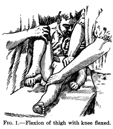
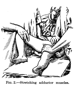
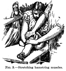
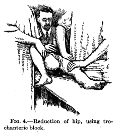
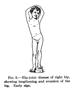
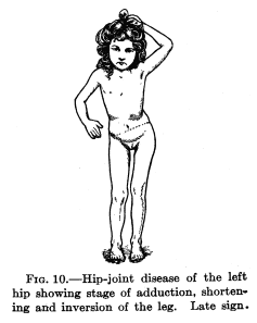
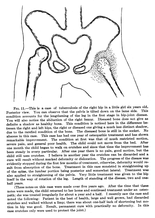
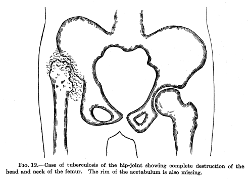
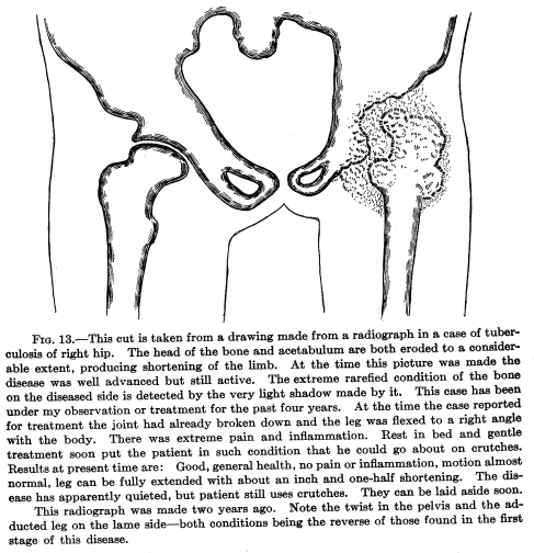
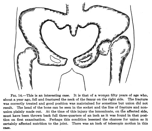
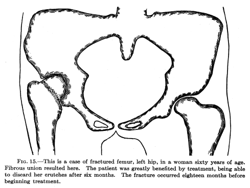
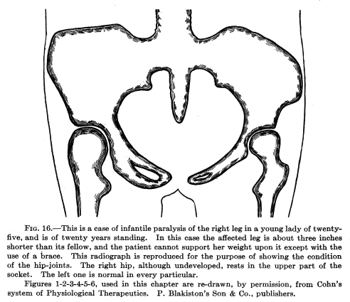