
The centers for the heart were determined in much the same manner as were the lung centers. The electrodes were placed first upon the different parts of the parietal pericardium. This stimulation was followed by contractions of the muscles near the fourth thoracic spine. The areas of the third and fifth nerves were also affected in some animals, but the muscles innervated from the fourth thoracic nerve were always affected, as were also the intercostals of the same area. The intercostals of the third and fifth nerves were more frequently affected than were the spinal muscles of the same area. The stimulation of the visceral pericardium gave the same reactions as did also the stimulation of the heart muscle. The direct stimulation of the heart muscle, however, initiated one reaction which was not found in connection with the stimulation of the pericardium. This was the contraction of the cervical muscles. The trapezius was almost invariably affected, and the scaleni very often. Other cervical muscles were reflexly contracted in reply to stimulation of the heart muscle in varying degrees in different animals. No difference in the position of the muscular contraction could be determined in relation with the stimulation of the different valves. Indeed, it was very difficult to secure any reactions in answer to the stimulation of the valves or of the endocardium, unless the strength of the current was so greatly increased as to affect the heart muscle also. The sensory nerves of the heart are evidently distributed to the cardiac musculature more freely than to the endocardium.
The stimulation of the tissues near the fourth thoracic spine initiated changes in the heart beat which are of two kinds. In some individuals the strength of the beat is increased, in others the rate, while in yet others both rate and force are increased. Whether this difference is due to some imperceptible difference in the manner of giving the stimulation, or whether it is due to some physiological difference in the metabolism of the neuron systems concerned, or whether it is due to structural peculiarities or to some other factor, we do not know. This is one of the points which awaits further investigation. The facts are as stated. Stimulation of the tissues near the fourth thoracic spine of an animal whose thorax has been opened under anesthesia affects the pulmonary blood vessels rather than the heart. The heart is quickened if it is affected at all. Without anesthesia or mutilation, the heart is always quickened, and usually the beat seems somewhat less forceful to the touch.
Direct stimulation of the sympathetic ganglia in the upper thoracic region caused great increase in the rate of the heart beat. Direct stimulation of the vagus above the superior cervical ganglion decreased the pulse rate. Stimulating manipulations of the vagus, in the absence of anesthesia or mutilation, lessened the rate if the stimulation were given high in the neck, but increased the rate if the stimulation were given just above the clavicle,--or rather, in animals, the place were the clavicle would be if there were one. The reason for this seeming anomaly is that the sympathetic fibers which reach the heart are carried with the vagus from their exit from the superior, middle or inferior cervical ganglia. The accelerators are more immediately effectual, hence the stimulation of the nerve trunk at a point where it carries both kinds of fibers causes the accelerator effect to be most evident. Stimulation of the vagus below the heart, as, for example, just above the diaphragm, initiates inhibitor influences to the heart. These are reflex, and are of the same nature as those impulses of gastric origin which interfere with the hearts action under certain abnormal conditions. Stimulation of the vagus nerve in the neck is rather an easy matter; its inhibition is rendered rather difficult by the presence of the pulsating carotid artery. Pressure upon the nerve increases the effect of the pulsations of the artery upon it, and the effect of this is the stimulation of the vagus, rather than its inhibition. The most satisfactory experiments upon the action of the heart centers were performed upon persons.
Stimulation of the tissues near the fourth thoracic spine caused an increase of as much as fifteen beats per minute in the pulse rate. In those persons in whom the rate was greatly increased, the force of each beat was somewhat lessened. The utmost efforts at stimulation were unable to increase the pulse rate at all in some individuals. In all, when efficient stimulation was given, the blood pressure was raised. This reaction was no doubt partly due to the simultaneous reflex stimulation of the pulmonary vaso-motors, and in part to the cardiac effects. The rise of blood pressure thus produced may amount to twenty millimeters of mercury in some individuals. In others, the effects are much less pronounced. Efficient stimulation always produces some change, however in a normal person. The effect of this stimulation upon the sphygmogram is usually very pronounced. In persons whose muscles are very heavy, and who have been of robust health for a long time, it requires a considerable amount of strength to effect the deeper muscles in sufficient degree to effect a perceptible change in the sphygmogram. 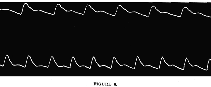 Upper
sphygmogram, normal, resting pulse. Rate, 71. Blood pressure,
125 m.m. of mercury.
 Upper sphygmogram, normal, resting pulse. Rate, 72. Blood pressure, 119 m.m. of mercury. Lower sphygmogram, after two minutes of the artificial lesion at the fourth thoracic vertebra. Rate 63. Blood pressure, 113 m.m. of mercury. It
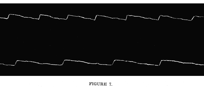
is usually easy to secure rather striking variations in the sphygmograms
in reply to the stimulation of the heart center by somato-sensory impulses.
These experiments were performed in the following manner:
The artificial lesion in the neighborhood of the fourth thoracic spine lessens the efficiency of the heart beat. This is due either to decreased rate, or decreased force, or both, at first, but after a few minutes persistent inhibition, or the maintenance of the lesion for a few minutes, the beat becomes quick and weak in all cases. The sphygmograms taken after prolonged treatment of this kind show the decreased power of the up stroke; it is not so high as before, and its slant is greater. The systemic arterial pressure is decreased at the same time. This decrease in blood pressure is no doubt partly due to the dilatation of the pulmonary vessels, but in part it depends upon the lessened efficiency of the hearts action. In some of the subjects of these experiments, the inter-scapular muscles were contracted in an abnormal manner, and the manipulations were painful. Any manipulations which were given secured the relaxation of the stiffened muscles and a temporary return to the normal condition of the pulse. The effect was usually only temporary, except in a few cases in which the muscles had been stiffened by some temporary condition. In these cases, the manipulations afforded the temporary relief which was all that the conditions required. The pressure upon the sensory nerves which is maintained by muscles which are kept contracted is exactly the same in kind as the pressure which is exerted by the fingers in the experiments in inhibition. The effects are the same, except that the contracted muscles persist in their pressure for days and nights, until the neurons concerned are exhausted. The relief of the abnormal contractions, however secured, stops the abnormal sensory impulses and permits the normal relations to be effectual as soon as the neurons are able to regain their normal metabolic conditions. The effects of abnormal conditions effecting the vagus are very irregular. The experimental pressure upon the trunk of the vagus in the animal produces at first a slowing of the heart. This is followed by a period of irregular rhythm, and this is maintained as long as the pressure is continued. The irregularity of the rhythm noted in these cases is probably due to the complexity of the factors which regulate the hearts action. It appears from these experiments that the action of the heart may be affected by somato-sensory impulses from the area of distribution of the fourth thoracic nerves, and that those movements are most effectual which affect the relations of the joint surfaces. Abnormal conditions affecting the somato-sensory impulses carried over the third, fourth and fifth thoracic nerves may exert a direct influence upon the hearts action. Any condition which affects the sensory fibers of the vagus may affect the action of the heart. No aid in the diagnosis of valvular lesions is to be expected from the existence or non-existence of reflex muscular contractions in the inter-scapular region. |