The Abdominal and Pelvic Brain
Byron Robinson, M. D.
1907
CHAPTER VIII.
NERVES OF THE TRACTUS URINARIUS
(NERVI TRACTUS URINARIUS). - (A) ANATOMY, (B) PHYSIOLOGY.
These are times which try men's souls. - Thomas Paine.
The object of research is not to know the truth merely but to discover
something that will benefit some one - relieve suffering and prolong life.
(A) ANATOMY.
To the urinary tract pass nerves from: (1) plexus
suprarenalis, (2) plexus renalis, (3) plexus ureteris, (4) plexus ovaricus,
(5) ganglia lumbales, (6) plexus communis arteriae iliacus, (7) ganglia
sacrales, (8) plexus hypogastricus (9) plexus vesicalis, (10) plexus urethralis,
(11) plexus mesentericus superior, (12) plexus mesentericus inferior, (13)
plexus arteriae uterinae, (14) plexus sacralis (spinal). The above
nerve plexuses solidly and compactly anastomose with each other and with
all abdominal sympathetic plexuses, thus connecting the tractus urinarius
intimately and profoundly through the nerve plexuses with all other abdominal
viscera.
(1) The Plexus Suprarenalis (Paired).
Bilaterally from the external border and proximal
angle of the abdominal brain depart from five to eight coarser and finer
nerves to supply the adrenals. These nerves are remarkably developed
in infancy. The strands of the suprarenal plexus possess many small
ganglionic masses in their course, and at the points of division.
For the small adrenal the nerve supply is enormous. In the plexus
suprarenalis may be found the ganglion suprarenale or nervus splanchnicus
minores. The plexus suprarenalis sends branches to the plexus renalis
and on the right side also branches to the plexus diaphragmaticus.
(2) Plexus Renalis (Paired).
Bilaterally from the external border and distal lateral
angle of the abdominal brain departs a wide meshed plexus of nerves along
the renal arteries to the kidneys. The renal plexus is composed of
larger and smaller ganglia with larger and smaller strands and it is extensively
fenestrated. Nerve branches from the renal ganglia course distalward
on the ureter and obliquely medianward to join the plexus aorticus.
The renal plexus is one of the richest in ganglia and strands. In
fact, the renal plexus frequently appears as a continuation of the coeliac
ganglion. There is a profound and solid connection between kidney
and abdominal brain. The renal plexus ensheathes the renal artery
with a network of ganglia and cords arriving at the kidney through
the hilum. The plexus renalis
receives strands from the second and third ganglia of the lumbar lateral
chain. The renal plexus is connected with the plexus mesentericus
superior and inferior. The renal plexus arises from: (a), the major
splanchnic; (b), the minor splanchnic; (c), the - first lumbar ganglion;
(d), the coeliac ganglion; (e), plexus mesentericus superior; (f) plexus
aorticus - six sources. Each renal plexus contains four to six ganglia.
A profound connection, anastomosis, exists between the renal plexus and
plexus aorticus, hypogastricus and ovaricus - i.e., the kidney and genitals
are profoundly and solidly connected or anastomosed, by nerve cords and
ganglia.
The renal plexus is practically all sympathetic.
Certain nerve nodes - ganglia renalia - remarkable for number and dimension
- are distributed in the plexus renalis. The largest renal ganglia
lie on the ventral surface of the renal artery, while several smaller ones
lie in the bifurcations of the arteria renalis and on the distal and proximal
border of the renal artery.
The plexus renalis receives some branches from the
plexus adrenalis and the plexus mesentericus superior. The nervus
splanchnicus minor supplies a branch to the plexus renalis which is frequently
strengthened by branches from the two proximal ganglia of the lateral lumbar
chain.
(3) Plexus Ureteris (Paired).
The ureter is supplied by a rich plexus of nerves
from many sources, as may be observed from its vigorous and brusque rhythm,
resembling cardiac contraction. The ureter consists of calcyces,
pelvis and ureter proper, and each segment is supplied in a degree from
different areas of the abdominal sympathetic, and lumbar and sacral chain
of ganglia, however, united into one unit of power in order that the ureteral
rhythm may be periodic and orderly from proximal to distal end. The
ureter is supplied by: - (a), plexus renalis; (b), plexus aorticus; (c),
plexus ovaricus (spermaticus); (d), lumbar lateral chain; (e), sacral lateral
chain; (f), plexus hypogastricus; plexus arteriec; (h), plexus mesentericus
superior; (i), plexus mesentericus inferior; (i), sacral nerves - ten sources.
By the silver method on fresh ureters of animals we could demonstrate rich
plexuses or networks of nerves on the walls of the ureter, with ganglia
at the union of junction of the anastomosing nerves. Three strong
and important points of rich anastoinoses of the plexus ovaricus and plexus
uterinus with the plexus ureteris occurs at (a), where the ureteris is
crossed ventrally by the vasa ovarica (spermatica) which solidly unites
the ureteral and ovarian (spermatic) nerve plexuses. This explains
the reflex pain of ureteral irritation (e. g. calculus) on the ovary or
testicle(retraction). (b), Where the ureter crosses dorsally to the arteria
uterina a strong and solid anastomosis occurs between the plexus ureteris
and plexus arteriae uterinae. Ureteral irritation (e. g,. calculus)
may be transmitted to the uterus (genitals) and bladder. (c), The plexus
ureteris and plexus comniunis arterioeiliacus solidly anastomose at the
point where the ureter crosses ventrally to the iliac arteries. This
explains the reflex pain in the thigh during ureteral irritation, e. g,.,
ureteral calculus.
(4) Plexus Ovaricus (Spermaticus-Paired).
The ovarian plexus arises from the plexus aorticus, extending
from the ganglion coeliacum, located at the arteria cceliaca, to the ganglion
hypogastricum, located on the promontoriuin. Its chief origin is
from the ganglion ovaricum. Immediately subsequent to its origin
from the plexus aorticus it presents about a dozen nerve strands which
gradually coalesce and converge into three main nerves trunks, studded
with ganglia, and accompany the vasa ovarica to the ovary. At the
point where the vasa ovarica crosses ventral to the ureter the accompanying
plexus ovaricus forms a rich anastamosis with the plexus ureteris.
This anastamosis of the plexus ovaricus with the plexus ureteris explains
the reflex pains of the irritated ureter (ureteritis, calculus) in the
ovary and uterus (testicle retraction). The anastomosis of the plexus
ovaricus with the plexus ureteris solidly and compactly connects the ureter
with the entire length of the plexus aorticus.
(5) Ganglia Lumbales (Paired).
The two proximal lumbar ganglia send branches to
the proximal plexus ureteris, as well as branches to the plexus renalis
and plexus ovaricus, thus supplying the proximal end of the ureter.
(6) Plexus Iliacus Communis Arteria, (Paired).
A small artery springs from the common iliac and
supplies the lumbar spindle of the ureter. This solidly connects
the plexus ureteris with the plexus of nerves that accompanies the iliac
and femoral vessels, accounting for the pain in the thigh during attacks
from ureteral calculus and ureteritis.
(7) Ganglia Sacrales (Paired).
The proximal sacral ganglia send branches to and
anastomose with the plexus ureteris, thus intimately connecting the pelvic
ureter with all other sympathetic pelvic plexuses.
(8) Plexus Hypogastricus (Paired).
This powerful plexus sends several branches to the
pelvic ureter, solidly anastomosing the ureter with the genital tract.
(9) Plerus Vesicalis (Paired).
The vesical plexus consists of a wide meshed network
of nerves supplying the bladder with greater and smaller ganglia studding
the plexus at the junction of the anastomosing nerves. The vesical
plexus arises from: (a), plexus hypogastricus; (b), ganglion cervicis uteri;
(c), nervi sacrales; (d), lateral sacral chain; (e), nerve plexuses following
the course of the three vesical arteries (superior, middle and inferior)
derived from the hypogastric plexus, (a large spinal nerve supplies the
bladder from the third sacral, thus making a mixed nerve supply to the
bladder). The rhythm of the bladder (systole and diastole) is not
so apparent as that of some other organs, as the ureter, heart, uterus
or enteron, being modified by the interference of the spinal nerves.
The vesical plexus is a leash of nerves which supplies
the distal ureter and bladder. So far as I can learn from dissection,
it originates in the pelvic brain (ganglion cervicale). The plexus
vesicalis solidly anastomoses with all other sympathetic plexuses in the
pelvis.
(10) Plexus Mesentericus Superior (Unpaired).
Sends some branches to the proximal end of the ureter.
(11) Plexus Mesentericus Inferior ( Unpaired).
Sends several branches to the ureter. Nos.
10 and 11 anastomose the ureter with the tractus intestinalis, and hence
when ureteral pain arises it will be diffused through the intestines, and
will confuse ureteral and intestinal colic.
(12) Plexus Arteriae Uterinae (Paired).
The uterine artery is accompanied by a strong nerve
plexus ensheathing it. At the point where the uterine artery crosses ventrally
to the uterer the nerve plexuses of the artery and ureter anastomose with
each other. This explains the uterine reflex pain during attacks
of ureteral calculus and ureteritis.
(l3) Plexus Urethratis (Paired).
The urethral plexus is a continuation of the vesical
plexus accompanied by the sympathetic nerves which arrive at the urethra
on the supplying blood vessels.
The above thirteen plexuses are sympathetic, hence
it is evident that the tractus urinarius is dominated by the sympathetic
nerve in its function (rhythm).
(14) Plexus Sacratis (Spinal).
The sacral spinal plexus sends nerves to the bladder,
and hence gives rise to a mixed nerve supply. However, the sympathetic
dominates, as it compels the bladder to assume rhythm (diastole and systole).
The chief spinal nerve to the bladder arises from the III sacral, and supplies
the body of the bladder.
(B) PHYSIOLOGY.
The establishment of the nerve supply to the tractus
urinarius serves as a foundation to an understanding of its physiology.
A complete nervous system comprises (a) a peripheral apparatus, (b) a conducting
cord, and (c) a ganglion cell. The object of the nervous system is
that the peripheral apparatus shall collect data (sensation), the conducting
cord shall transport it, and the nerve ganglion shall reorganize and utilize
the nerve forces.
The collection, transportation, and utilization
of nerve forces from and to the tractus urinarius is a matter of vast importance
in diagnosis and practice.
The function of the tractus urinarius is practically
comprised in four acts, viz.: - peristalsis (rhythm) secretion, sensation
and absorption. All visceral muscles, being under the sympathetic
nerves, must execute rhythm, contract and relax, or atrophy. The
object of the kidney is to secrete fluid while the object of the urinary
tract (ureter, bladder and urethra) is to conduct a stream of fluid to
the external body by means of periodic rhythmical movements. From
ureteral sensibility, i. e., from urine flowing on the sensitive ureteral
mucosa, every three to five minutes a brusque, peristaltic wave passes
from the proximal to the distal end of the ureter. The vesical and
urethral waves are more irregular, as the bladder is practically a reservoir.
The periodic ureteral peristalsis is due to the sympathetic ganglia located
within the ureteral wall. So long as the ureteral peristalisis is
not interfered and especially the ureteral stream is not obstructed, the
ureters perform their periodic rhythm. However, as soon as mechanical
obstruction to the ureteral stream arises (as from flexion, calculus, ureteritis
or stricture) the non-drainage induces residual deposits with resulting
accumulations of bacteria, whence the vicious circle occurs in the tractus
urinarius exactly similar to the vicious circles arising from obstruction
in the pylorus or the biliary ducts. The urinary ducts are independent
organs conducting the urine to the external body by means of rhythmic,
periodic waves, regardless of the bodily attitude or force of gravity.
The kidney is a composite organ, consisting of numerous secretary organs
- malpigian corpuscles and tubuli uriniferi - and no doubt these secrete
rhythmically, periodically, though the urine exists constantly in the ureteral
calcyces and pelvis - that being the accumulative results of secretion.
The sympathetic nerve, however, is a silent, ceaseless, painless agent,
unconsciously increasing its function - rhythm, secretion and absorption
- as food and fluid are offered.
It should be remembered that nerve forces travel
in the direction of least resistance, i. e., a nerve plexus containing
the greatest number of nerve strands. It is not multiplication of
ganglion cells that increases intelligence, it is multiplication of nerve
connecting cords that facilitates transmission. Hence in diseases
of the channels of the tractus urinarius, as calculus, stricture, ureteral
flexion, or ureteritis, the organs connected with the tractus urinarius
by the greatest number of nerve strands will suffer the most trauma.
For example, in ureteral calculus the pathologic irritation from the ureter
passes over the giant renal plexus to the abdominal brain, whence reorganization
and emission occurs on the plexus gastricus to the stomach, inducing nausea
or vomiting. Again, the plexus ureteris is profoundly connected or
anastomosed with the plexus ovaricus (spermaticus); hence during attacks
of calculus the testicle suffers pain and is retracted, also the ovary
suffers pain. In short, an irritation in the tractus urinarius will
induce the most pain in the viscera possessing the plexuses with the greatest
number of nerve strands.
The influence of the plexus ureteris is patent when
micturition is so urgent and irregular in the presence of calculus or ureteritis.
The plexus vesicalis is influential in indicating the line of pain in calculus,
and the plexus urethralis is a continuation of it, localizing the pain
in the glans penis (male) and the pudendum and clitoris (female).
Hence, as regards pain in the tractus urinarius, it aids in diagnosis by
manifesting the most prominent symptoms along the nerve plexus containing
the greatest number of nerve strands, such as the plexis renalis (stomach
- vomiting), plexus ovaricus or spermaticus (retraction of the testicle).
Since the nerve plexuses of the tractus urinarius
are solidly and compactly anastomosed with all the other nerve plexuses
of the abdominal sympathetic, the pain from ureteral disturbances is rather
diffuse. However, since the nerve plexuses of the tractus urinarius
are extensively and profoundly connected with the plexuses of the tractus
genitalis, ureteral disturbances are more intensely reflected over the
plexuses of the tractus genitalis, e. g,., in the nerve plexus of the ovary,
pudendum, clitoris (female), and of the testicle, perineum, penis (male).
As regards lithiasis, the chief manifestation from
the tractus urinarius is pathologic physiology, that is, disordered function,
rhythm, absorption, or secretion. Hence the clue to the local disorder
must be sought in the nerve plexuses suffering most intensely, associated
with the tractus urinarius. For example, in calculus there may be
the reno-uterine reflex, the reno-testicular reflex, all indicating intense
pain along the above-indicated nerve plexuses.
The stamping pain of Clement Lucas is where one
afflicted with a ureteral calculus stands on one foot and stamps, which
places the psoas muscle on a violent tension, and traumatizes, massages
the ureter, which, if it possesses a calculus, will induce vigorous ureteral
peristalsis and consequent ureteral pain, colic. Jordan Lloyd's method
of inducing pain in the ureter with calculus, by a blow on the erector
spinae muscles, is simply another process by which the lumbar muscles (especially
the psoas) massages the ureter, exciting vigorous ureteral peristalsis
and consequent pain and colic.
The explanation of pain intensified in different
regions of the body during attacks of calculus or other diseases of the
tractus urinarius must be sought in the line of the nerve plexuses and
their anastomoses with other nerve plexuses. For example, ureteral
calculus produces pain in the plexus spermaticus (pain and retraction of
the testicle) because the plexus ureteris anastomoses with the plexus spermaticus
where the ureter is crossed ventrally by it (vasa spermatica). A
useful suggestion for remembering the nerve plexus of the tractus urinarius
is to recall the arterial supply, as the ureteral nerve plexuses accompany
the arteries of the tractus urinarius.
The function of the tractus urinarius is rhythm
(peristalsis), absorption, sensation, and secretion. The rhythm keeps
its tract always full. It is a perfect system of waterworks whose
stop-cocks or sphincters are always in order and on guard.
|
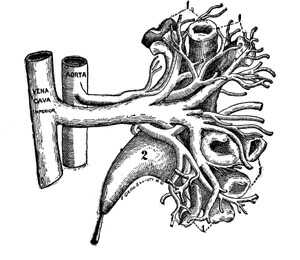
ARTERIAL SUPPLY OF THE TRACTUS URINARIUS
Fig. 18.
The proximal part of the figure is from corrosion anatomy. The nerve
supply to the tractus urinarius is perhaps best remembered by recalling
its blood supply, for the sympathetic nerves accompany the vessels, especially
the arteries. The arteries to the tractus urinarius are: (a) the
arteria adrenalis; arteria renalis; arteria ovarica (spermatica) (x) arteria
media ureteiis, (y) arteria uterina, (z) the three vesical arteries, observe
each of which is accompanied by its plexus of nerves. The tractus
urinarius is richly beset with vasomotor nerves. This anatomic fact
is evident from the violent symptoms induced by an ureteral calculus.
See also Fig. 2, for rich ganglia renalia. |
|
NERVES OF THE TRACTUS URINARIUS - CORROSION ANATOMY
Fig. 19.
This specimen presents quite faithfully the circulation, the kidney, calyces
and pelvis. The two renal vascular blades I present opened like
a book. The corrosion was on the left kidney and the larger vascular
blade is the ventral one. The vasomotor nerves accompanying the
tirinary tract may be estimated by the fact that a rich plexiform network
of nerves ensheath the arteries, the calyces, pelvis and ureter proper.
When the renal vascular blades are shut like a book their thin edges come
in contact, but do not anastomose. The edges of the vascular blades
are what I term the exsanguinated renal zone of Hyrtl, who discovered
it in 1868, and we, at present, employ it for incising the kidney to gain
entrance to the interior of the calyces and pelvis with minimum haemorrhage. |
|
CORROSION ANATOMY (Hyrtl's exsanguinated renal zone)
Fig. 20.
In this specimen of corrosion anatomy the renal vascular blades (ventral
and dorsal) are closed like a book. It presents (left kidney) on
the margin of the dorsal lateral surface the exsanguinated zone of Hyrti
- the line of minimal haemorrhage for cortical renal incision. A
rational method to estimate the quantity of nerves of the tractus urinarius
is to expose the number and dimension of the arteries and other tubular
ducts which are ensheathed in a plexiform networks fenestrated, nodular,
neural vagina of nerves. |
|
NERVES OF THE TRACTUS URINARIUS
Fig. 21.
The nerves of the urinary tract were dissected in this specimen under
alcohol. The ureters, which I term swan-shaped, were irregularly
dilated and contained valves (V), SP, abdominal brain, D, ganglia renalis
distributed over the dilated ureteral pelvis. C. plexus adrenalis.
The plexus ureteris is rich in plexiform network. B, great splanchnic,
Observe that the proximal ureteral isthmus (neck) lying in a groove in
the renal pole is not dilated. |
|
CORROSION ANATOMY
Fig. 22. This specimen
of corrosion anatomy presents the ureteral calyces, ureteral pelvis,
and proximal end of uteter proper together with the arteria and vena renalis.
All segments except the vein are ensheathed in a rich plexiform network
of the nerves governing peristalsis, absorption, secretion, sensation.
When a ureteral calculus becomes mobile in the ureter, Peristalsis (violent)
and sensation (pain) become evident. |
|
RELATION OF SPINAL NERVES TO TRACTUS URINARIUS
Fig. 23.
Illustrates the relation of the spinal nerves to the ureter, especially
its plexus lumbalis. The ureter is intimately connected with the
genito-crural nerve (A), hence the pain reflected in the thigh and scrotum
in ureteral colic and other ureteral diseases. (2) Ileo-inguinal nerve.
For illustration of ureteral nerves and legend of same, See fig.
24. |
