
(6th Edition) CONTENTS
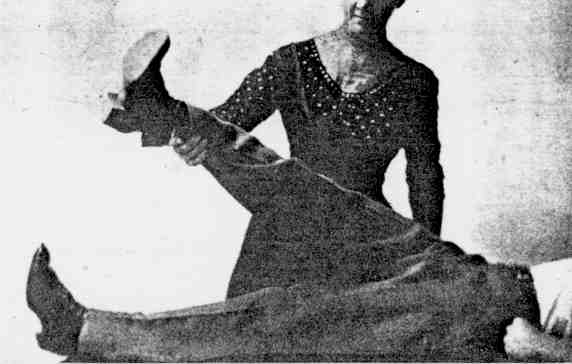
The most important part of osteopathic procedure is the examination of the patient, the determination of the lesion, the discovery of what is wrong in the human building. In some cases nature has taken care of a former abnormality of structure, and then what was once abnormal has become normal. An attempt to make a forcible correction of the apparent abnormality would lead to trouble. In some cases most serious results would ensue. In other cases, where the real lesion is not discovered, months of routine treatment would be worse than useless and might be a detriment rather than a help to the patient. So we say the first thing and the most important thing to do is to make a careful and painstaking examination of the patient. If there are no marked osteopathic lesions, the disease in question having been brought on by overwork or abuse of function, as overeating or drinking, there are certain definite areas in which the Osteopath works, and by securing a better nerve and blood supply hastens the recovery of the patient. This is especially true in cases of sickness induced by the various specific microorganisms which are the exciting agents of a long list of acute diseases, as pneumonia, la grippe, typhoid fever, scarlet fever, measles, chicken pox, mumps, whooping cough, diphtheria, erysipelas, tuberculosis, etc., etc. Even in many of these diseases there are predisposing lesions that weaken certain areas of the body and permit the entrance and growth of these exciting organisms. Yet in these very regions in which the Osteopath works to help recoveries there are, in a majority of cases, lesions of muscle, ligament, and of the bony structure as well, which have been induced reflexly. The abused organs or the diseased organs have sent nerve impulses to the cord, and they have sent them in such large numbers that they have in turn been sent out to the muscles and other structures lying in close proximity. A muscle is tense in proportion to the number of nerve impulses communicated to it. These abnormal nerve impulses, long continued, have produced contractures; these in turn, by pulling on the bones to which they are attached, have produced bony lesions. Some of these bring pressure to bear on the spinal cord and other structures, and in this manner aggravate the disease. For the beginner in Osteopathy it will be better to have the back of the patient bare. If the patient is a lady, a gown or kimono may be worn which opens in the back. A suitable kimono for taking the treatment is illustrated in Fig 2. After the operator has had some experience he will readily examine the spine of patients through the garments, the under clothing at least. As a general thing we know very little about our own bodies. Many are more conversant with the normal cow, horse or hog than with the normal human body. It will be an excellent thing for the one who expects to make a vocation or even an avocation of Osteopathy to study carefully the normal body. Study a number of them. Become thoroughly acquainted with the normal body, and especially with the normal spine. In this way you will be all the more readily able to detect the abnormal. The practitioner should study the various degrees of motility of spines, joints, necks, backs, etc., of normal individuals. He should learn the various degrees of tension, tenderness, and pliability of the various muscles of persons in health. A knowledge of anatomy and physiology will be very helpful. Lesions. It is not every vertebra that is out of line laterally, or deviates anteriorly or posteriorly, that may be said to be out of position in such a manner as to constitute a lesion. Before we can say that a deviation of such a character constitutes a lesion it ought to produce some pathological condition, or ill feeling of some kind. There should be some change in color and some temperature near the abnormality. There should be some contraction in the muscles and ligaments. There should be some inflammation, or a congestion bordering on inflammation, near the seat of lesion. There will be pain in nearly all recent lesions. Pain will be present on pressure in some conditions the muscles in close proximity will be slightly swollen and have a rigid feeling when worked over with the hand. It will be always safe and beneficial to manipulate the spine, ribs, ligaments, muscles, and other tissues, but do not attempt to correct a misplaced bone until it is known to be out of place in such a manner as to cause pressure, or is forming an obstruction that is causing some illness. Never manipulate a tubercular joint or spine. Do not cause pressure on lymphatic glands. Since the publication of the second edition of this book, doctors who are doing research work in Osteopathy are of the opinion that pressure on nerves resulting from vertebral displacements is not a cause of disease. This view is in harmony with the author's experience with one exception, and that is in those cases where a vertebra or vertebrae are misplaced anteriorly. This allows pressure to be exerted upon the spinal nerves as shown in Fig. A. In backward displacements the spinal foramen is made larger, and there can be no pressure exerted on spinal nerves. This is especially true in those cases where Pott's disease exists and there is a backward displacement of the vertebrae amounting to deformity. See Fig. B. Disease and disturbances of the circulation are not, as is generally believed by Osteopaths, caused by arterial or venous obstructions occasioned by misplaced vertebrae. Vertebral displacements and an obstructed circulation have been the two great points which Osteopaths have kept before the public. They have believed this, and acting on this belief, in attempting to adjust vertebrae have done much harm to suffering humanity. I want to show later how the best results may be secured from Osteopathy, or mechanical treatment, with no danger of injury to the patient. The real lesion, or spinal abnormality, when it exists, is in a tightened vertebral joint or series of such joints. The ligaments binding such vertebrae together have become shortened. The intervertebral substance becomes thinner and is frequently absent. In some cases it entirely disappears and ankylosis sets in. When a vertebral joint is not as free in its movements as it should be the adjacent microscopic tissues are involved and the flow of lymph and circulation in such structures is impeded. For this reason we claim that the real spinal lesion is immobility of spinal joints. Other joints may be affected in much the same manner. For the above reasons we would be more explicit in giving caution to avoid harsh and severe treatment and to refrain from adjusting or attempting to adjust bones of the spine or pelvis unless we are positively certain that such apparent maladjustments are the cause of diseased conditions. It follows that the best treatment will be directed to loosen the tightened joint or joints and so manipulate the surrounding tissues that circulation will be restored. Chiropractors have been doing their work, using a theory that is entirely wrong, but in practice they have only loosened such joints, and in that way have secured good results. They would secure better results if they paid some attention to surrounding tissues. A Chiropractor performs his work by placing both hands on a spinal vertebra, one over the other, enforcing it, and giving a quick thrust. The patient is in the recumbent position, and this thrust is given with the idea of adjusting misplaced vertebrae; but the only effect is to secure more mobility of the joints. Were force used sufficient to move the vertebrae serious injury would result. This has proved to be the case resulting from some treatment given by both Chiropractors and Osteopaths. Fig. No.51a illustrates the manner in which the Chiropractor thrust is delivered. Some have the elbow bent, and in straightening out the elbow the thrust is given. Other practitioners attempt to withdraw the hands after making the thrust to allow the vertebrae to recoil. The effect of working on a spinal articulation with sufficient force to loosen the joint, is to stimulate the segments of the spinal cord nearest that joint, and send nerve impulses in increasing numbers over the nerves that find their origin in that portion of the cord. This nerve stimulation will be caused by any mechanical force acting on the spinal column sufficient to influence the cord. The circulation of the blood, both arterial and venous, must be kept free. Tightened, tense muscles and ligaments, various abdominal organs out of position, as in enteroptosis, interfere with the proper circulation of the blood, lymph and nerve impulses. This quickly leads to disease. For example, when the tissues tighten in the neck from any cause, circulation is obstructed and inflammation of the tonsils, pharynx, and other structures takes place. And on account of the stagnation of the circulation, pathological germs find a lodging place and various diseases such as diphtheria, scarlet fever, etc., begin. When there is trouble with the ears, eyes, nose or throat we may be sure that there is interference with their blood and nerve supply somewhere, and oftentimes that obstruction is in a tense muscle or ligament. Spinal vertebrae, of course, are found out of position, but this is very seldom a cause of disease, unless such malposition is the result of a severe accident. Displacement of a vertebra sufficient to cause pressure on a nerve would be very serious, indeed, but is very rarely found. The founder of Osteopathy is very fond of claiming that " the Great Master Mechanic left nothing unfinished in the machinery of his masterpiece - Man - that is necessary for his comfort or longevity." But if spinal vertebrae are so easily misplaced as to cause pressure on nerves and blood vessels and thus cause disease, Deity has made a mistake in designing his masterpiece - Man. The reader must not think from the above that I undervalue the good results to be obtained in Osteopathy, or mechanical treatment, only to obtain better results than have been secured in the past we must perform our work with a view to loosen tight joints, relax muscles, ligaments and fascia, and not attempt to move bones that are not causing pathological conditions. Much good has been accomplished with the old theory as a basis, but where it has been carried to its logical conclusions harm has also resulted. All honor to the founder of Osteopathy, even if his theory was, in part, resting on false premises. Diagnosis, Methods of. The method of diagnosis most in vogue and the principal one which the Osteopath uses is palpation, the use of the hand or hands in determining the condition of nearly all the tissues of the body. With careful work and much practice, comparing the normal with the abnormal, the sense of touch becomes very acute, and the least difference in the density and the motility of the various tissues is readily determined. The patient should be requested to relax all tissues as much as possible and not to make any resistance to the various movements unless requested so to do. The other methods of diagnosis are inspection and percussion. With the beginner, inspection is also important, as by it he notes curvatures, unequal development of muscles, differences in the color of the skin, apparent age, height, weight, peculiarities of gait, manner of standing, sitting, etc. Percussion enables us to learn the condition, size, shape and position of various organs, the presence of cavities, gas, tumors, etc. This form of diagnosis calls into use a small hammer, but more generally the second and third fingers of the right hand are used to strike the middle finger of the left hand which has been placed over the part to be percussed. In examining a patient it may be well to begin with the neck. The important point in the diagnosis is to discover
the pliability and mobility of the spine. If it is too pliable
there is danger of curvature. If portions of the spine are
too stiff and there is not proper motion in the joints of the vertebrae
it is a cause for disease. This may be determined, for the
dorsal and lumbar portions of the spine, by examining as in Fig.
32; in the cervical region by examining as indicated in Figs.
14 and 15. The ease
with which this movement is accomplished, together with a rotation
of the head, determines the amount of pliability. By examining
a few persons who are in health you can determine what the normal
should be. EXAMINATION AND TREATMENT. In many of the descriptions of treatments which follow you might think that you were to put a bone into its proper position at once, but you will not, and it would not be best to do so. It often only results in making an articulation more pliable, which is the real object in view, looking to the ultimate result of restoring the patient's health. The utmost care must be taken not to injure a patient, which may be easily done in the case of a child or a weak person. Special Directions for Treating. In giving a general treatment, try to do the work in twenty minutes. When you begin to practice Osteopathy it will take thirty minutes or longer to give the general treatment, but after you have practiced for a while you will feel that you are wasting time if you do not give it in twenty minutes or less. In using the shorter time you will do the work very effectively. In treating many cases you will obtain better results to give a short and very specific treatment. Not more than five minutes is necessary for the entire operation. As the founder of Osteopathy used to say, "Do what is needed and then quit." He called those who spent time in going over the entire body "engine wipers." He wished Osteopaths to be mechanics, first-class engineers, and to fix what was wrong in a workmanlike manner. In nervous troubles and in many constitutional diseases Osteopaths have discovered that they get better results when they give the general treatment. This helps the circulation and makes a tired patient feel like new; and the treatment, after all, when there are no specific lesions to remove, is but little more than deep massage, in which nearly all the muscles of the body are manipulated. One may give this treatment, in such a manner that many patients come to look upon it as a luxury. And many will take it when they are only slightly indisposed. Some business men take the treatment as a means of relaxation. Many others take it when they are simply tired. In acute cases the Osteopath treats every day and sometimes more often. When the patient becomes better, three treatments, and then twice per week, will be sufficient. In treating chronic cases I have obtained good results by giving the treatment every day for a week and then treating three times per week. When the patient became better, treatment was given twice per week, later only once per week. In chronic cases I found it necessary to treat my patients for three to six months, though some did fairly well after one month's treatment. A number of cases I have found it necessary to treat from one to two years. That is a long time, but results justified the time spent. In treating such cases I have found it to be beneficial to let the patient rest from the treatment for from one to three months and then begin treatment again. Some patients do not seem to improve for the first six months; then they continue to improve until they are well. I have had patients who did not make any visible improvement in a year. They would quit the treatment and begin to improve from that time on. That is one reason why I have found it advisable to have patients rest from the treatments for a month or more. Office Examination. In outlining the examination of patients in the office, where by far the greater number of an Osteopath's patients are treated, I will give my own methods, which I tried in every way to simplify and was successful in doing so as time progressed. My reception room was well lighted and kept very clean. Everything about it was bright and cheerful. The hardwood floors were well covered with bright-colored Indian rugs. There were plenty of rockers. A good supply of up-to-date popular literature, including some bearing on Osteopathy, was always on hand. There was a good library of the latest medical works. There were four good-sized treating rooms in connection with the reception room. These contained a treating table, costumer, dresser with mirror attached, and a couch on which many of the patients rested after treatment. This added greatly to the benefit of the treatment in the cases of many nervous patients. For the ladies there were many full-length and full-fashioned kimonos, which were kept well laundered. In the treating rooms were running water and a good supply of towels and soap. The kimonos were open in the rear, which permitted of a thorough examination of the back, which I always did on first treating the patient. The gentlemen were nearly always examined the first time on the naked back. They removed only the top shirt, and I lifted the undershirt when I wished to examine them. This permitted of a thorough examination. The patient sitting, the examiner stands behind and notes any inequalities on either side of the neck. Sometimes one side bulges and on the other side there is a corresponding hollow. This condition indicates curvature in this region, with the convexity to the full side. On this side the tissues will be found to be hard, tense, and tender. When this condition is present it is frequently indicative of a curvature lower down. We now note whether there are enlarged tonsils, hypertrophied lymphatic glands, goiter, or any unusual pulsations or enlarged vessels. With the patient now reclining on the back in a comfortable position, with all muscles relaxed, we proceed with the examination by palpation. The neck may be gently manipulated by placing one hand on the forehead, for the purpose of rotating it by using the forehead as a lever, while the other hand manipulates the muscles in the back of the neck for the purpose of further relaxation. In examination, as in treating, the Osteopath never rubs. He never allows the hand to slip on the skin, but is concerned with moving all the deeper tissues. The Osteopath now with the points of the fingers examines the tissues just beneath the skull. Often these are found tense, and the patient complains of pain here during examination and treatment. The fingers are now allowed to travel down the mid line at the back of the neck, and find the spinous processes. The first one to be felt beneath the skin is the second, or axis. The position of the first, or atlas, is rarely determined by the spinous process, though in some cases, about one in fifty, it may be found. We are able to count the vertebrae in this manner and to note their position. The seventh, or vertebra prominens, has a very prominent spinous process. It can be differentiated from the first dorsal, the one just beneath it, by rotating the head when the patient is in a sitting position and noting that the spinous process of the seventh cervical moves perceptibly while the spinous process of the first dorsal does not move. Anterior, posterior, or lateral deviations may be determined by the examination of the spinous processes. The position of the transverse processes may be noted by turning the head to one side, moving the examining fingers up a little and to one side from the spinous process. A prominent projection will be found when the one on the other side will be found in the same manner and the fingers will then move down from one to the next process on both sides, determining the relative position of each with reference to neighboring processes. This enables us to determine lateral deviations, twists, or torsions of vertebrae; also posterior or anterior deviations from the normal. Osteopathic treatment of the neck is for the purpose of removing lesions, which may consist of any departure from the normal in any tissue and which is causing abnormality of function. In following the description let us remember that all patients are not to be subjected to the same routine. This is too frequently done, to the detriment of the patient. What follows is for the purpose of describing the work in detail and to describe the work necessary for the removal of the various lesions usually found. This section of the book will be very frequently referred to by the numbered paragraphs where the description of the specific treatment is referred to in the proper treatment of each disease. 1. While the patient is in a sitting position on the side of the table, the operator, standing behind, the right hand placed on top of the patient's head, rotates the head so as to bring the neck of the patient against the thumb of the operator's left hand. The left thumb is moved successively along against the arches of the vertebrae, as shown in Fig. 1. In this manner there is secured a free motion between the vertebra, and the movement also assists in relaxing the tissues preparatory to removing any vertebral lesion that may be present. 2. The patient is sitting and the operator is standing behind, and bends the neck of the patient as far forward as possible on the chest of the patient. This stretches the strong posterior neck muscles, including the ligamentum nuchae. 3. The patient sitting, the operator stands in front and puts his arm about the neck of the patient, so that the bend of the elbow comes beneath the chin of the patient, and the hand grasping the base to the skull. The other hand is free to manipulate any of the vertebrae in any desired direction, though this hold is more applicable to the atlas and the axis. The head of the patient may be given a lifting motion and moved over in the required direction. See Fig. 3. 4. The scaleni muscles, the deeper ones at the side of the neck, are often tight and contracted. Pressure may be made on the first rib on the contracted side, as in Fig. 4, while the other hand bends the head forcibly to the other side, at the same time rotating the head. 5. The patient lies on his back. The operator stands at one side with one hand on the patient's forehead, the other beyond to the other side of the neck. The hand on the forehead rotates the head from side to side, alternately relaxing and stretching the muscles at the side and back of the neck, while the hand placed at the side of the neck stretches the muscles toward the operator with each movement of the neck. The hand on the neck will be moved from one position to another as the tissues relax beneath it, and the hand may be brought down onto the shoulder during the process of relaxation. This same treatment may be applied to all the tissues in front of the neck down to the clavicles. See Fig. 5. 6. The hyoid bone may be found just above what is often called Adam's apple, the thyroid cartilage, the largest cartilage of the larynx. The bone feels as if it were shaped like the wishbone of a chicken, minus the protuberance at the angle. While it should be freely movable it is often held tight by the muscles, the supra hyoid and the infra hyoid, attached to it. This bone is frequently drawn backward and downward, and by pressure on nerves is the frequent cause of nervous coughing and may be responsible for complete loss of voice. By manipulating the neck from side to side the thumb and forefinger may be gradually insinuated under it, and it may be lifted up and forward. The tissues all about it should be thoroughly relaxed. See Fig. 6. 7. The pneumogastric nerve may be pressed upon, manipulated and stimulated by deep pressure behind the anterior border of the sternomastoid muscle on a level with Adam's apple, as in Fig. 7. This is a very important nerve, osteopathically, owing to its large distribution to important organs. 7A. The spinal accessory and glosso-pharyngeal nerves may be reached by deep pressure upward and inward behind the angle of the jaw. 7 B. The sub-occipital, great occipital, small occipital, and great auricular nerves may be stimulated as in cases of fever, headaches, etc., by deep pressure on both sides of the spine, just at the base of the skull, as in Figs. 8 and 9. The founder of Osteopathy, Dr. A. T. Still, when but a small boy, made pressure on these nerves by placing his head in a swing. See Fig. 10. He found that this pressure relieved his headache. This accidental discovery may have had something to do with his discovery, later in life, of Osteopathy. 7 C. By deep pressure of the tissues of the neck against the transverse processes of the second and third cervical vertebrae we may stimulate the superior cervical ganglion. See Fig. 9 A. 8. We will have occasion to refer to springing
the lower jaw by opening and closing the mouth
8 B. In connection with the above the hands may be used to draw up the tissues under the chin. Let the movement be circular and deep. The points of the fingers are used to execute this movement. See Figs. 12 and 19. 9. The head may now be twisted as far as possible to one side without causing inconvenience to the patient; then to the other side in the same manner. We often notice in executing this movement that it moves further to one side than to the other. On the side to which it turns the least we look for muscular or ligamentous lesions. See Fig. 13. 10. The head may be pushed as far forward as possible onto the chest, loosening the posterior muscles and other tissues far down the spine. See Figs. 14 and 15. 11. Some operators can use a very effective spiral treatment of the tissues of the back of the neck. One hand is placed on the forehead of the reclining patient, the other beneath the neck. The neck and head are both raised. The head is rotated in one direction, the neck in the opposite direction. Then the movement is reversed. See Fig. 16. 12. A number of movements have been devised to reduce atlas lesions. One has been given above as in No. 3. These movements may be used in a slightly different way for the other cervical vertebrae. The operator stands at the head of the table, the patient reclining. The operator grasps the head firmly with both hands and makes pressure with the fingers against the arch of the atlas behind. He raises the head slightly, supporting it against the body. Now as he rotates the head he presses the bone toward the normal position. See Fig. 17. 13. The neck tissues may be stretched as
in Fig. 18. In some cases
it will be of material assistance in the case of light patients,
to have an assistant to hold the feet of the patient, to afford
greater resistance. Many other movements in the treatment
of the neck will be developed in the regular routine of practice
by the skillful operator. This will be true of every portion
of the anatomy as well as of the neck. Most Of the treatment for the purpose of affecting the head is given in the neck, upper dorsal and other portions of the body. Nevertheless the Osteopath does some direct work on the head. 14. While the patient is lying on the back the palms of the operator's hands are passed from the center of the forehead each way, with varying pressure down over the temples and behind the ears. This movement has a quieting effect on the patient, soothing the nerves, and is frequently used in headaches. It affects branches of the fifth nerve on the forehead. See Fig. 20. 15. One palm is placed across the forehead and the other beneath the skull, or both palms may be placed on the forehead, one on top of the other, and great pressure exerted for a few seconds and repeated several times. See Fig. 21. This is useful in colds, headache, etc., as it helps to relieve the pressure in the longitudinal and lateral sinuses, large veins of the brain. 16. Treat along the midline of the skull, from the nose to the back of the neck, using the thumb in a circular, pressing motion with varying degrees of pressure for the same purpose as Nos. 14 and 15. See Fig. 22. 17. We may tap with the knuckles or percuss with one finger laid upon the center of the forehead in treating headaches, colds, etc. 18. Manipulate on each side of the nose and loosen all the tissues for the purpose of affecting the fifth nerve and freeing structures in close connection with it. See Fig. 23. 19. Use deep pressure, with a gliding movement of the little finger, to work over a portion of the fifth nerve, supplying the tear duct, for the purpose of opening it or keeping it open. Begin at the inner corner of the eye. See Fig 24. 20. The forefinger will find a little depression in the skull, just below the eyebrows, between the center and inner margins of the eye, where the supraorbital branch of the fifth nerve emerges from the skull. It is a nerve of nutrition to the eye, and passes outward over the forehead at an angle of forty-five degrees. Free the tissues about and in this little opening with a gentle, pressing, circular movement of the tip of the forefinger. See Fig. 25. Work along the nerve with the palm of the thumb. In some cases of neuralgia it will be found to be extremely sensitive, which will be greatly lessened as tissues are relaxed about it. In treating the eyes this nerve is often stimulated. The nerve may be felt beneath the skin. 21. The fifth nerve may be treated where
it emerges from the skull above the eye, in Figs.
25 and 20. Also
over both jaws, above and below the roots of the teeth. It
may also be treated below the malar cheek bones, as in Fig.
26. It may be treated along the sides of the nose, as
in Fig. 23. Thorough
treatment of this nerve is frequently necessary in cases of neuralgia.
22. The fingers must be very clean as they work inside of the orbit to tone up weak or contracted muscles in cases of strabismus. The finger may be inserted deeply, yet carefully, and worked around the eyeball, both to relax and free up the tissues and to promote a better circulation. 23. The nail must be thoroughly clean as it is used to break up the little blood vessels which form a network running into the pterygia, which, if let alone, will grow towards and cover up the pupil of the eye. 24. Granulations may be broken up by crushing them between the thumb and forefinger. For this purpose, folds of the lid may be lifted up, or one finger may be inserted beneath the lid. 25. The patient, lying on the back, the eyeballs may be pressed back into the orbit several times with the palms of the thumbs and held there for a few seconds. This helps in toning up the various structures and assists in the general circulation of the eyes. 26. The palm of one finger may be placed
over the eyeball and tapped, as in percussion, by the forefinger
of the other hand. This acts very much as No. 25, and is useful
in cases of cataract. See Fig.
27. 27. The patient sits on the side of the table and the operator notices any deviation from the normal in exaggerated curves, lateral curvature, or any number of vertebrae or a single vertebra which may be misplaced. He notes any tenderness, as he palpates with the fingers, that may be found in the tissues on either side of the spine or between the spinous processes. See Figs. 28 and 29 for fairly normal spines. 28. The tips of the spines, the spinous processes, may be noted by the red color brought out on them by swiftly passing the hand over them with some pressure. In this manner their position may be noted, thus disclosing any deviation from the normal. 29. A finger may be placed each side of the spinous processes and passed down, in this manner noting any lateral deviation from the normal. This may be done either on the bare spine or over the clothing worn in treating. See Fig. 30. 30. The palm of the hand may be passed down rapidly, from the base of the skull to the sacrum, for the purpose of noting any deviations from the normal, either posterior or anterior. Pressure exerted on the top of the head, as in Fig. 69, will frequently reveal tenderness at some point in the spine. 31. The patient rests in the prone position, lying on his stomach, while the operator, with the palms of the fingers, notes contracted muscular tissues, pulling the muscles away from the spine on either side. These contracted muscles often feel like small ropes beneath the fingers. See Fig. 31. 32. The limbs of the patient are flexed as he lies on his side, and the operator holds them in this position as he gently springs the spine, as in Fig. 32, noting its relaxed or contracted condition. 33. While the patient is lying on the side in a comfortable position the vertebrae are carefully examined by the fingers of the operator. Pressure is made between the spinous processes of each one, to note the condition of the ligaments and the approximation or separation of the various vertebrae. See Fig. 33. When the ligaments have grown too thick they fill the spaces and produce what is known as the smooth, stiff spine. 34. With the patient sitting on the side of the table, and the operator standing behind, he may begin the treatment of the back by placing the tips of his fingers on the patient's shoulders and with the thumbs loosen the muscular tissues in the upper part of the back. If the hands are long he can travel up and down a good portion of the back with the fingers in the above position. See Fig. 34. The fingers may now be used in relaxing all the tissues on the shoulders from the neck over the top of the shoulders. 35. While the patient is sitting the operator passes one arm over one shoulder and under the opposite arm in front. With the other hand he makes fixed points on the spine with the thumb, against which he rotates the body with the other arm, and in this manner thoroughly loosens the structures and replaces misplaced vertebrae. See Figs. 35 and 36. The position is reversed and the other side of the spine is treated in the same manner. The patient may be held, as in Fig. 68, one shoulder braced against operator's body. 36. The patient sitting, and the operator standing behind places one hand on the top of the patient's head and with the other hand makes fixed points with the thumb along the spine, using the head and neck as a lever. The use of the hands may then be reversed and the other side of the spine treated in the same manner. The operator will be surprised with the efficiency of this movement and the power that can be exerted at any given point along the spine against the thumb by making the right pressure on the head. See Fig. 37. 37. The patient sits, preferably on a stool, and places his hands behind his neck. The operator stands behind and passes his hands under the patient's arms and takes his wrists and places one side of the flat of his knee at the patient's spine and lifts the patient up and backward against the knee. This is an efficient treatment for loosening the various articulations and stretching the ligaments which have become tightened. It stretches the spinal, scapular and neck muscles. The lower ribs are raised. See Fig. 38. 38. The patient sitting, the operator stands to one side and behind, or kneels beside the patient on the table, and passes one arm back of the patient's neck and under the patient's arm on the other side, thus bending the patient's neck forward. With the free hand fixed points are made along the spine and the patient's body is rotated against the thumb of the operator. This movement is very effective in upper dorsal regions. See Fig. 39. 39. The patient sitting, the operator stands behind and places one knee beneath the arm of the patient in the axilla. This holds the shoulder and the ribs on that side in a fixed position. The operator may use one or both hands in raising one or more ribs or in stretching the opposite side. See Fig. 40. This may be used in combination with Figs. 46, 47. 40. The patient is sitting, and the practitioner stands behind to one side and reaching around the patient in front grasps the lower edge of any rib. With the other free hand he raises the arm of the patient on the same side as he lifts the rib. This will be helped by having the patient take a deep breath as the rib and arm are raised, then holding both as the patient expels the air. See Fig. 41. 41. The patient is sitting on a stool with the knees against the wall. The operator stands behind and places his knee on the appropriate round of the stool, with the knee against the vertebrae he wishes to correct, seizes the patient by the shoulders or under the arms and pulls the patient back against the knee, rotating the patient to make the necessary correction of the spine. Extreme care must be exercised in executing this movement for fear of injury to the patient. See Fig. 42. 42. The patient while sitting on the side of the table is bent forward so that his head is between his knees, the operator using forcible pressure on the upper dorsal region and head. This relaxes the ligaments of the lumbar and sacroiliac regions. See Fig. 43. 43. The patient is sitting on the side of the table. The operator is standing in front of the patient, with a pillow between himself and the patient. Both hands clasp the spine of the patient as in Fig. 44, when deep pressure may be made, sinking the vertebrae well in; then by rotating the body pressure may be made to the side wished. This is an excellent movement to correct a lateral curvature or any lateral or posterior displacement. In case of an anterior displaced vertebra the vertebra above and below may be brought forward in this manner, thus gradually correcting the one which is anterior. 44. The position is the same as in 43 for both operator and patient. By grasping the spine firmly on each side the patient may be lifted and the spine stretched, or correction may be made in this manner when only two or three vertebrae are approximated too closely. See Fig. 45. 45. Fig. 46 shows an excellent movement in cases of spinal curvature. The side which is shortened may be stretched by using the wrist under the patient's arm, and the hand, as shown in the figure, raises and stretches the side. The other free hand moves from point to point along the spine and forces it over into position. 46. The side may also be stretched, as in Fig. 47. Firm upward pressure may be made with the hand grasping the ribs while traction is made on the upstretched wrist of the patient. 47. The patient lies in as easy a position as possible, with the face downward, but for comfort the face may be turned to one side, with the toes extending over the end of the table. Let the arms hang over the side of the table. Have the patient relax as much as possible. The operator uses the palms of the hands in a circular, pressing movement to relax all contracted tissues of the back. See Fig. 48. With the cushions of the fingers he can pull the muscles away from the spine, as in Fig. 31. 48. With the patient lying in the prone position, the operator standing at one side of the table grasps the hip of the patient on the further side in front. The heel of the other hand can then travel up and down the spine, exerting considerable pressure while the other hand pulls the hip upward, giving the spine a torsion. The operator works from both sides. This movement is very effective in removing lesions and relaxing contracted tissues. See Fig. 49. 49. With the patient in the prone position, lying across the table as in Fig. 50, the operator stands at the head of the patient, and with the thumbs working each side of the spine lie can further relax tissues and stimulate the nerve through the spinal cord. 50. With the patient in the prone position, and the operator standing at the head of the table with the thumbs each side of the spine, as in Fig. 51, he can further relax the tissues and can also exert considerable pressure when necessary to correct posterior displaced vertebrae, or for the purpose of stimulating the spinal cord and through it the organ or organs which receive their nerve supply from any particular section of the spinal cord. 51. The patient is in the prone position, and the limbs are raised in the operator's arm, as in Fig. 52, and rotated while the other hand makes fixed points with considerable pressure on the lower part of the spine. 52. The patient is in the prone position, and pressure is made in the lower part of the spine, while first one then the other limb is raised, as in Fig. 53. This movement, as well as No. 51, assists the operator in relaxing the tissues and replacing posterior vertebrae. 53. The patient lies on his side in a comfortable position. The operator stands in front and grasps the patient's uppermost arm, as in Fig. 54. With the other hand he relaxes the tissues about the shoulders and down to the spine and pretty well down the back, using the arm as he holds it at the elbow as a lever, working it back and forth to aid in the manipulation. The spine is manipulated and any deviations are corrected at the same time and in the same manner. 54. The tissues may be so relaxed that the shoulder may be manipulated quite freely. One hand may pull up the scapulae, while the other presses on the shoulder, as in Fig. 55. The hand may also be insinuated under the scapulae, as in Fig. 56, and the tissues thoroughly manipulated and loosened. 55. While the patient is reclining on the side, and one hand has made its way under the patient's scapulae, the other hand grasps the patient on the shoulder and rotates the entire shoulder. See Fig. 57. 56. The patient is lying on his side on the table. The operator places one hand beneath the patient's neck and grasps the occiput, as in Fig. 58. The operator brings his chest against the other side of the patient's head. In this manner considerable traction may be made on the neck and upper dorsal region, and very effective corrective work may be done with the spine with the free hand as shown in the cut. 57. The limbs of the patient may be flexed and braced against the operator, who bends over and grasps the spine thus brought into relief, as in Fig. 32. The spine at any point may now be manipulated by pulling it toward the operator. 58. With one elbow on the hip, the other on the shoulder, as in Fig. 59, as the patient is lying on the side, the operator's arms may be extended, thus stretching the hip away from the shoulder while the hands are free to manipulate tissues and the spine as well. 59. The patient lies on the side, and the operator, with one hand in front of the hip and the other hand behind the shoulder as in Fig. 60. By pulling on the shoulder and pushing against the hip the spine may be twisted for the purpose of relaxing tissues, including muscles, nerves and ligaments. With the same motion the side of the back upper-most may be stretched by separation of the operator's arms. 60. Fig. 61 is the exact opposite of the above, and the torsion is applied by reversing the position and motion. This is very useful for stretching and relaxing the various tissues of the back and spine. 61. The patient lies comfortably on his back. The elbow of the patient is held by the operator with one hand, while with the other he reaches across the body of the patient and grasps the muscles of the back, as in Fig. 62. The arm may be stretched as movements are made to loosen the tissues manipulated with the free hand. The spine may also be manipulated with the free hand its entire length. 62. The patient lies on the back, and the operator stands at the side of the table and reaches across the body of the patient and grasps the spinal edge of the scapulae, pulling it out as he brings the arm of the patient across the chest, as in Fig. 63. 63. A small patient lies on the back. The knees are grasped in one hand and flexed, as in Fig. 64. The free hand is introduced under the patient, grasping the lower part of the spine and manipulating it as the knees are made to describe a circle. This is a very thorough method of manipulating the lower dorsal and lumbar regions of the spine. 64. The patient lies on the back. The knees are flexed on the thighs and the thighs are brought forcibly against the abdomen. The pressure may be relaxed, then increased. This movement relaxes the ligaments and muscles of the lumbar and sacro-iliac regions. See Fig. 65. 65. The fifth lumbar vertebra is frequently found posterior. With the patient on his back, the clinched fist of the operator is placed under the vertebrae and one limb at a time is taken by the ankle and forcibly put through the motion of external circumduction, straightening the limb out with considerable force, thus bringing the weight of the body on the vertebra. 66. In case the fifth lumbar vertebra is anterior the lesion is more difficult to reduce. The ligaments may be loosened, as in Nos. 63 and 64, and the vertebra moved from side to side, as in Nos. 35 and 57, thus gradually bringing it into its proper position. 67. In case we desire strong inhibition, for the purpose of lessening the number of nerve impulses passing from any section of the cord to any given organs, we may hold the spine with a strong grip, partially lifting the body from the table, as in Fig. 32; or we may place a book under the spine, requesting the patient to recline heavily upon it, as in Fig. 66. The position of the book, as indicated in the cut, is for the purpose of quieting the peristaltic action of the bowels in cases of excessive diarrhoea. 68. When the sacrum is found to be posterior it may be moved to its proper position in many cases by properly relaxing the sacroiliac ligaments as in Nos. 47, 51, 52, 64. Then have the patient lie on the side of the table. The operator then places his knee against the sacrum and pulls back on the hip and shoulder, thus gradually forcing the sacrum into position. See Fig. 67. 69. Another movement for restoring to its proper position a posterior sacrum is for the patient to sit on a stool. The operator places his knee against the sacrum, and holding the patient about the body rotates it as he pulls it backward, thus bringing pressure to bear on the sacrum. See Fig. 42. 70. The coccyx is frequently misplaced. It can be manipulated after relaxing the tissues about it. Frequently it is found necessary to insert the forefinger into the rectum, when the coccyx may be grasped by the thumb and finger and moved in the desired direction. This latter treatment should not be given oftener than once per week, and must be carefully done for fear of injuring the delicate tissues. 71. The finger, anointed with vaseline, is inserted into the rectum for various purposes by the Osteopath. In doing so, time must be given for the sphincter muscle to relax. In piles, the finger presses the blood out of the congested veins, and with a circular, sweeping motion with the palm of the finger smooths out, frees and stimulates the action of nerves and blood vessels. In case the rectum is prolapsed it may be pushed up into position. 72. The clavicle, or collar bone, is frequently found displaced. The sternal end, when out of position, affects the tissues of the throat and is a prominent factor in diseases affecting this region, including goiter and circulatory and nerve disturbances in the arms. The operator stands at the side of the table and takes the elbow of the patient, who is reclining, and inserts the fingers of the other hand under and above the clavicle, near the sternal end. The elbow is now brought over the breast of the patient and the fingers inserted more deeply under the clavical. This movement brings heavy pressure onto the fingers by the clavical, which results in raising the latter, when the sternal end can be placed in or toward its normal position. See Fig. 70. This treatment may be applied at either end of the clavicle as the case may require. 73. The clavicle may also be raised and
placed in its normal position with the patient sitting, as in Fig.
71. The elbow is grasped and raised, which raises the
clavicle, when the thumb may be inserted above and under it.
By bringing the elbow upward and across the chest the clavicular
ligaments may be stretched and the clavicle properly replaced.
This treatment is effective for either end of the clavicle. In treating the thorax we must remember that the ribs are connected with the vertebrae, and that when the spine has a curvature, or one or more vertebrae are twisted or displaced in any manner, the ribs connected with those vertebrae are very frequently misplaced as a result. These combined lesions cause secondary lesions of the muscles, ligaments and cartilages. In correcting any maladjustment of the ribs advantage is taken of the muscles attached to them, especially the pectoral and latissimus dorsi muscles. 74. The vertebral end, or head of the rib, is nearly a fixed point. Pressure exerted at the angle of the rib tends to move the rib about that fixed point. This movement is made more effective by the forcible elevation of the arm and in some cases by the rotation of the shoulder. 75. The first rib is often raised near the sternal end. It may be depressed after the scaleni, the muscles of the neck, are thoroughly loosened. It may be depressed by pressure as the operator stands behind the sitting patient, as in Fig. 4. The head is bent forward and rotated, as in the cut. This stretches the scaleni muscles attached to the rib. The pressure is still applied as the head is turned toward the rib. 76. The first rib may be also corrected as the patient is in the recumbent position, as in Fig. 72. The head is raised and rotated away from the rib, while the pressure is applied to the rib. Later the head is rotated toward the rib as the pressure is again applied. 77. A further treatment for the first rib is applied as the patient is in the recumbent position, as in Fig. 72. With the head turned toward the affected rib and slightly elevated, pressure is made on the rib with the thumb, and the elbow on the same side is grasped and carried across the chest to the opposite shoulder. 78. When the first or second rib is displaced downward it can be raised; the patient sitting, the operator standing behind. The operator places the tips of the fingers at the lower border of the rib and lifts on it as he rotates the head, with the free hand, forward and then strongly backward. This uses the neck muscles, the scaleni, to pull the rib upward. Should the anterior cartilages protrude they may be pressed into position as the above treatment is being given. 79. Should the first and second ribs need elevating, nearer the spine, the operator may stand in front of the sitting patient while he presses and lifts up near the head of these ribs, while he rotates the head forward and to the opposite side with considerable force. See Fig. 73. 80. The first rib may be depressed, with the operator sitting at the side of the patient with the patient's arm across the operator's shoulder. This elevates the structures oil that side. Pressure is now made on the upper border of the rib, just above the clavicle, as the head is rotated to that side and forward. See Fig. 74. 81. The practitioner stands in front of the sitting patient. One arm reaches around the patient, a little past the spine to the angle of the ribs, as in Fig. 47, while the arm of the patient is raised on the same side. The arm is stretched up and rotated back and down while the operator lifts on the angle of the rib. This serves to lift the rib in front as well as at the angle, because of the traction exerted on the pectoral muscles in front by the elevation of the arm. This motion should be carefully executed, yet sufficient strength should be used to make it effective. It may be repeated several times if necessary. The hand which holds the ribs may select one after another, as may be necessary, as the arm is stretched and rotated. In giving the above treatment the muscles, nerves, and ligaments are strengthened and toned. If one rib is very much lower than it should be the patient should take a full breath when the rib is pulled up, and as the breath is expelled the rib is still held for a few seconds. 82. Fig. 46 represents another method of elevating and separating ribs. The patient sits and the operator stands in front. The operator forces the spine to the side in question, which helps to separate the ribs. The arm of the patient is raised by the wrist on that side, which raises the ribs. A combination of 81 and 82 is frequently effective. 83. The patient sitting and the arm of the patient across the shoulder of the operator, as in Fig. 75. This gives more of a hold on the lower rib while the patient's head is bent away from the side being treated, curving the spine and throwing the rib more into relief. 84. The patient sits on a stool and the operator stands behind, with his knee against the angle of the rib. With one hand the operator elevates and rotates the arm of the patient, and with the fingers of the other hand, beneath the rib in front, lifts upon it as the patient takes a deep breath. The entire treatment may be reversed and the rib depressed. See Fig. 41. 85. The patient is sitting. The operator standing in front grasps the head of the rib in question, with one hand passed about the patient, while the other hand presses on the sternal end of the rib. By rotating the body of the patient the rib may be sprung into position. 86. The patient is reclining on the back. The operator, standing to one side, reaches over and takes the arm of the patient on the other side. With the free hand he takes the rib at or near the angle, as in Fig. 62, and as he pushes it into place he lifts up on the outstretched arm and rotates it backward. This calls into play the anterior muscles attached to the rib or ribs and assists in the movement. The patient may further assist the operator by taking a deep, full breath as the rib is lifted up, which the operator holds for a few seconds as the breath is exhaled. 87. The patient may lie on the side. The operator uses the same general movements as in No. 86. This of course applies more to the upper ribs. 88. As the patient lies in the prone position the heads or angles of the ribs may be replaced, as in Figs. 76, 50, 51, by working directly over them. 89. When the patient lies in the prone position
many cases of luxated ribs may be treated by pressure of the thumbs against
the angles of the ribs, throwing the ribs either upward or downward, as
indicated by the needs of the condition.
THE ELEVENTH AND TWELFTH RIBS. 90. These ribs are very frequently found displaced. The surrounding tissues must be fully relaxed. When the rib or ribs are displaced upward we have the patient lie on the side, with the limbs flexed on the abdomen, when the ribs may be grasped by the operator and pushed into position, forcing them upward. 91. The patient sits on the side of the table, while the operator holds each end of the rib. The patient takes a full breath, and as the patient exhales, the rib is pushed into position. 92. When the rib is displaced upwards the patient takes either position, lying on the side or sitting, and the rib is manipulated, as in Nos. 90 and 92, only in the opposite direction. 93. Frequently we find the ribs flattened
over the liver or stomach. They may be pulled out, as in Fig.
77. Then they may be rounded into shape by using pressure
on the ribs at the sides, as in Fig.
78. Repeated treatment in this manner will materially
affect their shape. In treating and manipulating affected
ribs or misplaced ribs we find it necessary to repeat the treatment.
In some cases it takes considerable time, a number of treatments
being necessary for permanent results. The ribs are so prone
to slip back into their old positions when they have been out of
position for a long time. The Osteopath in examining the abdomen uses inspection, percussion and palpation. The latter conveys the most information concerning the condition of this important region and its contents. We note displacements of various organs, new growths, tumors, relaxed or tense conditions of the muscles, differences in temperature, enlarged or pulsating vessels, muscular contractions, distended or contracted walls, etc. 94. A general treatment of the abdomen is frequently very helpful, either for relaxing or toning muscular tissues, for increasing or decreasing the amount of blood in the abdominal vessels, and for its general effect on the nerves. The patient lies on the back, with the knees flexed. The operator stands at one side of the table, and with the palm of the hand, not the tips of the fingers, relaxes the muscles of the abdomen. The operator may begin low down to one side and work up on that side, then on the other side in the same manner. The ribs may be slightly sprung inward to assist in the relaxation of the abdominal walls as they are undergoing manipulation, as in Fig. 79, which shows the general position of the hands. The abdomen may be spanned by the hands, with the thumbs on one side and the finger tips on the other side, and the abdominal contents moved in this manner from one side to the other. 95. In treating any part of the body, but especially the abdomen, the hands should be of such a temperature as not to be disagreeable to the patient. Cold hands used here will cause a contraction of the tissues, and thus interfere with the work. Direct pressure may be made with the flat of the hand, as in Fig. 80, over the center of the abdomen well below the umbilicus. Pressure may be gradually increased, with some side pressure to force the contents of the small intestine toward the caecum, the lower part of the large intestine which lies low down on the right side. 96. It is often advantageous to lift up the intestines, as in Fig. 81. With the patient reclining, the knees should be flexed to allow of more thorough relaxation of the abdominal muscles. This movement should be repeated several times and the abdominal contents held for a minute or so each time. 97. The patient is sitting, and the operator is standing behind. He places the sides of the palms of the hands deeply beneath the abdominal contents, as in Fig. 82. The patient is requested to bend forward as the operator twists his wrists, so that the sides of the hand next to the patient are turned inward and upward. Then request the patient to straighten up as the operator lifts the contents, and holds for a moment. 98. The patient lies on the side, permitting a relaxed condition of the abdominal walls. The operator stands behind, as in Fig. 83, and lifts the abdominal contents upward, and may also lift them toward the median line, thus straightening out the caecum, or sigmoid flexure, as the position may allow. 99. The operator may straighten out the sigmoid, and at the same time tone up the muscular tissues, by insinuating the palms of the hands deeply and low down on the left of the abdomen, and suddenly lifting the contents. The movement must be carefully and cautiously performed. 100. It is often of advantage, as will be indicated in various treatments, to tone up the solar plexus. This may be done by deep steady pressure, with a slightly circular motion just below the sternum. Pressure should be directed backward and upward, as in Fig. 84. 101. The liver, spleen, and stomach and other upper abdominal viscera may be toned by placing one hand on either side of the ribs, as in Fig. 85. Pressure, alternating with a few seconds of rest, is made as indicated. 102. The liver and adjacent abdominal viscera may be toned by alternating pressure and relaxation directly over that organ, as indicated in Fig. 78. 103. The gall bladder lies underneath
the anterior portion of the liver, just beneath the points of the ninth
and tenth ribs on the right side. The bile duct leaves it at this
point and proceeds to the duodenum, in the shape of a reversed letter "
S " when it enters that portion of the small intestine, about one and a
half inches below the umbilicus. In cases of gallstones, or inflammation
of the bladder or duct, we may assist nature in emptying the bladder and
pass stones along the duct by manipulation.
In the treatment of diseases peculiar to women, and some diseases peculiar to men, lesions of the pelvis play a most important part. One may never expect to effect a cure while these lesions are permitted to remain. To remove them is of the greatest importance. Pelvic lesions will also be considered here because they affect the limbs, causing sciatica, paralysis, enlarged veins, errors in circulation, etc. These lesions are often accompanied by spinal lesions, which also affect the spine and through it other internal organs. The discovery of these lesions requires considerable anatomical skill, and an attempt to correct lesions here should only be made when the operator is absolutely sure of his diagnosis. The whole pelvis may be tipped backward, in which case the superior posterior iliac spines will be found to be too prominent. In case the whole pelvis is on a torsion, one side back, the other side forward, one superior posterior iliac spine will be found prominent and the other less prominent than normal. In case one side of the pelvis is higher than the other, the superior posterior spine will be higher on the high side. The limb on the high side will be shorter than the limb on the low side. This may be determined by slightly manipulating both limbs to relax all tissues, and then comparing the internal malleoli of the tibia with each other. This may be done by placing them close together. Another way is to have the patient hold a tape line between the front teeth, and measure to the anterior superior spines of both ilia, and also to each internal malleous of each tibia. In case the upper portion of the pelvis has moved forward, the superior posterior iliac spines will be found to be less prominent than in the normal pelvis. In making the examination the operator will best determine the condition by having the back bare. Great care must be exercised in making the examination. The points of tenderness in case of luxation will be found over the sacroiliac articulation, both in the muscles and in the ligaments, interfering with nerves in the same region. 104. When the pelvis is tipped backward patient sit on a stool or table. The operator stands behind and places his knee against the upper part of the sacrum, while he takes the patient beneath the arms and pulls upward and backward on the trunk with a rotary motion to first one side and then the other. The assistant sits in front and draws the pelvis forward. See Fig. 42. 105. For backward tipping of the pelvis the patient may recline on the table with the face down. With one hand the operator makes pressure on the upper part of the pelvis, while he lifts the limb on that side with considerable force. This treatment is given first on one side then the other. 106. For a backward tipping of the pelvis the patient lies on the side on the table. The operator stands behind. 'The knee is placed firmly against the sacrum, and the shoulder and the leg are both drawn backward. This forces the pelvis forward. See Fig. 67. 107. When the pelvis is tipped forward the patient may lie on the side. The operator stands behind, and as he presses against the lower part of the sacrum and pelvis with one hand he pulls back with the other hand from the upper part of the front of the pelvis, the anterior superior spine of the ilium. 108. The patient sits on a stool, while the operator stands in front. The assistant is stationed behind the patient and holds the pelvis in front and draws it backward, while the operator with his arms under those of the patient manipulates the body of the patient forward with a lifting, rotary motion. 109. Should the pelvis tip upward on one
side, the quadratus lumborum muscle may be stretched, as in Figs.
59, 60, 61.
Then No. 108 may be applied, with the assistant holding down the
high side of the pelvis. We frequently have lesions which affect only one side of the pelvis. We often find one of the innominates backward and downward at the same time; again one will be upward and forward. The former will lengthen the limb on the same side, the latter will shorten it. These lesions are the most common, but we may find their exact reverse. We may find both innominates luxated in different ways or in similar ways at the same time. In order to determine these lesions we must dependon
the position of the posterior superior iliac spines as indicated in lesions
of the whole pelvis. We must compare the length of the limbs.
Make measurements between the coracoids of the scapulae and the anterior
superior spines of the ilium. Look for tension and tenderness in
the sacroiliac ligaments; also at the pubic symphysis.
110. When the innominate is luxated backward have the patient lie on the back. The operator places his fist beneath the posterior superior spine of the ilium. The other hand grasps the ankle and flexes the limb on the patient's abdomen, when the limb is rotated outward and downward with considerable force. In this manner the weight of the patient helps to force the innominate into position. 111. The operator may grasp the crest of the ilium, and also the tuberosity of the ischium, and by alternately pushing on the one and pulling on other may set either a forward or backward luxation. This is done with the patient either lying on the back or on the side. 112. Combinations of the above movements, 110,
111, or the work used in correcting the whole pelvis, may be used in correcting
any lesion of an innominate.
The general treatment of the limbs may be modified in various ways for the treatment of different and definite lesions. 113. The limb may have various tissues relaxed preliminary to other work, by manipulating the various tissues by seizing the limb in both hands, as in Fig. 88, and with a rotary movement of the hands move and relax all the tissues to the bones. 114. Both internal and external rotation and circumduction of the limb may be performed, flexing the calf of the leg on the thigh and the thigh on the abdomen, then straightening the limb out with the proper rotation, with more or less force. See Fig. 89. 115. The sciatic nerve may be stretched by extending the limb, as in Fig. 90, at the same time bearing down considerably on the foot. 116. The patient reclining, the knee may be lightly flexed and the operator works under it with both hands, stretching the muscles outward and working in quite deeply. See Fig. 91. 117. The foot may be flexed, extended and rotated with considerable force on the ankle. Ligaments and other tissues are thus relaxed and the circulation is promoted. See Fig. 92. 118. In treating the feet the arches may be sprung to increase the arch, and pressure and traction may also be applied to it. This relaxes ligaments, permits of replacing misplaced small bones and tones up the muscular structures of the foot. See Fig. 93. 119. The toes may be stretched and rotated with force at the same time. In some cases they must be handled with care. The treatment assists in the circulation by freeing all tissues, nerves and blood vessels. 120. The saphenous opening, through which the long saphenous vein passes, lies one or two inches below the lower end of Poupart's ligament, on the inner side of the thigh. This opening is often practically closed by tense muscles. It may be made free by external and internal rotation of the limb, as in Fig. 89. Then the opening itself may be manipulated. 121. The extended limb may be seized by the foot when the patient is in the reclining position, and turned outward as far as is comfortable for the patient, then be allowed to come to its normal position, See Fig. 94. This may be done, say ten times. Then it may be turned inward the same number of times. This movement puts the muscles on a strain by torsion, tones them up and assists in the circulation. Old subluxations cause considerable trouble. A slight misplacement of the hip is often treated as disease of the knee, neuritis, etc. These misplacements are not always discovered by the ordinary physician, but when the real cause is removed the case is cured. Reducing these luxations, when the case is an old one, requires a course of treatment to relax all muscular tissues, to render the parts flexible and the ligaments more pliable. 122. When we discover that the hip is dislocated, with the head of the femur up and back on the dorsum of the ilium, in which the limb is short and the toes turned inward, we flex the knee and rotate it inward. This frees the head of the femur. Then we rotate outward and make extension while we press on the great trochanter to force the head into the acetabtilum. See Fig. 95. This luxation comprises one half of all hip dislocations. 123. The head of the femur is sometimes down and back near the sciatic notch. This also shortens the limb and turns the toes inward. The treatment is the same as above, as one writer puts it, "bend up, turn in, roll out and extend." Use one hand to manipulate the great trochanter. See Fig. 95. 124. Sometimes there is a thyroid dislocation of the hip, in which the head has been forced downward into the obturator foramen. In this case the knee is flexed and the toe points either inward or outward. To make the proper reduction the leg is flexed on the thigh and the thigh on the pelvis. The knee is rotated inward as far as possible, followed by extension. Pressure is made at the same time to force the head of the bone into its proper position. See Fig. 95. 125. When the head of the femur is forward and onto the pubis the toe turns outward. The head of the bone can be felt and seen. Treat as in No. 124. Should this prove unsuccessful place the patient on the side and draw the limb backward with considerable force, stretching all the muscles about the head of the bone; then lift the head over the pubis and place it in its proper position. 126. Ankle dislocations are reduced by simple traction. The knee is flexed on the thigh, the thigh on the pelvis. An assistant holds the knee and the operator holds and pulls the foot, giving it at the same time a slight rocking motion. See Fig. 96. 127. Dislocations of the knee are reduced by strong traction. Pressure to force the tissues into place may be made at the same time. The knee joint may be sprung by placing arm under the knee and pressing down on foot. See Fig. 97. 128. In treating the shoulder and arm the shoulder may be rotated as in Fig. 57. The operator grasps it before and behind the shoulder. 129. The shoulder joint may be sprung, as in Fig. 98. The forearm of the operator is placed in the axilla, and the arm of the patient pushed toward the side. This relaxes tissues and frees up the circulation. It may be freed of adhesions and the circulation promoted by seizing the wrist, bringing it back and up under axilla and stretching out in front with force. See Figs. 103, 104. 130. The elbow may be flexed on the same principle as indicated as in No. 129, by placing the clenched fist of the operator on the patient's arm just above the elbow and bending the arm upon the hand so placed. See Fig. 99. 131. In some cases it is well to work carefully down the arm, rotating the muscles on the bones and working carefully on the forearm between the bones. See Figs. 103 and 104. Fig. 105 represents the stretching of the long head of the biceps, after which the arm will be flexed and the tendon pressed into its groove. 132. All dislocations of the shoulder may be reduced by having the patient recline. The operator places his stockinged foot in the axilla and makes strong traction on the arm of the patient. The knee may be used instead of the foot, as in Fig. 100. 133. In cases of elbow dislocations, where both bones are displaced backwards or displaced externally or internally, or the ulna is backwards, the operator places his knee in front of the elbow joint, pressing against the ulna and the radius with the knee and bends the forearm. This plan uses the muscles to pull the bones into place. When the radius is backward the above or simple pressure, with manipulation, may be used. For forward dislocation bend the elbow over the knee. Use extension and manipulation. See Fig. 101. 134. The various wrist dislocations are all reduced by traction. Some manipulations may be used. 135. Should the ulna be dislocated at the wrist, pressure will be sufficient. 136. For the various dislocations of the hand use pressure, traction and rotation. In many cases of old dislocations a prolonged course of treatment is necessary. 137. A table and stool are represented in Fig. 102. The stool is fourteen inches square on top and twenty-one inches high. The table is a folding one with a steel frame and thin wood top. It can be carried easily to the patient's home. One for regular office use would be padded and more substantial in design. The table represented in the cut is five feet and ten inches long, and twenty inches wide and twenty-six inches high. 138. A General Treatment is given by a great many Osteopaths in connection with the specific treatment needed for the ailment for which the patient is being treated. General treatment is an advantage in a number of cases. It is given for nerve troubles and for the general circulation. The treatments will vary greatly with different Osteopaths but the following is a sample: 139. The patient reclines on a table, lying on his side. The Osteopath begins by loosening up the tissues of the back, as indicated in Nos. 53, 56, 58. The shoulder is manipulated, as in Nos. 54, 55, the lower limb as in No. 113. The spine is sprung from one end to the other, as in No. 57. The patient then lies on the other side and the treatment is given as before. The patient then takes the reclining position, when further treatment is given the back as in Nos. 47, 48, 50, 51, 52. Afterward the patient lies on the back and the treatment is commenced at the head, as in Nos. 14, 15. The neck is treated as in Nos. 7B, 9, 10, 11, 13. The arms are treated as in Nos. 130, 131. Further treatment is given the back, and the ribs are raised as in Nos. 61, 62, 86, 64. The abdomen is treated as in Nos. 94, 95, 96, 100, 101, 102. The lower limbs are treated as in Nos. 113, 114, 115, 116, 121. The patient now sits on the side of the table and the back and spine are further treated as in Nos. 1, 2, 5, 34, 36, 37, 43, 44. 140. A treatment that has come into use, recently among Osteopaths, is shown in Fig. No. 108. It is spreading the ischia. It is used in case of enlarged prostate gland. The founder of Osteopathy claims that spreading these bones invariably helps this condition. In sciatica, the pyrafomis muscle which crosses the sciatic nerve is frequently contracted and causes pressure on this nerve. This muscle may be stretched by forcibly carrying one leg over the other as seen in figure No. 90B. The muscles and other structures of the leg may be stretched as indicated in figure No. 88A. This movement helps to reduce contractions and assists circulation in the lower limbs. The muscles in the anterior portion of the leg may be stretched by extending the limb as in figure 89B [Note: As there is no figure 89B in the original text, I assume that he is referring to figure 89A - D. McMillin]. A good method of raising the ribs is illustrated
in figures 47A and 77A.
In the first mentioned illustration we see by holding and stretching
the arm upward we bring into play the pectoralis major and minor
muscles which help in raising the ribs. In figure 77A
it will be well to have the patient elevate his arms and while he
lowers them to spread and raise the ribs. This movement acts
in stretching the diaphragm which is attached to the ribs.
Another method is illustrated in figure 76A.
Figure 69A represents closing
and spreading the knees against resistance. In figure 29A
we have represented an abnormal spine. This patient suffered
a severe attack of typhoid fever, and was very badly constipated.
Notice the lateral swerve, but what is worse the anterior condition
of two lumbar vertebrae. The spine, in its normal condition, is a chain of flexible nature. It is formed of a number of bones called vertebrae, from the Latin vertere, to turn. There are thirty-three of these vertebrae, of which seven are in the cervical, or neck region, twelve are in the dorsal region, which occupies the space from the neck to where the small of the back begins, five are in the lumbar region, the small of the back to the sacrum, five are in the sacral and four in the coccygeal region, which is the coccyx. The twenty-four upper vertebrae should remain separate and flexible through life. Those in the sacral and coccygeal region, in the adult are firmly united and form two bones, the sacrum and coccyx. The spine has four normal curves, the cervical, dorsal, lumbar, and pelvic. The first, or cervical, extends from the base of the skull to the second dorsal vertebra. This curves inward. The dorsal curve extends from the second dorsal vertebra to the first lumbar vertebra. This is an outward curve. The lumbar curve extends from the first lumbar to the sacrum. This curve is a forward one. The sacral curve begins at the union of the last lumbar vertebra with the sacrum and extends to the tip of the coccyx. This is an outward curve. In very young children the cervical and lumbar curves are absent, and when the child is sitting the spine presents only one curve from the base of the skull to the end of the spine, and this curvature is an outward one. The cervical and lumbar curves are called compensatory, and develop a little later. The curves of the normal spine should be frequently examined, until the operator has a good idea of the normal spine. He should be acquainted with the normal flexibility of the spine as well. |