
|
Let us go deeper into the study of the life-saving powers of the lymphatics. Possibly less is known of the lymphatics than of any other division of the life-sustaining machinery of man. We lay much stress on the uses of blood and the powers of the nerves, but have we any evidence that they are of more vital importance than the lymphatics? If not, let us halt at this universal system of irrigation, and study its great uses in sustaining animal life. These quotations from The Philosophy and Mechanical Principles of Osteopathy are given to show the importance in Dr. Stills mind of a thorough understanding of lymph and the lymphatic system. Those who have followed his teachings and writings know that Dr. Still always urged this point. Any considerable attention for to the subject will
show the need of an intimate knowledge of the origin, properties and movements
of the lymph, of the structure of the lymph vessels and glands, and of
the relation of the entire lymphatic system (including the lacteals) to
the metabolic processes and the general economy of the body.
Importance of The Lymphatics In support of this statement I submit the following
propositions:
2. Since the blood does not come into direct contact with the tissue cells (except in one organ -- the spleen), one of the main functions of blood circulation is to supply and renew the lymph to all tissues. As has been tersely said, the blood feeds the lymph, and the lymph feeds the cells. 3. The excretion of the cells, the waste products of metabolism, are carried by the lymph back into the blood-stream, for elimination through the several emunctories. 4. Through the lymph channels metastasis frequently occurs, especially of malignant tumor cells. 5. The lymph glands or nodes afford (through their product, the phagocytes) a very considerable protection against various forms of infection. 6. By the use of consistently osteopathic procedures, manipulative and otherwise, the lymph currents may be made to serve as an exceedingly great factor in the abortion of and the recovery from a wide range of diseases. The practical value to the physician of a study of the lymphatic system lies in the last statement given above. It is true that in the mechanism of the recovery from disease the lymphatics automatically or functionally play an important part, and the osteopath usually increases their functional activity even though he does not realize how he accomplishes it. If, however, he can ssist natures reparataive efforts by controlling and utilizing the forces which are contained in that all-pervasive fluid, the lymph, he is so much the better able to cope with disease. It will be necessary to dwell briefly on the anatomy of the lymph vessels and glands. To obtain a fair conception of the relation of the lymphatic system with the blood circulation, the following from Gage will be of assistance: A tolerably correct pictorial idea of the entire vascular system may be formed by considering the blood vascular part as made up of a great tree, the heart forming a short trunk, and the arteries, veins and capillaries the branches; but there is present the untree-like character of the direct union of the terminal twigs of the arteries and veins, that is, the venous and arterial capillaries are continuous. The lymphatic system may then be represented by two vines of unequal size, but which together follow all the blood vessels to their ultimate ramifications, and in many places even send minute twigs beyond them. The analogy with a vine is further borne out by the lymphatic vessels, as they remain of a more uniform diameter than the blood vessels; and finally, the terminal twigs, like those of a real vine, end freely or blindly, often in slight expansions like leaves, thus forming a marked contrast with the terminal twigs of arteries and veins, which cannot properly be said to terminate at all. In a word, the blood-vascular system forms a complete circle or circuit in itself, while the lymph-vascular system joins the blood-vascular system at its central or trunk end, but ends blindly at the periphery. One might with propriety carry out Gages picture
by imagining the tree and vine enclosed in a huge bag. In that case
the terminal twigs and leaves and tendrils of the vine would not only fill
all of the space between the tree branches, but would push out into the
substance of the bag as well. For lymph ducts penetrate nearly all
the structures, including the corium (beyond which the lymph passes into
the rete Malpighii), and many perivascular spaces and other interstices
are filled with loose connective tissue which is permeated with lymph.
The total volume of the lymph is consequently enormous.
Experimenters, through different methods of determination and different
conditions of the body, have variously estimated the amount of lymph to
be from 1.6 up to 1.3 of the total body volume. The volume of the
blood ranges from 1.15 to 1.11* of that of the body. If from these
several estimates we use as a mean ratio 1.4 to represent the volume of
the lymph, and 1.12 that of the blood, we find that there is about three
times as much lymph as blood. Even if it performed no important function,
the mere mass or bulk of this fluid would compel us to view it as having
no small influence in the economy of the body. And since its mission
is so vital to all the structures, we cannot regard it lightly.
This immense body of fluid constitutes a medium of exchange so universal that man has been described, not inaptly, as an aquatic animal. Every cell is bathed in lymph. Every cell depends upon the lymph for nourishment. Every cell is afforded drainage by means of the lymph. As Byron Robinson says, the lymph performs an import and export duty: carries on a commerce with every other tissue. Development of Lymphatics A series of studies made on pig embryos by Dr. Florence
R. Sabin of Johns Hopkins University demonstrated the fact that the lymphatic
vessels are developed from the veins. Her highly interesting reports
of these studies are embodied in three papers contributed to the American
Journal of Anatomy. Injection of large numbers of embryos in various
stages of development** showed that the lymphatics begin to bud out from
the veins ayt four points--two in the veins of the neck, and later two
in the inguinal region. As authority for the statement that the lymphatics
do not develop as a separate system, but are an outgrowth or appendage
of the venous system, I quote Dr. Sabin:
The proof that the lymphatic ducts bud off from the veins is as follows: It has been established that the ducts invade the skin from four points, two anterior and two posterior, and that the growth is from center to periphery. Starting from the time when the ducts have completely covered the skin, every stage has been followed backward until the ducts are extremely small and extend only a short distance from the vein. In this stage the opening into the vein is just as perfect as in the later stages. Morover, previous to the stage in which this bud connected with the vein is found, there is no trace of a lymphatic duct or sac, as there would be if the sac formed first and subsequently joined the vein. Anatomy of the Lymphatic System For anatomical distinction three sets of lymphatic vessels and glands are noted--the eetal or superficial, the ental or deep, and the visceral or those of the various organs. In general, the superficial or subcutaneous vessels follow the veins, while the deep or subaponeurotic set follows the arteries. In all of them the course of the lymph is the same as the venous blood-flow, from periphery to center--though in emergencies this may be reversed (through anastomotic collateral vessels) so as to afford cell nourishment and drainage.
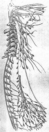
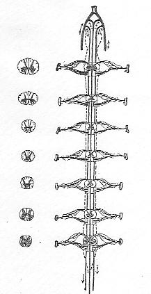
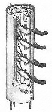
There are from 500 to 600 of these lymphatic nodes or glands in the human body. They are small bean-like nodules developed from a plexus or network of the vessels, and are usually surrounded by loose connective tissue. In childhood they are reddish-gray in color, and on section are quite translucent; in advanced life they generally become atrophied and much darker in color. They occur singly in some positions (solitary glands), but generally in groups or chains. They are so placed in loose connective tissue and in the flexures of joints--as to be freely movable. In consequence they admit of considerable enlargement before occasioning pressure symptoms. The glands usually receive two or more afferent vessels. Generally before entering the gland each vessel breaks up into several smaller ones. The efferent vessels are larger and fewer in number. From the ease with which they may be observed, the cervical, axillary and inguinal glands are oftenest noted. From the osteopathic standpoint, the cervical glands receive the most serious consideration, but those of the axilla and groin may by no means be ignored. A knowledge of the relations of the deep and superficial cervical glands, their afferent and efferent vessels, and the different structures drained by the different glands, is of the utmost importance to the osteopath. Lymphoid or adenoid tissue, similar in structure
and function to the nodes, occurs in many situations. It has not
the organized glandular form of the nodes, but consists of a fine network
of anastomosing cells. Where this tissue is clearly defined it is
spoken of as a follicle. In other places it is quite diffuse.
It is abundantly found in the diffuse form in the entire digesetive tract,
while in the follicular aggregations it occurs in the tonsils and in the
small intestines. In the latter location the follicles are known
as Peyers patches or agminated glands.
Lymphatics of the Intestines The lymphatics of the small intestines, while structurally
and functionally identical with the rest of the system, psses the additional
function of absorbing the chyle through the intestinal stomata. There
are really two distinct sets of intestinal vessels. Those of the
mucosa absorb and convey the chyle, and they alone can be correctly spoken
of as the absorbent, lacteal or chyliferous vessels. Those of the
muscularis convey lymph only. Sappey holds that only the vessels
of the intestinal villi absorb chyle, so that they are the only true lacteal
vessels. No such distinction is usually made, it being the custom
to speak of the vessels which have their origin in the small intestines
as lacteals. Except during digestion, the lacteals carry lymph precisely
as do the other lymphatic vessels.*
The Nerve Supply Before taking up the physiology and pathlogy of the lymphatic system, we must inquire as to its nerve supply. Until recently it was thought that the flow of the lymph was due solely to mechanical forces. These are respiratory movements, intra-abdominal pressure, muscular contraction, the difference in pressure between the lymph capillaries and the terminal ducts (pressure at the opening of the ducts being very low or even negative), the inherent contractility of the vessel wwalls, VIS A TERGO, etc. The thirteenth edition of Grays Anatomy contains the following statement: The lymphatics are supplied by nutrient vessels,
which are distributed to their outer and middle coats; but no nervees have
at present been traced into them. In the latest edition the last
clause is replaced by the following: and here also have been traced many
non-medulated nerve-fibres in the form of a fine plexus of fibrils.
Concerning the glands, the following appear in Gray: Little is known of
the nerves, though Kolliker describees some fine nervous filaments passing
into the hilum.
On the other hand, Landois says: The nervous system
has a direct influence upon the movement of lymph through innervation of
the muscles of the lymphatics. In addition there are still other
special effects of the nerves upon the absorptive activity of the lymphatic
radicles. Landois also mentions Golzs experiment, which was as
follows. He injected a dilute salt solution into the subcutaneous
lymph spaces, and found that it was readily absorbed. The absorption
was retarded by division of the nerves to the extremities, and the destruction
of the central nervous system caused the solution to remain unabsorbed.
Hazzard in his Practice of Osteopathy quotes Dr. Stills views on the innervation of the lymphatic system and especially of the thoracic duct, with particular reference to the causation of obesity. He says Dr. Still points out spinal lesion to the full length of the thoracic duct, acting through the various spinal sympathetic connections, splanchnics, etc. He mentions especially lesion at the 4th dorsal, which he calls a center for nutrition, and at the 7th cervical, opposite which the duct ends. He has called attention to lesion in the upper dorsal region, just below the cervical, giving rise to the growth of a fleshy cushion, a condition of affairs that seems to influence the lymphatic system and cause a deposition of fat. He also works high in the cervical region, opposite the transverse processes of the vertebrae, for nerves controlling the caliber of the duct. In his last book, The Abdominal and Pelvic Brain, Byron Robinson employed the following language to show the vasomotor control of the lymphatics: The functioning of the tractus lymphaticus (sensation, peristalsis, absorption and secretion) is controlled by the nervus vasomotorius (sympathetic). The tractus lymphaticus is richly supplied by a plexiform, nodular network, a fenestrated anastomosed meshwork of the nervous vasomotorius which controls its physiology. In view of the findings of many investigators, the
authority of Dr. Still (which is no doubt somewhat empirical, being based
probably on clinical experience), and the positive assertion of so great
an anatomist as Robinson, we are justified in accepting the vasomotor control
of the lymph vessels, subject, however, to the mechanical influences alluded
to above.
The Movement of the Lymph Let us revert to the general movement of lymph, beginning with its transudation through the walls of the blood capillaries. At the outset we are confronted by conflicting views of the origin of the lymph plasma. By some it is thought to be solely an infiltration of blood plasma by osmosis through the capillary walls. A larger number believe it contains in addition a secretion of the capillary endothelium -- a product of cellular activity. The fact that it differs in chemical composition from the blood plasma seems to prove the latter point to be correct. It is a slightly viscid, alkaline fluid, nearly colorless and odorless, varying somewhat in different locations, and carrying variable numbers of leucocytes. The leucocytes are regarded as casual guests of the lymph, and not an essential part of it. Passing into the intercellular lymph spaces beyond the capillaries, bathing all the cells and supplying them with the nutrient properties it contains, and receiving their excretions, the lymph begins its journey back to the blood-stream. Laden with katabolic products, it enters the lymph capillaries, thence passes into the larger vessels, and is carried along to a lymphatic node or so-called gland. It is in these nodes that some of the leucocytes (at least those known as lymphocytes) are generated. The progress of the lymph is somewhat retarded in passing through the nodes, and foreign substances, whether living germs or inert matter, are caught and imprisoned. This accounts for the tenderness and hypertrophy of the nodes in so many pathological conditions. Emerging from the gland, the lymph passes on--generally through several glands--until it reaches the lymphatic duct on the right or the thoracic duct on the left. Through one or the other of these terminal vessels it enters the blood-stream at the junction of the subclavian and internal jugular veins. Before again traversing the arteries, the lymph is modified by entering the pulmonary circulation, where it is charged with oxygen. While we must not overlook the lacteals, which during digestion pour the chyle into the thoracic duct, with that exception we see in the lymphatics a system of closed ducts. In this respect the lymphatic system as a whole resembles the spleen and other ductless glands. This inadequate, imperfect and necessarily brief
sketch of the lymphatic system is presented in order that the salient facts
be before the minds eye. Much is omitted that could profitably have
been introduced did the limitations of this chapter permit. The composition
of the lymph and chyle, a fuller discussion of the lacteals, the question
of selectivity of the absorptive epithelium, the structure of the walls
of the vessels and the nodes, the evolution and degeneration of the leucocytes
and other cellular casual guests of the lymph -- these and many other points
could be dwelt upon, all having a direct bearing on our main subject.
I trust that the readers interest will have been sufficiently aroused
to induce them to pursue the study further.
Functions of the Nodes An important function of the lymphatic nodes has
been only touched upon. I refer to the formation within them of lymph-cells.
Strictly speaking, the lymph nodes are not glands in the ordinary meaning
of the term, but they may be classed with the organs known as ductless
glands -- those forming an internal secretion. In the case of the
lymphatic glands the internal secretion is the lymphocytes. That
these are most important factors in phagocytosis is seen by the large number
of pathological conditions in which the glands become inflamed and swollen.
Who has not seen inguinal glands enlarged and tender from an inflamed corn,
or axillary glands exquisitely painful from a boil on the forearm?
These simple forms of lymphangitis are quickly abated with the subsidence
of the original inflammation. It is amply proven that in these, as
well as in graver conditions, every measure which aids the formation and
circulation of lymph helps to relieve the inflammatory state.
Lymph and the Endocrines All of the foregoing portion of this chapter was
written several years ago, and the author sees no occasion to change or
revise it thus far. He desires, however, to direct attention to the
next two paragraphs, which were also written at the same time.
Again, may it not be true that in the diseases of the ductless glands a great help toward establishing a cure is to be found in agencies that tend to increase lymph pressure and flow? For example, take Graves disease and the serious symptoms it presents (tachycardia, digestive disorders, extreme prostration, exophthalmos, tremors, etc.) and the well-demonstrated fact that osteopathic treatment causes marked improvement. Can we show that correction of the lesions which are found in these cases directly produces the benefit manifested? Not always, I believe. May it not then be reasonably assumed that those symptoms at least which are referable to the auto-intoxication caused by a modified distribution of the thyroids internal secretion are due in part to failure of the lymph to properly transport the secretion? In such event, may not the treatment administered to correct the anatomical faults give the lymph the needed impetus? The above suggestions, though made empirically, seem to be justified by recent research into the functions and relations of the ductless glands. Again quoting the axiom that the blood feeds the lymph and the lymph feeds the cells, we must see the supreme importance of free lymphatic activity. In this connection permit a further quotation: Now that we know the important relation existing between the various secretory glands, and among these the ovary and the testicle are not the least important, we can understand that in dementia precox menstruation is delayed or that there is sexual precocity, for menstruation is a pluriglandular cyclic process. We can understand sexual excesses, vagaries and perversions. It is easily understood why the symptoms are brought out or accentuated by menstruation or brought on by pregnancy, repeated pregnancy, or by miscarriage. Hence DEMENTIA PRECOX IS A SERIOUS AND EXTREME TYPE OF ENDOCRINE ABERRATION OR ABNORMALITY EVIDENCING ITS PRESENCE BY PSYCHIC RATHER THAN SOMATIC ALTERATIONS. So as we delve into one mental aberration after another, the internal secretory glands seem to be more and more related to conditions characterized by psychic manifestations. The physical and mental development of the individual are dependent on the action and interaction of the ductless glands. The nutrition of the body, of the mind and of the sex organs, as we are learning more and more, are dependent on the trophic stimuli of the ductless gland system. Long before the trophic relation between the various glands and the ovary is evidenced by menstruation and development of the secondary sex characteristics, these glands are concerned with the body growth. The physical and mental development of a growing
child is dependent upon the activity of the hypothysis gland and particularly
the thyroid gland. Bony growth is of course related to calcium letabolism
and here the thymus and the parathyroids and adrenals are of importance.
The thymus and the parathyroid glands are particularly concerned with calcium
metabolism and, to this degree and probably in ways which we do not yet
understand, they are intimately associated therefore with bony growth and
the development of the skeleton. We do know that hypothymism causes
short bones, thin bones, fragile bones. We know the lack of physical
and mental development in cretinism. We know that dwarfs may also
be due to an underactivity of the hypophysis gland.*
I would direct attention to that common affection of the cervical nodes, tubercular adenitis. So frequently does it occur in childhood that one writer says 96 per cent of all children become infected at one time or another with tuberculosis of the cervical lymphatics. The fact that so often a tubercular gland or group of glands will remain quiescent for a long period may lead to the belief that there exists a simple adenitis, and local treatment may light up the tubercular process, with very bad results. If there is any likelihood whatever of tubercular infection all work in the cervical region should be done in the most guarded manner, to avoid causing inflammation which may terminate in suppuration. Glands which have softened can be dealt with only surgically. The general treatment of the patient in all such tuberculous cases is the same as in pulmonary and other forms of tuberculosis -- outdoor life, an abundance of nutritious, easily digested food, together with appropriate osteopathic treatment. Transubstantiation in the Lymph-Stream In searching for the elusive, mysterious seat of
that transubstantiation whereby the assimilated portion of the ingested
bread becomes the actual body; in seeking to uncover that stage of the
anabolic process at which protein is transformed into protoplasm, it is
through the lymph-stream that we shall, if ever, discover the ultimate
metamorphosis.
The Lymph and Nutrition To the inquiring mind countless channels of thought
are opened by questioning the influence of lymph formation and flow in
relation to many diseases. How can we direct nutrition to impoverished
cells? How can we through the lymphatics reach and stimulate the
emunctories and accelerate excretions? If we learn to accomplish
these and other results, in some measure, then is this study not wholly
in vain, and it behooves us to hearken to Dr. Still as he bids us to go
deeper. In the present chapter I can do little more than indicate
some possibilities. The questions may be taken up in a later article,
but in the meantime each must elaborate the subject for himself.
The lymphatic system is as necessary to the body as the quartermaster department is to an army. The army may have the finest uniforms, the best ammunition, and all the accompanying impediments, but if provisions are not forthcoming there is speedy disintegration. The whole mechanism is upset. So with the body. Without the continual supply of lymph to all the parts we would have wasting, disease, and death. Every living cell must have its continual supply of nutrition, or it will cease functioning. When the army is going through a battle the supplies
must still keep coming, in greater abundance and with greater efficiency
than before. If the depletion in the ranks is too great, sometimes
the deficiency in the fighting force is filled by members of the quartermaster
corps, who fall into the breach and are quickly developed into fighting
men. They may not perform their new duties so well as the trained
regulars, but after a time it would be almost impossible to distinguish
them from the veterans.
The Lymph in Orthopedic Surgery Here we can draw a very fair comparison between an army and the body. The lymph plays an all-important part in the metabolic processes, through its double role of nutritional supply and filtering process in the nodes, but in regeneration of tissue it takes on a third function, namely, cell forming. This function is very necessary in repair cases, following operation, and so must be of great interest to the orthopedic surgeon, who looks for the formation of new tissues and structures. Here it is that the lymph throws its cells into the breach to fill up the gaps in the ranks of the regulars. Take the simple operation of subcutaneous tenotomy, so necessary in many cases of talipes, particularly those of the equinus types. It is not unusual in the authors experience to sever the tendo Achillis without the external loss of two drops of blood, by making an incision parallel to the tendon and turning the knife so as to cut squarely across the tendon. What follows? There is rapid exudation of lymph from both cut ends, and new cells are formed to fill the interspace, though it be an inch or more in extent. Within eight weeks new tendon and sheath fill the gap. The external wound is scarcely visible and the new portion of the tendon is fully as strong as the old. In any process of regeneration there must be the regular line tissue which leads in the action. We look back to the embryo, even back to the blastula and gastrula stages in the development of any animal, and we find that development took place in regular lines and layers of cells, uniform and homogeneous. Later, some of these cells, or groups of cells, were cut off from the rest of the regular layer, and then began to take on new characteristics. They became specialized, and lost all apparent connection with their primitive brethren. In the main, however, the greater part of these first cells developed, and in the mature human we find them divided into two great classes, epithelium and endothelium. The epithelium refers to the layers of cells which line all systems which open into the exterior, such as the skin, digestive tract, respiratory tract, and the uro-genital tract. Endothelium means the layers of cells which line all closed systems -- blood vessels, peritoneum, pleura, etc. These are in reality primitive cells, and it is these cells which are most easily replaced. In regeneration, then, the organs which are most quickly repaired must necessarily be those which have epithelial or endothelial connections. A wound in the skin, for example, if it heals by first intention, will very quickly be restored to normal. The epithelial cells in the immediate vicinity react with astonishing rapidity. An extra supply of lymph is rushed in, and lymphocytes are ingested in great quantities by the epithelial warriors. They dont even wait to take food from the cells; they take cells and all, and in their increased metabolic activity transfer everything into their own protoplasm. Thus active karyokenesis is established, the epithelial cells become spindle-shaped as they go on developing, fibroblasts are formed, and soon we find that these primitive cells have made themselves into the next number in the series, connective tissue. Some authors say that connective tissue is the most primitive, in that it never develops very highly, but from an embryological standpoint, surely, epithelial tissue has every claim. The exception, of course, is nervous tissue, which comes directly from the ectodermal layer, but it goes through so much differentiation, this master tissue, that it is hardly just to classify it with epithelium. This newly-formed tissue, unless it is very superficial, will not become exactly like the old structure, but a cicatrix will be left, composed of white fibrous connective tissue, formed from the epithelial fibroblasts. Even before this process is finished, to illustrate how important is nutrition, the lymph capillaries in the region surrounding the cicatrix bring increased pressure to bear by dilating with their fluid, and actually push their endothelial linings through the hard, heterogeneous mass in order to rush in supplies to the newly-formed member. The blood vessels, of course, are doing likewise, because there is always harmony of action between the blood and lymphatic circulation. When big gaps are made in structures, as in the case
of an operation when part of a structure is removed, the parenchymatous
cells are not regenerated. Specialization has gone on too far for
new cells to be regenerated from primitive tissue. The veterans have
been destroyed. They cannot be replaced. The space is filled
with a debris of blood cells, epithelial and endothelial cells, and a great
deal of lymph. At first there is only a mass of material, an indistinguishable
conglomeration. Gradually, however, the forces of nature begin to
pick and choose, the chaff is separated from the wheat, and organization
begins to come out of chaos. The live cells ingest food and undergo
karyokinesis. The lymph stream carries off the products of metabolism
and other waste and poisons collected in this region, connective tissue
if formed, and soon the regeneration has taken place, the structure is
once more complete. In tissues where great specialization has not
taken place, as in tendon, cartilage and bone, practically complete restoration
of the original tissue is the result, providing that the germinal epithelial
linings (i.e., periosteum and perichondrium) have been preserved.
In organs where great specialization has taken place, as in the kidney,
restoration of the parenchyma is not effected.
To Stimulate Lymph Flow Regarding the FLOW Kirkes says: The flow of lymph may be increased by increasing the capillary pressure. This may be done by injecting a large amount of fluid into the circulation, or by the injection of such substances as sugar and salt into the blood. Various observers hold that the receptaculum chyli undergoes rhythmical contractions, in which case we may conclude that the pressure and flow of lymph are greatly increased after the ingestion of food. Some drugs (as curare) increase the flow of lymph, and it can be done locally by ligation of the veins. Biers constriction method of inducing local passive hyperemia deserves mention. However, with none of these means have we at present any concern. Among the noteworthy methods are: (1) Deep breathing.
With each inspiration the flow of blood through the innominate veins causes
a suction at the openings of the thoracic and right lymphatic ducts.
This may be augmented by intre-abdominal pressure if the abdomen is forcibly
drawn in. (2) Manipulation of the extremities by flexion of the joints
and compression of muscles. This may be either active or passive.
(3) Raising intra-abdominal blood-pressure by direct work over the abdomen
and by compressing the ribs. (4) Restoring normal tone to the diaphragm
if it is prolapsed or relaxed. Dr. Still suggests that such prolapse
may cause embarrassment to the thoracic duct. Hazzard elaborates
this suggestion in a chapter in his Practice entitled An Osteopathic Study
of the Diaphragm, which is well worth pondering. (5) Drinking hot
water, or preferably hot salt solution, or injecting the same per rectum
and retaining it.
The VOLUME of lymph may be increased in various ways, among which may be mentioned (1) Active and passive muscular movements. Landois says: Muscular activity causes increased lymph production as well as a more rapid escape of the lymph. The tendons and fasciae of the skeletal muscles, which possess numerous small stomata, absorb lymph from the muscular tissue, (2) Increase of blood-pressure by any of the manipulative means noted above. In this connection readers are commended to carefully study an address given by Dr. Hazzard at St. Louis in 1904, on Osteopathic Manipulation of the Blood-Mass. (3) Quantities of hot water or salt solution per os or per rectum. The reason for advising the use of hot water rather than cold lies in the fact that heat dilates the blood vessels, and absorption takes place more rapidly, while cold water causes contraction of the vessels. It will be remembered that Byron Robinson held that the lymphatic vessels, through their vasomotor innervation, possess the four functions of peristalsis, absorption, secretion and sensation (easily remembered by the word PASS, formed by the initial letters), and he ascribed the cause of most diseases in which the lymph and its flow are concerned (and does not this embrace much of pathology?) to either excessive, deficient or unbalanced activity in these functions. He strongly urged the importance in constipation and other chronic conditions which the osteopath is frequently called to treat, of giving regularly large quantities of water -- better hot -- and salt. Seemingly afraid that his patients would scorn or neglect to take plain salt, he gave them NaCl tablets, flavored to disguise the taste, directing that they be placed on the tongue and swallowed with the water. Salt is especially beneficial in that it stimulates the epithelium of the salivary, pancreatic and hepatic glands, the entire digestive tract, the urinary organs, etc. (The one condition in which it is contra-indicated is parenchymatous nephritis, as it is irritating to the inflamed kidney cells.) Both blood plasma and lymph plasma contain more than one-half of one per cent of salt. All of the glandular secretions contain salt. Salt is an important digestant, especially of vegetables. It is certainly a rational procedure to promote cellular activity by making use of this universal stimulant in the manner indicated above. Why should we seek to increase the volume and flow of lymph? Because only by having an ample fluid medium can the maximum energy of the cells be attained. It has been proved by Metchnikoff, Bizzozero and others of Virchows school that the normal individual cell is endowed with and exercises a power of self-defense and self-preservation against invasion. Only with a sufficient volume of lymph is normality of the cells assured -- through irrigation of the lymph spaces, maintenance of the nutrition of the cells, and free drainage of the excretions. And only through the continuance of maximum cellular activity can perfect health be maintained. |