
|
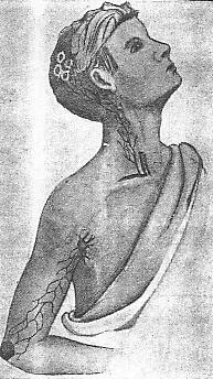
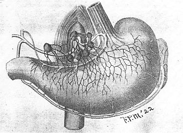
The purpose of this chapter is to show the relation between lymph glands and infections, and also the relation between lymph glands and malignant diseases in various parts of the body. A focal infection may spread throughout the body along the lymph channels or through the blood stream. Where the infection travels along the lymph channels, we usually find the lymph glands involved, which involvement is characterized by an enlargement of the glands. This is probably a part of our defensive mechanism against the spread of an infection. The same thing also applies in cases of malignant disease. The cancer cells traveling through the lymph spaces attack the lymph glands in close proximity to the seat of the disease and this involvement is characterized by an enlargement of the glands. Cancer cells are also carried through the blood stream. I herewith report a number of cases that have recently come under our observation wherein the involvement of the lymph glands in the neighborhood of an infection or in some cases at a considerable distance from the primary infection was manifested by an enlargement of the glands and in some cases by their destruction, but in all cases of infection herein reported the infection was limited to the original focus and the neighboring lymph glands which, no doubt, performed the function of preventing a further spread of the infection even though the lymph glands were in some instances destroyed. In all cases of malignant disease where the lymph nodules were involved secondarily, there was no improvement except where the malignant focus was removed altogether with all lymph glands involved. CASE 1. A lad eight years old was brought to the hospital with a mild infection involving the right elbow. There was limited motion but not any great amount of pain or swelling. An X-ray picture did not show any involvement of the bone. A careful examination revealed a very active infection of both tonsils. These were enucleated and the adenoids removed cleanly. The infection in the arm became quiescent after a week or ten days. In this case undoubtedly the infection in the arm was secondary to the infection in the throat, and the infection reached the arm through the blood stream. The arm was given no treatment of a manipulative character except a very gentle massage. Hot applications, however, were applied for a few days. The special feature of this case, however, was an involvement of the lymph glands in the back of the head, four or five of the mastoid and occipital glands were enlarged to the size of a large hazelnut. They were not especially tender, however, on pressure, and were quite hard. There did not seem to be any relation between the enlargement of these glands and the infection in the throat, but there were several patches on the top of his head, some of them almost as large as a silver dollar, where there was an active infection with a sticky excretion which caused the hair to mat. These sores on his head were healed after about two weeks treatment, which consisted of clipping the hair close to the scalp and cleansing the sores with soap and then putting on zinc oxide ointment. Undoubtedly this infection in his scalp was responsible for the enlargement of the glands in the occipital and mastoid regions, as the lymph vessels range from the top of the head downward. Following the healing of the sores on the boys head, the enlarged lymph glands gradually receded. The accompanying illustration shows the lymph glands involved and the source of the infection. In cases of this kind, the enlarged lymph glands require no direct special treatment where the glands have not broken down. Where the primary focal infection is cleaned up, the glands will recede to normal size. Where the glands have suppurated, drainage by incision is necessary. PLATE LXX. -- Case 1. CASE 2. The patient, a girl about 15 years of age, had a swelling on the right side of her neck nearly as large as a hens egg. The lump was immediately in front of the sternomastoid muscle and the top of it was about on a level with the angle of the jaw. This swwelling persisted for a number of months. It appeared to be a single lymph gland greatly enlarged, although there may have been sseveral glands which had coalesced. There seemed to be no tendency for the enlargement to recede. The patient gave a history of having had a considerable amount of throat infection, although at the time the case came under our observation the throat seemed to be fairly clean. The question arose as to the nature of the swelling and as to the best treatment to apply to reduce it. As the parents of the child were adverse to operation, an attempt was made to reduce the swelling by treating the neck osteopathically, and also bythe use of applications such as antiphlogistine, hot water, etc. This treatment, howevert, was carried on without success. Next the swelling was incised and a considerable amount of pus evacuated. A drain was introduced and this treatment for the time being reduced the swelling to a considerable extent. After a time the drain was discarded an the incision healed, but in a short time the gland swelled up again as large as it was before treatment. We next opened the abscess and after draining out a considerable quantity of pus, injected the cavity with bismuth paste. This treatment afater being tried for a couple of months proved to be unsuccessful. Finally, under an anesthetic, the swelling was opened freely and the wall of the abscess dissected away. The wound was drained for a few days only and healed readily without any further recurrence of the trouble. I do not believe this infection was tubercular in character as the infection was limited to one area. The glands in this case were secondarily involved from an infection in the throat of a pyrogenic character. PLATE LXXI. -- Case 2. Tuberculosis of the lymph glands is characterized usually by the fact that a considerable number are involved, and in the beginning the glands are very hard and do not enlarge rapidly. I might add in this connection that as a rule tubercular glands, if the proper hygienic treatment is carried on, will recede without surgical interference. As a rule, enlarged lymph glands in the neck in front of the sternomastoid muscle and below the jaw, where the condition is due to acute or chronic tonillitis, will recede to a normal state after the condition in the throat is cured, whether by operation, as the removal of the tonsils, or by other means. We do not consider it wise as a rule to manipulate an enlarged lymph gland. The treatment should be directed to the cause of the trouble and the gland itself requires no treatment of any character except perhaps local applications, unless the enlargement is considerable and persists for some time or unless the gland suppurates and breaks down. In the case of suppuration, usually drainage is all that is required. Occasionally excision as in the present case will be necessary. CASE 3. A lad 10 years of age injured his leg
about six inches above the ankle by striking it against the tongue of a
cultivator. The injury at the time was considered trivial, although
it was somewhat painful for a few minutes. No attention was paid
to the bruise for a week or ten days, when the lad developed a considerable
swelling at the place of injury and also complained of pain. An attempt
was made at treatment by home remedies and bandaging, but there was no
improvement, and after about a month the boy was brought to the hospital
at which time he was running a little temperature and the pain in the leg,
although not severe, was enough to cause him considerable annoyance.
There was a swelling at the site of the injury and the skin was considerably
discolored for an area of several inches. Although the skin was not
broken at the time of injury, there was evidently an infection here which
was characterized by a soft swelling, discoloration of the skin and temperature.
The infection was evidently carried to the involved tissues through the
blood stream, gaining entrance to the body probably through the throat,
although there was no history of any special throat trouble. The
infection proved to be quite superficial, lying between the fat and down
upon the fascia overlying the muscles just a little outside of the tibia.
This was incised for a couple of inches and a considerable quantity of
pus escaped. The cavity was curretted and drained. It healed
in about two weeks.
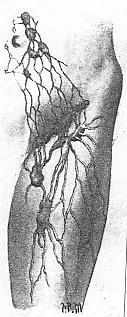
The special feature of this case, and for which it is principally presented, was the involvement of the inguinal lymph glands. These glands were enlarged, some of them to half the size of a hens egg. One gland had suppurated and required incision for drainage. Following the drainage of the abscess in the leg and the incision for drainage of the lymph gland that had suppurated, the balance of the glands receded to normal size in about ten days. The only treatment was rest in bed. Undoubtedly infection from the leg in this case followed along the lymph channels until the lymph glands in the groin were reached, and here the infection was limited. The accompanying illustration shows the route the infection travelled to reach the lymph glands in the groin. It is not uncommon for an infection in the foot, particularly an infection from an ingrown toe-nail, to cause an involvement of the lymph glands in the groin. CASE 4. I wish next to report a case of involvement
of the deep lymph glands along the external iliac vessels. The case
was unusual for the reason that the glands were so large that they had
been mistaken for a fibroid tumor of the uterus. This patient, a
young woman about 30 years of age, had given birth to a child about three
months before she came under our observation. There was a history
of unsatisfactory convalescence following delivery. The patient had
never been well enough to nurse her baby. On examining the case after
she was brought into the hospital, we found a large solid growth extending
a little past the mid-line of the abdomen on the right and up to within
two inches of the umbilicus, and then extending clear over to the left
side forming an immovable mass. The superficial lymph glands in the
inguinal region were also enlarged. There was a slight discharge
of pus from the uterus; she was running an irregular temperature.
An incision was made over the mass but without opening the peritoneal cavity,
as the mass below was attached to the abdominal wall. A finger was
inserted down into the mass and a large quantity of pus escaped.
A drainage tube was then introduced and left in the wound until drainage
ceased and the mass reduced. This required about three weeks.
The patient made a very satisfactory recovery although the wound was slow
in healing, a little discharge occurring for four or five weeks.
The entire mass, however, entirely disappeared after six weeks.
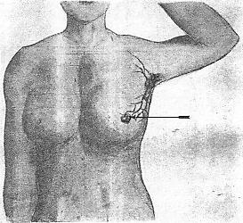
The infection in this case undoubtedly came from the uterus and occurred in connection with delivery, due probably to the introduction of unclean hands or instruments into the vagina or uterus. When I first saw the case I was somewhat in doubt as to whether or not there might be a malignant condition. When the mass was incised and pus escaped, I was then satisfied that we had only an infection to deal with. The subsequent history of the case demonstrated this to be true. The accompanying illustration shows the lymph drainage from uterus and the lymph glands along the iliac vessels. The reason cancer of the uterus is so frequently ultimately a fatal disease is on account of the involvement of these lymph glands, which cannot always be entirely removed, even though wide dissection of the parametrium is made at the time of operation for the removal of the uterus. Where malignant lymph glands remain after the removal of the uterus, the disease recurs. CASE 5, is that of a young man about twenty, an ex-soldier. He was brought to the hospital for treatment for a disabled hip. He was unable to walk due to the disability and it was thought that perhaps the condition was one which might be relieved by some sort of treatment. The history was not clear except that the hip had grown progressively worse following a slight injury. At the time we saw the case the hip was enlarged from the crest of the ilium down to six or eight inches below the great trochanter. There was an especially large swelling in the gluteal region. Both the deep and superficial lymph glands in the groin and abdomen on the same side were extensively enlarged. There was a firm mass here which extended well into the abdomen. The young man had lost considerable weight, but so long as he was quiet in bed there was little or no pain. There was no temperature. An incision was made over the large mass in the gluteal region and a large quantity of broken down, flesh-like material removed. The object of the operation was to determine the character of the growth. No pus was present. An examination of the tissue proved it to be sarcoma which probably had originated in the region of the hip, either in the soft tissues or from the bone. The involvement of the lymph glands, of course, was entirely secondary in this particular case. The wound was sutured and healed without any trouble. The disease, however, progessed steadily and the young man was sent home. He died about two or three months later. PLATE LXXIV. -- Case 5. This case serves to illustrate the involvement of lymph glands in malignant disease. The glands involved are those adjacent to the seat of disease. The object of the incision was to determine whether the disease in the hip was malignant or due to infection. It was quite evident, however, even before operation, that the condition was malignant, but as there was no risk in making the incision it was done with the hope that the condition might prove to be an infection, in which case an improvement could have been expected from the treatment. CASE 6. This case is given for the purpose of illustrating the involvement of axillary lymph glands. The patient, Mrs. M., 55 years of age, was examined only recently for an enlargement in her breast. It was a single lump about the size of a walnut just a little to the outside of the nipple. The lump had given her considerable pain, and there had been a discharge of blood and serum through the nipple for a couple of months. The lump, however, was not attached to the skin nor to the muscle beneath. Upon examining the axilla, we found a few lymph glands slightly enlarged. The patient was considerably distressed about the condition and thought it might be malignant. I was inclined to believe that it was non-malignant, but recommended operation. The breast was removed and the axilla cleaned out, stripping out the fat and the lymph glands abong the axillary vessels. The tumor was cut open and proved to be a broken down cyst. The cyst had become infected and the lymph glands were enlarged secondarily to the infection. There was no evidence of malignancy, although condition of this kind if allowed to run often become malignant, and for that reason the operation was advised. PLATE LXXV. -- Case 6. In this case it was not found necessary to remove the pectoral muscles. The patient, of course, has experienced a great deal of relief both in mind and body following the operation and may, of course, expect permanent relief without fear of recurrence. I wish in this connection to report a case of carcinoma of the breast in a woman of about 60 years of age where the condition had existed for over a year. The skin for about an inch about the nipples had broken down and there was a large lump as big as ones fist in the middle of the breast. The lymph glands in the axilla were palpable, some of them as large as a small sized hickory nut. The lump, however, was movable. In this case, a very wide incision was made and the breast and pectoralis major and minor muscles were removed down to the ribs. The fat and lymph glands were removed from the axilla for a considerable distance. Whether or not the operation will eventually prove successful depends, of course, upon whether or not all of the lymph glands containing cancer cells were removed. There did not appear to be any involvement of the tissue outside of the breast itself excepting the skin about the nipple and the lymph glands. The wound healed readily and the patient regained good use of the arm. The operation was performed only a year ago and to date there has been no recurrence. We are not safe, however, until a number of years have elapsed in saying that the disease has been entirely eradicated. I recall one case of cancer of the lung which developed 10 years after the removal of the breast. I recall another case in which the breast and lymph glands had been removed and where the disease recurred several years later in the axillary vessels and nerves, resulting in obstruction of the circulation to the arm and intense pain due to the encroachment of the disease upon the nerves. In cancer of the breast unless the operation is comparatively early and the breast and underlying fascia and axillary lymph glands are completely removed, the disease is very apt to recur. The most common site of recurrence is in the axillary tissue, but not infrequently metastatic cancer appears in the lung, pleura, spinal column or brain. The accompanying illustration shows the lymph drainage from the breast and the way by which the lymph glands in the axilla become involved at a comparatively early date following the appearance of malignancy of the breast. CASE 7. This case, Mrs. C., aged 58 years, came to the hospital for examination giving a history of having had stomach trouble for about a year. Her general condition seemed fairly good although she had lost some weight. There had been more or less distress for a number of months after eating and she had for some months vomited quite a good deal, although for several months just preceding the time of examination there had been very little vomiting. A barium meal was given and the patients stomach examined with the fluoroscope. There was quite an extensive filling defect along the region of the lesser curvature. As the patient was rather heavy, nothing of a very definite nature could be determined upon palpation except that the stomach was tender. An examination of the stomach contents was not made. The diagnosis of obstruction was made from the fluoroscopic examination and it was thought that the condition was either cancer or obstruction due to old ulcers. An exploratory operation was advised and accepted by the patient. An incision was made in the mid-line between the ensiform and umbilicus and the stomach examined. There was an extensive cancer which involved practically all of the lesser curvature and extending well down toward the great curvature. The stomach could not be brought out through the wound. The lymph glands, particularly along the lesser curvature, could be palpated. On account of the extensive nature of the disease, no attempt was made to resect the stomach. There was no evident involvement of the liver. The wound was sewed up and the patient recovered from the operation and left the hospital in three weeks. PLATE LXXVI. -- Case 7 This case is reported to show the futility of attempting to resect the stomach except where the operation is done early and while the disease is still confined to a small area, which, together with the adjacent lymph glands, can be removed by resection. The accompanying illustration shows the blood supply to the stomach, its lymphatics, and adjacent lymph nodules. Along the lesser curvature the lymphatics run in a direction away from the pylorus to terminate in the nodes along this border of the stomach. Along the greater curvature, the drainage is towards the pylorus. CASE 8. Mrs. M., age 60 had ben under a doctors care for three months previous to the time she was referred to me. The nature of her trouble had not been determined, although she had lost considerable weight and had suffered a great deal from distention of the abdomen, vomiting and constipation. Her condition became rapidly worse for a week preceding the time she was brought to the hospital. Upon examination we found her abdomen distended and upon palpation found a small lump low in the abdomen on the right side which could be moved about. The patient had not been able to retain food for almost a week. There was evidence of obstruction of the bowel, the nature of which wew were unable to determine at the time of examination. Operation was recommended which was performed as soon as the patient could be prepared. The abdomen was opened with a right rectus incision and upon exploring we found an obstruction in the small intestine which proved to be a growth inside of the gut which almost completely obstructed it. The lymph nodes in the mesentery were also involved. About six inches of gut were resected and a V shaped section of the mesentery removed back of its posterior attachment. Both ends of the bowel were closed by purse-string suture and a lateral anastomosis performed. The patient made a very good recovery and has been well up to date, two years following the operation. An examination of the tumor proved it to be carcinoma. The probabilities are that the patient will have no recurrence, but we cannot positively state this to be true until several years more have elapsed. This case is presented for the purpose of showing how cancer of the colon or small intestine involves the lymph glands of the mesentery, and an operation, unless it is sufficiently wide in resection of the gut and mesentery to include all of the involved lymph glands, there is certain to be early recurrence of the disease. The accompanying illustration shows the extent of the involvement of the lymph glands in this particular case and the rather wide resection necessary for their removal. PLATE LXXVII. -- Case 8. |