
|
The manner in which the cells of the blood have been developed, both ontogenetically and phylogenetically, is of great interest. Many problems presented by the cells of the blood in the leukemias and in patients with abnormal inheritance are explained by a knowledge of the biological relations of the blood. An accurate understanding of the structure and the function of the red bone marrow is especially desirable, and much work has been done in the study of this important tissue. Unfortunately we have only begun to pick up a few facts and these have not yet been properly classified. The importance of the red bone marrow in physiological economy is indicated by the fact that this is one of the last of the tissues of the body to lose its blood supply as a result of bleeding or of any other cause of anemia. Animals which have been bled to death retain a fairly
adequate blood supply to the bone marrow as well as to the heart, lungs,
digestive tract and respiratory muscles long after the muscles of locomotion,
the skin, spleen and omentum have become apparently completely bloodless.
In the anemias the bone marrow retains its blood supply long after other
tissues become very pale. Yet the marrow is affected by many abnormal
conditions and the effects of these may be very severe.
The marrow is seriously affected by vertebral and costal lesions which affect the innervation of the blood vessels and the cells of the hematopoietic tissues. Such lesions and their effects have been studied in osteopathic institutions. When vertebrae or ribs are lesioned there is a condition of strain in and around the affected joints. The joint surfaces are abundantly supplied with sensory nerve endings and these are subjected to abnormal pressure due to this tension. The swelling and edema which always follow any strain of joints increase the synovial fluid, and this also irritates the sensory nerve endings. The central relations of the sensory nerves distributed to the joints are interesting. Normally sensations from the joint surfaces are not carried to the cerebral centers, but they exert an important influence upon the nerve centers of the spinal cord. Impulses derived from any joint surface affect the spinal nerve centers which control the skeletal muscles moving that joint and the viscera which were derived, embryologically, from the same somites. When vertebrae or ribs are lesioned the abnormal sensory impulses affect the centers which control the small, deep spinal muscles and the intercostal muscles. These muscles are stimulated constantly and are thrown into a condition of hypertonicity, which is called contracture by several pathologists. This constant hypertonicity of the spinal and intercostals muscles tends to perpetuate the lesion. Several interesting changes occur in muscles subjected to this abnormal form of stimulation and in time a condition of rigor is present. Still later fibrosis occurs and after this time complete recovery of the normal condition of the muscle is probably impossible. The visceral centers affected at the same time by the abnormal sensory impulses from lesioned articulations include the centers governing the red bone marrow of the bones concerned in the lesion. Again, when vertebrae or ribs are lesioned there is some swelling or edema of the surrounding tissues. This edema is not yet well understood, but the effects produced by a strained ankle or a bunion are too well known for the facts of the case to be doubted. This edema is always present around lesioned vertebrae or ribs. Edematous fluids are not quite identical with normal tissue juices; in edema there is some acidosis, some accumulation of carbonic acid and other katabolites and other effects of the disturbed circulation through the affected area. The tissues around the thoracic vertebrae and the ribs are so constructed that in lesions of these bones the intervertebral nerves and the adjacent sympathetic ganglia are subjected to considerable pressure, due to the edema. The nerves passing through the intervertebral foramina to and from the sympathetic ganglia upon the heads of the ribs pass through the edematous tissues and are thus subjected to pressure and to the effects of the abnormal tissue juices. Medullated nerves are not quite so seriously injured as are the non-medullated nerves and the latter carry impulses from the sympathetic ganglia to the blood vessels and to the viscera innervated from the same spinal segment as that concerned in the lesion. The nervous control of the red bone marrow is not yet well understood. The nerve fibers entering the bone marrow in any part of the skeleton include medullated fibers, which seem to be sensory, and non-medullated fibers derived from the sympathetic system. Of the sympathetic fibers at least two groups are present, vasomotor nerves which terminate in plates upon the walls of the blood vessels, mostly the arterioles, and other nerves which terminate in fine brush-like endings which lie free among the intrinsic cells of the bone marrow. The sensory nerves are distributed chiefly to the periosteum and to the region of the red bone marrow nearest the bony walls. In animals, a bony lesion affecting the circulation through any area of red bone marrow is followed by atrophy of the marrow cells of that area. Within two or three years after such a lesion has been produced, the interior of the bone shows pale, hardened areas in which bony spicules and connective tisssue are associated with only a few pale areas of remaining marrow cells. These latter areas contain little or no evidence of active blood development, but are made up chiefly of blood vessels, connective tissue cells and a few atrophic remnants of blood-forming tissues. The effects of various diseases and poisons upon the bone marrow have been studied in many laboratories, and the discussion of the fats in this connection is best associated with the diseases in which such marrow changes are of interest. A brief review of the structure of the red bone marrow
may be of interest.
The blood of adult marrow includes all the cells present in fetal blood, but the proportion of younger cells is greater in the marrow from younger individuals and in lower animals. Since the bone marrow is the chief site for the manufacture of red cells and the granular white cells, it is easy to find immature cells of the two groups abundantly present in the red marrow. Hyaline cells are abundant in the red bone marrow, especially in younger subjects and fetuses. Cells which cannot be differentiated from true lymphocytes by any method of staining now in general use are abundant. Larger cells which resemble large lymphocytes are present in about the same relative proportions to the small hyaline cells as is the case in the blood. There is much difference of opinion as to the origin, functions and destination of these cells. (Plates V, VI.) There are also hyaline cells which are definitely myeloid in origin and which seem to be derived from the reticular cells. These cells may or may not give the oxidase reaction. The hyaline myeloblast (non-granular marrow cells; lymphoid mother cell; undifferentiated leucoblast; hemocytoblast; lymphoid hemoblast; microlymphoidocyte; macrolymphoidocyte; stem cell; indifferent lymphoid germinal cell) is a cell which is rounded, occasionally somewhat irregular in form, with hyaline, basophilic protoplasm and a nuclear structure which is characteristic. The myeloblast seems soft and has relatively great protoplasm. There are no true granules but the protoplasm is faintly and irregularly granular in structure. Vacuoles are found within the protoplasm frequently. The nucleus has a fine, delicate reticular appearance with one to several fairly large nucleoli. The lymphoblast differs from the hyaline myeloblast in being somewhat firmer in structure and in having a smaller nucleus with coarser chromatin masses and a heavier nuclear membrane. Azur granules are sometimes found within the protoplasm of the lymphoblast but not the myeloblast. The oxidase reaction is of little or no value in distinguishing these primordial cells, because immature forms do not always give the oxidase reaction even though they are of the myelocytic group. The nucleus of the myeloblast has a definite nuclear membrane which is very delicate and very thin; it is sometimes difficult to see this membrane but it can be shown by careful study in all cells of this type. The chromatin is rather evenly distributed throughout the nucleus, but may be somewhat denser around the nucleoli. The parachromatin is abundant and the nucleus generally appears pale on this account. Under pathological conditions the parachromatin is often relatively diminished so that the nucleus appears much deeper in color. The chromatin granules are distinctly demarcated from the parachromatin (oxychromatin) in these myeloblasts, in which respect they are differentiated from lymphocyte nuclei; in the latter the chromatin granules have indistinct outline. In the myeloblasts which seem to be younger the chromatin granules present a stippled appearance. With further development the granules tend to arrange themselves in strands with node-like masses at the intersections. From two to five nucleoli are present within the myeloblast nucleus. These nucleoli within the same nucleus present differences in staining reactions, indicating that they have different chemical structure. These are smaller than the nucleoli which may be found in sections of lymphoid tissue. Variations in the structure of the myeloblast are found in the leukemias and under other pathological conditions. Such cells may be found undergoing mitosis in the circulating blood, especially in the leukemias. Cells with relatively more abundant protoplasm are sometimes found in the circulating blood in splenomedullary leukemia. Another cell found in this disease has its nucleus indented deeply in several areas, thus producing a lobulated appearance; this is called a Reider cell. These cells are occasionally found in the circulating blood in sarcoma of the bone marrow. They are not found in normal bone marrow at any time. The myeloblastic nature of the Reider cell has been called in question, chiefly on account of the presence of fine granules in its protoplasm. In two of our fulminating cases of leukemia we have found two different cells of this type. In one fine azure granules were present and the chromatin was arranged in coarser masses; no doubt these were immature monocytes. In the other type no azur granules were present and the chromatin was arranged in extremely fine and delicate granules; these Reider cells were certainly properly included with the myeloblasts though the nucleus was deeply lobulated. From the hyaline myeloblast all the cells of the bone marrow, and perhaps the lymphocytes are, or may be, derived. From the myeloblast are developed myelocytes, from which are developed the adult neutrophiles, eosinophiles and basophiles of the circulating blood; megaloblasts, from which are developed the normoblasts and the erythrocytes and also the plasma cells, Turck cells and monocytes. They are increased under abnormal conditions. Under very abnormal conditions de-differentiation seems to occur; leucocytes appear to become transformed into myelocytes or myeloblasts. In one of our cases of aplastic anemia the cell-structure indicated this type of anaplasia. Lymphocytes which are apparently of normal adult type, sometimes dividing, are found in the circulating blood and the daughter cells are morphologically identical with the microlymphoidocytes, or smaller myeloblasts This relation was noted very plainly in one of our cases of sarcoma of the bone marrow associated with marked anemia. Whether the cells from which lymphocytes arise are identical with the myeloblasts or not has not yet been definitely determined. One group of hematologists, called Unitarians or monophyletists, emphasizes the identity of the stem cell which is the progenitor of lymphocytes and other blood cells. The other group, called polyphyletists, recognizes the marked similarity of the primordial cell of the lymphocytes and the primordial cell of the myeloytic series but denies their identity. According to the latter group the cells derived from bone marrow elaborate oxidizing and, perhaps, other ferments, and these cells can be recognized by the oxidase reaction and by other specific methods of staining. Inasmuch as the argument is based on a study of acute or fulminating forms of the leukemias and other seriously abnormal conditions it is evident that the actual facts can be determined only with great difficulty. More spherical than the microlymphoidocyte in the stained smear and somewhat more deeply staining, are other cells not more than five microns in diameter. The protoplasm of these cells may be so thin as to be invisible; possibly it may be absent. These have been supposed to develop into normoblasts. Cells which resemble these, but which have a thicker rim of protoplasm, with a nucleus which is eccentrically placed and occasionally lobulated, are present also. These differ from ordinary lymphocytes in the eccentricity of the nuclear positon and in the finer chromatin structure. Megakaryocytes are hyaline cells with basophilic protoplasm. These have a single, long, ribbon-like, coiled nucleus of characteristic form. They may form red cells by budding off fragments of their peripheral protoplasm; probably this is not a normal source of red cells. There is much reason for believing that they form platelets by budding off very small masses of their protoplasm. Osteoblasts are usually found in the bone marrow. They do not seem to be concerned in blood formation. Large phagocytic cells are sometimes found. They disappear very quickly during the process of preparing the slides for examination. They ingest large numbers of senile or abnormal red cells and other debris. These phagocytic cells are especially over-filled in pernicious and in sickle-cell anemias. They have a single round nucleus and are not readily distinguishable from other monocytes unless they contain fragments of red cells. Similar cells contain droplets resembling myelin glycogen or some related carbohydrate substance, and various other granules which seem to be deutoplasmic. These cells are supposed by Osler and others to develop into normoblasts. Erythroblast is a term applied to several types of cell by different hematologists, but always with the understanding that the cell so named is intermediate between megaloblast and normoblast. These cells may be eight to twelve microns in diameter. The protoplasm stains rather feebly and the nucleus shows a small amount of chromatin which has a typical wheel-like arrangement. The protoplasm contains no granules and scanty or much hemoglobin. Intermediate forms are found in series between any type of cell described as an erythroblast and the cells which are commonly called normoblasts. (Plates VII, VIII.) Normoblasts are nucleated red cells. They are very abundant in the red bone marrow. The protoplasm contains hemoglobin in varying amount; the less the amount of hemoglobin the greater is the basophilia of the protoplasm: with increasing hemoglobin concentration the cells show increasing avidity for eosin. The nuclei of the normoblasts vary greatly. Extrusion of the nuleus is common; so also is gradual dissolution of the nucleus. Intermediate forms are abundant between normoblasts and adult erythrocytes. Normal erythrocytes are also found within the red bone marrow. Normoblasts have been divided by Howell into mature and immature forms. The immature forms are a little larger than normal adult erythrocytes; they stain variably according to their degree of maturity, from basophilic to acidophilic; the chromatin fibers are arranged radially, and they divide by karyokinesis, very rapidly. Mature forms are eosinophilic, as are adult red cells; they are of the same size as adult cells; the nucleus is very small, is deeply staining, often vacuolated, without chromatin structure. Other cells present a peculiar rosette-like arrangement of chromatin within the nuleus. These nuclei are recognizable as erythrocytic even when they seem to have no protoplasm. The bone marrow is very abundantly supplied with granular white blood cells in various stages of development. Eosinophilic forms are not very abundant except under pathological conditions. Eosinophilic myelocytes are larger than adult eosinophiles. They have a basophilic, hyaline, intergranular protoplasm with large round granules which are deeply eosinophilic. The large, round, pale nulei are placed near the periphery of the cells and may be bare for half their circumference. Cells smaller than those with nulei which present indented, saddle-shaped and polymorphic shapes, are present in series leading to the normal adult eosinophiles which appear in the circulating blood. The basophilic, intergranular, hyaline protoplasm gradually diminishes with increasing maturity and is barely visible in the eosinophiles of normal adult human blood. Basophilic granular cells are scanty in normal marrow. Cells which resemble polymorphonuclear neutrophiles except that the fine granules are definitely basophilic are present. Typical mast cells are rare; they may be mononuclear or polymorphonuclear. Amphophilic myelocytes are present in small numbers. Atypical granules in any of the younger forms of basophiles, eosinophiles and neutrophiles are occasionally found even in normal marrow, and they may be so abundant under certain abnormal conditions as to render the differential count of the various forms almost or quite impossible. Neutrophiles of adult forms, younger neutrophiles
which are approximately of adult type, neutrophiles with larger and merely
indented nuclei and all intermediate gradations are present in the bone
marrow. The typical neutrophilic myelocytes are large, sixteen microns
or more in diameter when fresh, and they may reach twenty microns when
spread out in a thin smear. Their nulei are very large, pale, without
marked chromatin structure and they often appear naked because of the thinness
of the protoplasm around them. The protoplasm is scanty, filled with
very fine granules which do not take any stain with avidity but are feebly
neutrophilic. The very early and young myelocyte (Cornils marrow
cell) disappears quickly and it is rather difficult to find good stained
specimens of this form. Many names have been applied to the intermediate
stages of development of the neutrophiles, (metamyelocyte, promyelocytes,)
but intermediate gradations between every stage and the next are present;
there is no logical demarcation between the types. The mononuclear
neutrophile with its round or slightly indented nucleus is often called,
incorrectly, a transitional cell. It is more abundant in the normal
bone marrow than in the normal blood, no matter at what age of the subject
enumerations are made. These cells, which are usually included with
the monocytes of the circulating blood, include several types which differ
only slightly in their nuclear structure and which probably have somewhat
different lines of development. Among these are cells which have
a very fine and delicate arrangement of chromatin in a perfectly round,
pale nucleus; other cells with larger masses of chromatin, a nucleus which
is sometimes indented or even saddle-shaped, and still other cells in which
the chromatin is in large masses connected with delicate fibrillae.
Much further study must be made of the cells of normal human adult bone
marrow before the relations of the different cell types can be accurately
determined.
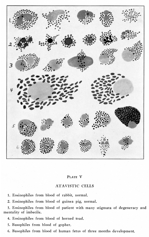
Human adult blood cells show very peculiar and puzzling changes under abnormal conditions. Some of these changes suggest very strongly a condition of atavism or reversion to earlier forms. Atavistic traits are those qualities not normally present in an individual of a certain race or family, but present normally in some distant ancestor. For example, oval blood cells are not present in normal human blood at any stage of development, nor in normal human bone marrow. Oval non-nucleated blood cells are normally present in the blood of the camel and certain related animals and are found, though rarely, in the blood of human beings. When they are so found in human blood, this is considered atavism. This condition of atavism, that is, reversion of human tissues to some ancestral form, is very puzzling. It was formerly supposed that atavism proved direct inheritance, -- that is, that the occurrence of oval non-nucleated cells in human blood, for example, proved that the human race descended from some ancestor whose blood contained cells of this type. It is now supposed that this is not necessarily the case but that the presence of oval red blood ells indicates an ancestry from a race in whose hematopoietic tissues a potentiality of oval forms existed. In other words, the reversion is to some primordial cell structure in which were present the possibilities of development of red cells of different forms, and for some unknown reason in certain human beings whose development has been adversely affected in some way the blood-forming tissues followed the line of development leading to oval red cells rather than the line leading to round red cells. While oval red cells are rare in human blood, several cases have been reported in which they are present, apparently as a developmental anomaly. The ontogenetic development of the mass of the blood cells, as well as of the individual cells, follows the phylogenetic development in many respects. This statement applies to the relationships of the various classes of leucocytes, as well as to their absolute numbers, and it applies also to the variations in the blood cells under physiological or pathological conditions. Under abnormal conditions of the circulation, the nutrition, or the metabolism of the body, the blood, as a mass, tends to revert to its primeval appearances. It is not possible to determine whether the cells themselves actually assume primeval appearances, or whether the formation of new cells, under the abnormal conditions, becomes imperfect and of a more or less embryonic type, and thus the cells found on examination present the characteristics of phylogenetically and ontogenetically immature cells. The latter view is inherently more probable, and it is further supported by the fact that certain irritants in the circulating blood seem to being about first an increase in the relative numbers of cells showing the characteristics of old age, while the continued presence of the irritant is associated with increasing numbers of phylogenetically younger cells. In one of our cases, a young woman apparently healthy, though not robust, had blood with oval cells. The first blood examination was made as a routine procedure in the clinic of The Pacific College of Osteopathy. The oval cells were recognized immediately. During the three years following the first examination her blood was examined many times and the cells always included a large proportion of oval forms. During most of that time she was in ordinarily good health. The oval blood cells showed no changes, and the condition was evidently one of a developmental peculiarity; an instance of atavism. Other cases of oval red cells in human blood have been reported in various periodicals. Developmental anomalies in the blood cells are often associated with developmental anomalies of several other parts of the body, especially those of the nervous system. The recognition of atavistic blood cells may differentiate a developmental and therefore an essentially incurable nervous or mental condition from an acquired neurosis or insanity with similar symptoms. Atavistic cells are present in the blood of paranoiacs, morons, idiots, feeble-minded children, and in various developmental or inherited abnormal states These cells are not present in the blood of persons suffering from nervous or mental symptoms which are due to trauma, acquired diseases or bad training. It is to be remembered in this connection that even those neuroses based on abnormal development may be greatly improved by suitable methods of treatment. Embryonic but not usually atavistic cells are present in the blood after extremely serious demands have been made upon the hematopoietic tissues, as, for example, after repeated hemorrhage, or long infection with virulent pathogenic organisms. Atavistic cells are often present in the leukemias and in erythremia, chlorosis, pernicious anemia and malignancy of the bone marrow. They are rare in aplastic anemia and are not commonly found in ordinary infections nor in ordinary secondary anemias, though these may be extremely serious. A very brief review of some of the cells characteristic
of the blood of certain lower animals, together with the conditions under
which such cells are found in human blood, is interesting, though the relationships
which are concerned in these instances of atavism cannot be explained at
this time.
Most invertebrates carry their oxygen-bearing chemicals in the blood plasma. Certain mollusks and tunicates have colorless globulin-like substances which hold oxygen in a loose combination and carry it to the tissues; these are called achroglobins. Other mollusks and certain crustaceans use copper instead of iron in a pigment called hemocyanin; this also carries the oxygen, feebly bound, to the tissues. Hemocyanin is blue in tint, in arterial blood, and is almost or quite colorless in venous blood. Certain worms carry oxygen by means of an iron-containing green pigment (colorless when reduced) which is called chlorocruorin. Echinoderms and certain other marine animals have a red pigment, also iron-containing, called echinochrom; this is colorless when reduced. The hemoglobin percentage varies rather irregularly. In general, among all animals the hemoglobin increases in ascending scale of vertebrates and from infancy to maturity. In most diseases affecting the blood the hemoglobin is decreased, though in certain diseases in which the amount of water in the blood is diminished, the hemoglobin is relatively increased. The color index is high in lower vertebrates, since
these have extremely large erythrocytes. Among mammals, the color
index increases generally from the lower to the higher forms, and from
infancy to maturity. In most diseases the color index is lower than
normal; in pernicious anemia the color index is high, because of the presence
of many abnormally large red cells. The saturation index is never
increased by any abnormal condition.
The power of the erythrocyte cytoplasm to carry a relatively large amount of hemoglobin is thus one charateristic of the higher development, the more nearly perfect specialization. Vertebrates carry hemoglobin in red blood corpuscles, and all carry small amounts of oxygen free in the plasma. The efficiency of the red cell as an oxygen-carrying structure increases with a fair degree of regularity, though many diversions from the direct line of ascent are found. Vertebrates below mammals have their red cells usually nucleated, though non-nucleated red cells are occasionally found in the blood of all those examined in our laboratories. In all vertebrates below mammals the red cells have either a cell wall or a definite external limiting layer which can be demonstrated by careful staining. This cell wall is not present in normal adult mammalian blood though a delicate peripheral condensation of the stroma is present. In severe anemias, such as may be due to intestinal parasites, gastric ulcers or old, severe, chronic infections with some hemolytic bacteria, human red cells show a marked thickening of the stroma at the periphery and this may somewhat resemble a cell wall. Erythrocytes in the blood of fishes are usually round or roundish, though long ovals are characteristic of some species. Except for occasional cells all are nucleated. The nuclei vary from round to rod-shaped, but are never polymorphic and are rarely lobate. The cells vary greatly in form and in size, and may be as much as five times the diameter of the normal human red cell. The counts are low and the oxygen-carrying efficiency is always far below that of human blood. Cells apparently identical with the megaloblasts of human blood in pernicious anemia are abundant in certain normal fishes, such as the minnow and the cod, but in other fishes, such as the perch, these large red cells are absent or very scanty. The hemoglobin is often present as granules within the red cell protoplasm, in the blood of fishes. The development of the red cells in the fish is of interest. The first stage is a small round cell resembling a lymphocyte but with characteristic wheel-like nucleus. Between this and the large nucleated hemoglobin-carrying erythrocyte there are all intermediate forms, including hyaline cells with more abundant protoplasm than that of lymphocytes, then basophilic granules or basophilic reticulation with thickenings at the intersections appear, then hemoglobin appears, at first very scantily, then more and more abundantly until the adult form is reached. The nucleus grows progressively smaller but remains present and apparently active throughout the life of the red cell. Amphibian blood contains erythrocytes which are oval in nearly all genera and in other genera are round or roundish. In some species the red cells are large enough to be visible to the naked eye. They are nucleated in all species though non-nucleated individual cells may occasionally be found in any blood. Irregular non-nucleated masses of hemoglobin-containing protoplasm are occasionally found. Nuclei are usually oval, occasionally irregular in outline, usually deeply staining. The nuclear structure of all amphibian erythrocytes presents one peculiarity,--the linin network arises from the nuclear membrane in masses somewhat resembling feet. Threads from these masses form the linin network of the nucleus with meshes rather regular in size and form except where the nucleoli and chromatin masses interrupt the continuity of the netlike arrangement. This peculiarity is rarely found in human blood. In our laboratory two cases have been found,both during pernicious anemiaoccurring in men between forty and fifty years of age, both of whom presented rather plentiful stigmata of degeneracy and both of whom had been of subnormal mentality always. Cells resembling the megaloblasts of human blood in pernicious anemia and related conditions are extremely rare in amphibian blood. Polychromasia is almost universal. Seasonal variations in blood formation are more marked in amphibia than in other animals. Blood counts taken from the same animal at different seasons varies more widely than does blood from different genera at the same season. Reptiles have only oval red cells, biconvex and nucleated. They are smaller than amphibian cells, generally. Their nuclei show a linin network which touches the nuclear membrane in slender, rather pointed processes. They carry the hemoglobin chiefly around the edges of the red cells, and in masses near the ends of the ovals. Cells resembling human megaloblasts are scanty and usually have nuclei which stain less avidly than do human megaloblasts. Seasonal variations in blood formation are generally less marked than in amphibia. Birds have oval, biconvex, nucleated red cells. Rarely round or roundish, non-nucleated red cells may be found. Birds have much larger red cells than do reptiles, and, generally speaking, their erythrocyte protoplasm is more efficient as an oxygen-carrying mechanism. The hemoglobin is carried mostly at the periphery of the cell. Cells with very scanty hemoglobin located mostly in the periphery of the cell were found in one of our cases of aplastic anemia, a baby seven months old. None of the red cells was nucleated, and no immature or myelocytoid cells, either red or white, were found in this blood. The red cells of birds present rather long ovals and the nuclei are oval of about the same general form as the cells. The cells are more uniform in size, form and staining than is the case with lower vertebrates. The genesis of avian red cells differs somewhat from that of red cells in other vertebrates. In the red bone marrow of nearly all species of birds the capillary walls are entire, and the capillaries are greatly dilated into sinus-like spaces. In these spaces the red cells are formed from mother-cells which lie next to the capillary walls. As the cells assume progressively more nearly adult traits they are pushed toward the lumen of the sinus and are washed out into the blood stream. In the red bone marrow outside the sinus walls there also are active masses of marrow cells, and from these masses the cells enter the sinuses by diapedesis between the endothelial cells. The endothelial cells themselves seem to form red cells by dividing into daughter cells of two different characters,--one is an endothelial cell, the other develops into a typical red cell. The tendency of endothelial cells to form red blood cells is not present in normal mammalian tissues, but after repeated hemorrhages in experimental animals this form of red-cell development has been reported, denied an again reported. We have found no evidence of this form of development in our cases of abnormal blood. Normal adult mammalian blood has only round, non-nucleated red cells, except that in the case of the camel and related species the red cells are oval and non-nucleated The finding of oval cells in human blood has already been discussed in this chapter. Oval nucleated red cells have been described in human embryoni blood, but these have not been found in any of the human embryos studied in our laboratories. Among mammals, the lower forms generally show the more distinct stroma, with greater irregularities in size, shape, and staining, and of greater instability under slightly abnormal conditions, as well as after removal from the vessels. Cabots rings and other structures abnormal to human blood are often found in the blood of lower mammals. The erythrocytes of the lower mammals become distorted more easily after removal from the vessels than do those of the higher forms, and the erythrocytes of the younger individuals, both human and animal, are more easily distorted. Under abnormal conditions affecting the nutrition of the human body, the erythrocytes become more fragile. The remarkable variations of form found in sickle-cell anemia (a peculiar developmental anomaly of blood cells found especially in negroes or in persons with some negro inheritance) should be considered in this connection. Among the lower animals also and among the young
in any genus, slighter nutritional variations, such as fatigue, poor nutrition
and starvation, affect the appearance of the erythroytes more seriously
than is the case in older individuals. In human children comparatively
slight metabolic disturbances produce very marked changes in the appearance
of the erythrocytes; conversely, in the presence of anemias appearing severe,
even slight improvement in nutrition is followed by very speedy improvement
in the blood picture.
Spindle cells are peculiar structures not found in the blood of mammals. They are very abundant in the blood of birds and are present in small numbers in the blood of reptiles, amphibia and fishes. They seem to function in much the same manner as do the platelets of mammalian blood. A spindle cell, as its name indicates, has a spindle, almond, or elliptical form, with the ends often rounded. It is four or five microns in length by two or three microns in width, and it has usually a thickened area near its center around its nucleus. The protoplasm is faintly fibrillar with the fibrillae arranged in irregularly concentric rings around the nuleus. In some animals a faintly granular appearance is visible. The protoplasm contains no hemoglobin. The nucleus stains feebly and contains chromatin in fine masses. The network is delicate and has slight thickenings at the intersections. After the blood leaves the vessels the spindle cells tend to become rounded, to throw out fibrin threads and to form net-like masses. Their development is still uncertain, though they are known to be formed in the bone marrow. These cells have not been reported for any human
blood. In our laboratories they have never been found in any mammalian
blood.
The total number of leucocytes per cubic millimeter of blood varies more for different individuals, and for the same individual at different times, among lower mammals than among members of the human race. Seasonal variations and daily variations are more marked among lower mammals. The same environmental and pathological conditions cause the same general changes in the total leucocyte count among lower mammals, and these changes are more pronounced than in adult human subjects. Childrens blood shows more extravagant reactions to pathological and environmental changes in both the leucocyte and the erythrocyte counts than is the case with adults. The maintenance of a fairly constant level of leucocyte count under varying pathological conditions cause the same general changes in the total leucocyte count among lower mammals, and these changes are more pronounced than in adult human subjects. Variations in the cells themselves and in the relative
numbers of different cell groups present many peculiarities which suggest
atavistic relations.
The earliest form of blood cell is a hyaline, basophilic, mononuclear cell bearing some resemblance to the small lymphocyte of normal adult human blood. It does not give the oxidase reaction. This has been called a primordial cell or a stem cell. It has been found in the blood or the tissues of nearly all forms of metazoa. Even such highly developed invertebrates as crabs and caterpillars have only this form of blood cell though it occurs in varying sizes. The bone marrow of marsupials contains only this type of cell, and from it other forms of blood ells are developed. Wandering cells capable of differentiating into these hyaline cells and also into tissue cells are present in the tissues of nearly all metazoa, even those as low as sponges. Crustaceans have a mass of cells near the stomach in which large hyaline cells are abundant, and are rapidly dividing. This mass seems to be the chief hematopoietic tissue of animals of this type. In all vertebrate blood are found plasma cells, large and small hyaline cells resembling human lymphocytes, and large mononuclear cells resembling human endothelial cells. In fishes the small hyaline cells have a delicate spongioplasm which is more finely meshed at the periphery of the cells, so that the cell often seems to have a cell wall. The protoplasm is scanty, the nucleus relatively large, but it is practically always possible to find some enveloping cytoplasm. Cells with extremely thin cytoplasmic covering of the nucleus, so often seen in human lymphocytes, are not found among the small hyaline cells of fishes. Granules which vary somewhat in staining are sometimes found, very scantily, in the hyaline protoplasm. The nucleus is always round or oval, and never shows any marked irregularity of contour. Large, mononuclear, basophilic, hyaline cells which are phagocytic are mportant in the blood of fishes. They surround foreign objects and they digest and utilize as food suitable foreign substances within the body. These cells often contain scanty granules which are feebly basophilic, feebly eosinophilic or amblychromatic. These cells sometimes contain lobed nulei and are probably the precursors of the granular phagoytes of higher forms of life. Both large and small hyaline cells are occasionally spindle-shaped or oval in certain genera of fishes and in these cells the nucleus also is oval. In the cod, the triton and certain related forms a large mononuclear cell is present which is irregularly triangular, with well rounded angles. The nuleus is relatively small and occupies one corner of the cell. The cytoplasm shows a web-like structure which presents some faint resemblance to granulations. This cell is very feebly basophilic and does not contain true granules. Cells presenting identical structure are occasionally found in human blood as a developmental anomaly. In ganoid fishes the hyaline cells resemble human lymphocytes but they possess a very distinct nuclear membrane. Lymphocytes , or similar cells, with distinct nuclear membranes were found in one of our cases of late lymphatic leukemia, a few days before death. Amphibian blood contains several types of hyaline basophilic cells, and these are generally rather more primitive in type than is the case in the blood of fishes. Azur granules are occasionally found in the cytoplasm, sometimes arranged at the periphery, sometimes near the nucleus. Amphibian blood also contains extremely large hyaline cells with deeply basophilic protoplasm and feebly staining nuleus which almost entirely fills the cell. The chromatin is in peculiar radiating masses. Cells of this type are often found in the blood of human beings as a developmental anomaly. A rather small hyaline cell shows unusually eccentric
nuclear site; this permits half or two-thirds of the nuclear outline to
be perfectly naked while there is an amount of cytoplasm almost or quite
equal to the nuclear area upon the opposite side of the nucleus.
This cytoplasm is feebly basophilic and it does not contain granules.
This cell is not found in normal human blood or marrow at any stage of
development, but it does occasionally appear in human blood after long
and severe infectious processes.
Reptilian blood contains many small and large hyaline cells, and also the extremely large cells with very large nuclei which have been mentioned for amphibian blood; they may be thirty or even forty microns in diameter. The large hyaline cells are the most important phagocytes of reptilian blood. The blood of birds includes all three sizes of hyaline cells described for reptiles and amphibia, and also a large cell with scanty, deeply basophilic protoplasm and a large round nucleus with large masses of chromatin which show marked avidity for stains; these resemble the Turck cells of human blood. These large hyaline cells are the most important phagocytes of avian blood. Birds also carry large mononuclear cells resembling the endothelial cells of human blood, especially during the active stage of some inflammatory process. Birds possess very little lymphoid tissue in their bodies and the hyaline cells are chiefly produced in the red bone marrow and in the spleen. All mammalian blood contains hyaline cells very like
those of human normal blood, except that very large hyaline cells with
large round nuclei, such as are present in the blood of birds and lower
vertebrates, are present in some of the lower mammals. (Plates V,
VI, XIII)
Progressive differentiation is more marked among granular cells than among hyaline cells. Reversionary traits are, for this reason, somewhat more easily recognizable. Basophiles (mast cells) are not found at all in the blood of invertebrates or of fishes. In amphibia and in higher vertebrates several varieties of basophilic granular cells are found. One type, especially, seems to be a precursor of the human neutrophile. The granules are abundant and are very fine, and they are sometimes only feebly basophilic. The nucleus of this type of basophile may have two lobes though usually a single nucleus is round or roundish. These cells are especially abundant in amphibian blood. Another type of basophile has a rather small nucleus, deeply staining, and a scanty, basophilic, hyaline, intergranular protoplasm. The granules are very large, very deeply basophilic and are arranged mostly around the periphery of the cell. The granules of this type of cell are so large, so deeply stained and so brilliant that they present an unusual and vivid picture. Other types of basophile show different forms of nucleus and different arrangement of granules; many intermediate forms are present. These forms appear in the leukemias of human beings. Reptilian blood contains a basophile with a large nucleus which occupies at least two-thirds the area of the cell, abundant, deeply staining basophilic granules of moderate size, and scanty, hyaline, intergranular, feebly staining basophilic protoplasm. The nucleus of this cell stains rather feebly with ordinary dyes. This type of cell is rarely found in the blood of birds, and is never found in normal embryonic or adult human blood or bone marrow. It occurs in human subjects with late lymphatic leukemia, though rather rarely. In our records such cells have been three times reported,--one man with very late lymphatic leukemia, one woman with an atypical leukemia following typical Hodgkins disease, and one late myeloid leukemia in which reversionary traits were unusually abundant. Basophiles of the type found in normal, adult, human
blood are not found in the blood of lower mammals, and are scanty in any
blood except human.
Eosinophiles are not found in typical form in the blood of invertebrates. The blood of fishes contains a few atypical eosinophiles. The granules are usually round, they stain feebly and are most abundant near the center of the cell. Reptilian blood contains eosinophiles abundantly. The granules vary considerably in size and in form in different species, and are sometimes definitely polygonal. Extremely large nuclei which stain feebly are present in many eosinophiles. In others there are smaller and sometimes bi-lobed nuclei containing definite chromatin masses which take stain with avidity. Rather smaller round nulei with indistinct chromatin are found in other species. Polymorphonuclear forms are also present; these cells are phagocytic and ameboid, and they behave much as human neutrophiles do. In certain snakes eosinophiles occur which have rather scanty, rod-like granules and very noticeable large nuclei in which the chromatin material is arranged in tigroid masses. The nucleus is placed at one side of the cell and often is completely bare of protoplasm for almost half its periphery. It has an abundant, intergranular, basophilic, hyaline cytoplasm. This cell has not been found in any human blood or marrow. The blood of certain other reptiles contains eosinophile granules which are quite long and rod-shaped, and these rods are arranged in the basophilic cytoplasm in radiating lines forming a star-like structure. The nucleus is smaller and tigroid markings are less marked than is the case with the cells just described. Similar star-like arrangement of rod-shaped eosinophile granules has been found in one case of aplastic anemia, in our records Many other stigmata of degeneracy were present in that case. In the blood of birds eosinophiles are less abundant. Two forms are rather common. The type most abundant contains granules somewhat finer than human eosinophile granules, and the nucleus is often bi-lobed. These cells divide by mitosis while circulating. Another form is less abundant. The granules are rod-shaped and are closely and densely packed together; no hyaline protoplasm is visible. The granules are intensely eosinophilic and the nucleus varies from round, notched and bilobate to polymorphonuclear. Thse cells are ameboid and phagocytic, and they seem to be rather closely related to the neutrophiles of human blood. They increase during inflammatory conditions, as do human neutrophiles, and they surround foreign bodies. Among mammals the eosinophiles are often very large
or very small, oval and rod-shaped granules are frequently found.
These oval granules are never found in normal embryonic or adult human
blood or marrow, but sometimes in human blood after long and severe infectious
processes with evidences of exhaustion of the hematopoietic tissues the
granules of the eosinophile become rod-shaped.
Really typical polymorphonuclear neutrophiles are found only in human blood. Very similar cells are found in the blood of primates, and cells which are polymorphonuclear but whose granules are feebly basophilic, feebly eosinophilic or feebly amblochromatic are present in the blood of nearly all vertebrates. Amphophilic cells which are phagocytic and ameboid, which are developed from the small hyaline cells previously mentioned, are found in many of the lower metazoa and are present in the blood of many vertebrates. Fishes have peculiar granular cells which are spindle-shaped or round. The granules seem to be concerned in the coagulation of the blood. They are acidophilic and stain deep red with Giemsas stain. The granules are much smaller than eosinophile granules of human blood. Granular cells in the blood of fishes often show vacuoles, and there is much reason to believe that these cells have some sort of secretory function. The granules are formed within the cell in increasing numbers so that the nucleus may be crowded into a very eccentric position. The granules then seem to dissolve, leaving vacuoles, and these seem to discharge their contents into the blood plasma. The cell again fills with granules, and the process is again repeated. In some fishes the cells of the lymphoid tissue form granules very abundantly during the digestion of food, and these are lost during sleep. These cells do not seem related to any human cells and have not been reported for any human blood or marrow. Amphibians have many amphophilic, basophilic and eosinophilic cells. All three forms of granular cells are ameboid and phagocytic, and they ingest many forms of pathogenic bacteria. These cells include both mononuclear and polymorphonuclear types In these cells the granules are often rod-like or oval rather than round. Birds have also eosinophilic, basophilic and amphophilic ameboid and phagocytic cells. True neutrophilic granules are not present in their blood. Among mammals fairly definitely neutrophilic granules are occasionally found. Apes blood contains neutrophiles something like those of human blood. With certain stains neutrophilic granules of varying sizes are found in the polymorphonuclear leucocytes of the goat, dog, mouse and a few other animals, though with other stains these granules are eosinophilic or amphophilic. The polymorphonuclear leucocytes of the rabbit have amphophilic granules which include a few rather large neutrophilic granules. The granules of the polymorphonuclear leucocytes of the guinea pig are very fine and are faintly eosinophilic. The granules of certain polymorphonuclear cells of the horse are neutrophilic and are extremely fine. The polymorphonuclear cells of the cow, pig, rat and sheep contain faintly eosinophilic granules. Under many conditions of exhaustion of the hematopoietic
tissues and in the leukemias, the neutrophilic granules diminish or disappear
and amphophilic, feebly eosinophilic or feebly basophilic granules appear
in the polymorphonuclear cells of human adult blood.
Differences in the numerical relations of the different classes of blood cells of mammals are of interest in this connection. In the laboratory of The A. T Still Research Institute in Chicago the blood of normal monkeys (macacus rhesus) was studied during the autumn and early winter months. Ten examinations were made of the blood of each of ten monkeys, all in good health, all about two years old. The hemoglobin was determined by means of Dares hemoglobinometer, and this was checked with the Meisner modification of Fleischls instrument. For the differential count a modification of Wrights stain was used and for each count 1,000 cells examined. The actual counts were based on cells found in 200 small squares, for the red cells, and 4,000 small squares, for the white cells. The blood was taken about four hours after the last meal, in each case, and at about ten clock in the morning. The following findings were thus secured: lowest highest average Hemoglobin 73% 82% 75.4% Red cells 4,030,000 6,000,000 4,643,000 White cells 8,290 12,650 11,200 Polymorphonuclears 32% 50% 42% Eosinophiles
.01%
12%
3.7%
Basophiles 0 .6% .24% Monocytes and
0
2.7%
.8%
Small hyaline
63.3%
37.7%
53.6%
Instances of atavism are often noted in connection with various deformities and developmental anomalies, in many tissues of the body. The blood cells share in this tendency of abnormal development to follow the line indicated by some remote progenitor. When atavistic cells occur in the blood of a human being during the course of some disease it may safely be concluded that the hematopoietic tissues of that individual were not quite properly developed during his embryonic life. Immature forms may appear in the circulating blood of any person as a result of disease affecting the bone marrow, but true instances of atavism occur only in human blood when other tissues of the body also show evidences of abnormal embryological development. The presence of these reversionary forms is to be included with other stigmata of degeneracy. The frequent occurrence of atavistic forms of white blood cells during the course of the leukemias suggests the possibility that these diseases have a developmental origin. In pernicious anemia atavistic forms are fairly common. The developmental abnormalities associated with pernicious anemia are discussed elsewhere. The study of the atavistic cells occurring during the course of other diseases may show that abnormal developmental conditions may be important factors in lowering immunity and in delaying or preventing recovery from accidental injuries or poisonings. |