
|
The part of the nervous system which is concerned in those activities perceived in consciousness is that which occupies the cerebral cortex. The cells of the cortex represent the highest development of the neuron. They are best developed in man, and they are the first sufferers when the nutrition of the nervous system is interfered with, or when poisons are found in the circulating blood. It is true that certain poisons have a selective action upon other cells of the nervous system, rather than upon the cortical neurons; but, as a rule, those abnormalities which affect the body at all, affect first the cells of the highest development, the most specialized function, and the most rapid metabolism. These cells are chiefly found within the nervous system, and the cortical neurons are most highly developed, most perfectly specialized, and most energetic in metabolic processes of all nerve cells. Since consciousness is affected through the intermediation of the cortical neurons, it follows that, so far as our present knowledge goes, the physiology of the cortical neurons is the physiology of consciousness. The functions of the cerebral cortex are localized
to a certain extent. The motor areas are very exactly definedso exactly
that in certain cases this knowledge may be safely used in surgical procedures.
The sensory areas are less exactly defined. Sight, hearing and the somesthetic
areas are fairly well recognized; smell is less definitely limited upon
the cortex, while there is very little known concerning the localization
of taste. Other sensations seem to have no exact representation upon the
cortex, so far as our present knowledge is concerned.
The placing of the different functions upon the cortex rests upon some law which is not yet recognized. The beginning of the cortical representation of somatic functions is found in the olfactory cortex. The other somatic functions became represented at later periods of development, but during the development among many classes of animals the progress has followed practically identical lines, and the areas in which certain functions are represented are the same for all classes of mammals, except as the varying degrees of functional development of certain areas may be associated with certain structural variations. It is not known what relationship exists between functional development and structural relationships. Apparently the different parts of the cortex are practically alike at early stages of ontogenetic as well as phylogenetic development. Yet areas which are practically identical become differentiated into areas of specific function, or specific nerve energies, and these areas are practically identical for all mammals. The problem thus presented is not limited to nervous phylogeny and ontogeny, but it presents more baffling puzzles, perhaps, in regard to nervous development than in the development of the other structures of the body. Given the placing of the primary sense areas and the primary motor areas of the cortex, the development of the other part s of the cortex seems logical enough. The areas adjacent to the primary sense areas, the overflow areas, receive the effects of the stimulation of the cells of these primary areas, and increasing complexity of neuron relationships follows naturally enough. As the result of the increased activity of the overflow areas, with the associated complexity of coordination of nerve impulses, the stimulation and development of the intermediate areas follows with equal facility. The progressive assumption of the functions of coordination by the overflow and intermediate areas is thus a matter simply of functional relationships of the primary sensory and motor areas, and of the morphology of the cortex itself. The conditions which modify the development of the overflow and intermediate areas are apparent from the consideration of the physiological relationships. In order that the overflow areas may be well developed it is, of course, first necessary that the physiological requirements of the cells themselves for normal nutritive conditions shall be met; good blood, flowing freely under normal pressure, the efficient removal of the wastes of metabolism, no less than the efficient feeding and oxygenation of the nutrient fluids, are essential first of all to the normal development of these areas. Given these physiological requirements, the development of the primary areas depends upon their activity, and this is dependent upon their stimulation. This is accomplished by means of the sensory apparatus associated with each cortical primary area. The development of the visual cortex depends upon the nerve impulses from the retinae; the development of the auditory cortex depends upon the impulses from the cochlea, of the somesthetic area, upon the impulses from the body, and so for each of the primary sense areas of the cortex. The development of the overflow areas depends upon the activity of the primary areas, much as the development of the primary areas depends upon the stimulation from the sensory apparatus. As the result of the activities of the overflow areas the intermediate areas are stimulated, and their development becomes possible. The localization of the various functions of the
intermediate and overflow areas thus depends upon the structural and functional
relationships of the primary sensory and motor areas.
There is thus a certain localization of the associative memories upon the cerebral cortex. The overflow areas of the sensory and motor areas are functional in retaining within themselves, probably as the result of variations in their functional activity, the effects of any given stimulation. The effects produced upon these cells by stimulation seem to be peculiarly far-reaching, so that subsequent stimulation in much less degree suffices to cause the same or similar activity to occur again. The activity thus initiated is associated with the conscious phenomenon called memory. Memory is the consciousness associated with the activity of neurons which repeat metabolic changes. The overflow areas are especially adapted to the retention of the effects of metabolic changes, probably partly because of their structural and physiological characteristics, and partly because the overflow is larger than the primary area, and contains cells variously related to the cells of the primary area. The overflow areas are thus concerned in the associative memories of the activities of the adjacent primary area. Between the overflow areas there appear other areas which give no response to cortical stimulation in animals, and whose injury does not produce any marked symptoms in persons who are of deficient mentality, or whose powers of classification are not well developed, whether they are not capable of education, or whether they merely have failed to receive education, in the broader sense. These areas have been called negative areas or
silent areas for these reasons. It is probable that the term intermediate
areas is better, since this morphological term has no psychological significance.
There is reason to believe that these areas are capable of performing certain
functions when the demands of the higher civilization are made upon the
individual. Probably no person uses all of these areas. If the part of
the cortex which is unused by a certain person should be injured, no localizing
symptoms would follow the injury. But another person, in whom those particular
cells might happen to be well developed, suffering from that same injury,
would lose a large proportion of his powers of mentality, might become
insane, in fact. The fact that certain parts of the brain remain unused,
probably throughout life, accounts for the different result s of brain
injuries and diseases. The following cases illustrate the possibility of
serious brain lesions without recognizable localizing symptoms:
Sachs and Berg, Medical Record, January 23, 1909: Case of otitic brain abscess in woman. Symptoms were, first headache, nausea and vomiting, not projectile. After that she seemed more talkative than usual, but always apparently rational. About two weeks later she complained of headache, and that day seemed to have forgotten the names of the people in her home. She seemed able to talk freely enough, but was unable to understand things said to her. There was at that time slight rigidity of the neck, some right facial paralysis, some weakness of right leg and arm. On the third day after this there was found some paraphasia, but she was able to speak fairly well; she was not able to understand the meaning of questions or commands. There was headache and some somnolence. At operation an abscess containing more than two ounces of pus was found about an inch below the surface of the left temporo-sphenoidal lobe. R. D. Rudolph gave this case report before the Association of American Physicians, Washington, D. C., 1909: Woman of 46; symptoms had been those of neurasthenia. There were compression attacks during the last six months of her life. These were accompanied or preceded by blood-pressure rise from about 120, the normal, to 200. The attacks were characterized by vomiting and profound coma. She died in one of these attacks. At autopsy two tumors were found, one growing from the pia mater over the left occipital lobe, the other over the left brain just behind the ascending parietal convolution. There were no localizing symptoms at any time. Sachs, at the same meeting, gave this report: A patient, a young man, had attacks of convulsive seizures at intervals of three or four months. There were no other symptoms. He died suddenly, and at the autopsy a large glioma was found occupying almost the entire left hemisphere. It seemed hardly possible that no symptoms should have appeared. The glioma had grown very slowly. Many other cases are recorded. Injuries slowly produced may be associated with compensatory activity of other parts of the cortex. For the most part, however, the clinic records seem to indicate that injury of any part of the brain is followed by a loss of the function associated with that part of the brain. The lesions found associated with those parts of the brain of more general function, and lesions of parts of areas of broad extent, give symptoms of less exact localizing significance than do lesions of small and definitely-located functional areas. The functions which are performed by areas of broad cortical extent are, for the most part, those of the earlier phylogenetic development. The olfactory cortex is one of these areas. The lack of localizing symptoms in cortical or ganglionar
lesions is due sometimes to the existence of pressure symptoms. The growth
of a tumor in any part of the brain increases the intracranial pressure
and lessens the blood supply to the entire brain. The pressure and the
lack of nutrition affect the functions of practically the entire nervous
system, since the abnormal condition of the brain affects the spinal centers
to a certain extent. Thus, the most prominent symptoms of brain lesions
may be characteristic of no particular area, and there may be even nothing
indicative of the brain lesions at all. Especially in the earlier stages
of the slowly-growing tumors, tubercles, etc., there may be difficulty
in making a diagnosis of brain lesion at all.
The primary visual area occupies the cuneate and
lingual gyri. In the human brain the visual area is placed rather more
upon the median aspect than is the case with animals. Because of the partial
decussation of the optic tracts, the retinal projection upon the cortex
is partially crossed in the human brain. The left occipital lobe receives
impulses from the left halves of both retinae; the right occipital lobe
receives impulses form the right halves of both retinae. The fovea of both
retinae is represented upon both sides of the brain. Thus, the injury of
the optic tracts at any point posterior to the chiasma is associated with
homolateral hemianopsia, with the loss of the contralateral fields of vision
and the retention of the field of direct vision of both retinae. The retina
is projected upon the cortex in an inverted manner, so that the lower right
half of each retina is projected upon the upper part of the right half
of the primary visual cortex , and the lower right half of each retina
is projected upon the upper half of the right primary visual area.
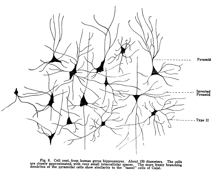
The structure of this part of the cortex displays certain peculiarities. The external layer of cells is not particularly well developed. The external layer of large pyramids, found in practically all parts of the cerebral cortex, are here represented by a layer of large stellate cells, among which a few large pyramids, mostly atypical, appear. The internal layer of large pyramids is present, and these pyramids are really giant cells. The seventh layer of polymorphic cells is rather well developed, both in the size of the cells and, in a certain degree, their number. The association tracts from the primary visual area are very intricate. This relationship of visual area is indicative of the important place in life which the visual coordinations fulfill. Fig. 8. Cell nest, from
human gyrus hippocampus. About 150 diameters. The cells are closely approximated,
with very small intercellular spaces. The more freely branching dendrites
of the pyramidal cells show similarity to the tassel cells of Cajal.
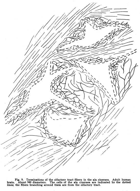
Whether there is any cortical area for the perception of colors has not yet been demonstrated. The phenomena of color vision present peculiarly baffling problems to the physiologist. The differences between colors, from the physical standpoint, is comparable to the differences between musical tones; that is, it should be perfectly proper to speak of blue as a higher or a faster shade of red, or of yellow as a lower or a slower shade of violet. Such ideas are primarily absurd in consciousness, in which these colors are recognized as qualitatively different. The nerve impulses carried by the optic tracts and the resulting activities of the cortical neurons must be identical. How qualitative differences can be based upon quantitative variations in vibration rate is most puzzling. Visual impulses originate in the retina. The structures of the eye are complex, and may be functional in causing considerable variations in the nature of the impulses sent from the retina and from the essential qualities of the objects seen; in other words, while the peripheral sense organs are supposed to translate environmental qualitative and quantitative variations into the language of nerve impulses, the eye seems to use considerable latitude in the translation, so that a very free and idiomatic translation may be made. Thus we have sent to the cortical areas the impulses concerned in the consciousness of qualitative color senses based upon the quantitative variations in vibration rate, that puzzling problem to which reference has already been made. Fig. 9. Terminations
of the olfactory tract fibers in the ala cineraes. Adult human brain. About
940 diameters. The cells of the ala cineraes are indicated by the dotted
lines; the fibers branching around them are from the olfactory tract.
Primarily, vision is of a flat, plane surface. The
ideas of distance, form, size, space in three directions, and the other
ideas ordinarily supposed to be primarily derived by means of visual impulses,
arise as the result of the activity of the overflow and intermediate areas.
The Cheselden case was reported in 1727. A child
born blind was couched when he was between thirteen and fourteen years
of age. When the bandages were removed from his eyes he thought things
seen touched his eyes. He saw only plane, colored surfaces. Later, having
forgotten the name of a certain animal, he picked it up and said, Puss,
so I shall know you next time. The visual impulses seemed not to be associated
at first with any ideas of solid form, or of the names of things. Evidently,
the process of relating the activities of the visual areas to other areas
is a matter of a certain length of time.
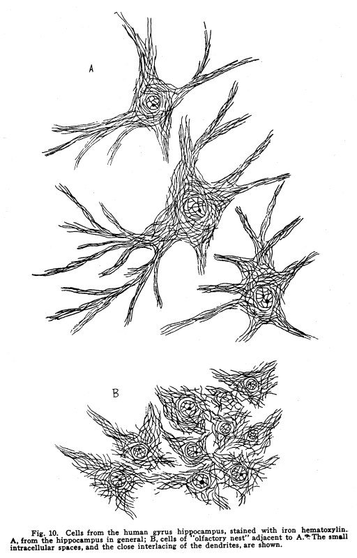
A man, having been couched, began to see for the first time. He experienced great difficulty at first in learning to eat. For some time he found it difficult to restrain the fear associated with the sight of an approaching fork or spoon toward his face as he fed himself. Another patient, under the same circumstances, displayed the most active delight in the colors first seen. Red, especially, filled him with a sort of joy. He saw red roses upon a bush in the yard. He did not, of course, recognize them, but he did so greatly admire their color that he was with difficulty restrained from climbing out of the window in order to examine them more closely. His incomplete coordinations failed to warn him of the danger of climbing from a second-story window into a rosebush. Fig. 10. Cells from the human gyrus hippocampus, stained with iron hematoxylis. A. from the hippocampus in general; B, cells of olfactory nest adjacent to A. The small intracellular spaces, and the close interfacing of the dendrites, are shown. Such instances illustrate the fact that primarily
a colored surface only is seen, and that the complex knowledge we think
ourselves to receive by sight is, in fact, the result of the primary visual
sensations qualified and modified by the activities of the cells of other
areas of the cortex.
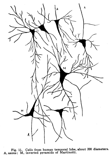
The visual overflow surrounds the primary visual area completely. Thus, the visual overflow is as extensive in comparison with the primary visual area as it possibly could be. It neighbors the auditory overflow toward the inferior part of its extent, and the somesthetic toward its superior extent. The structure of the visual overflow presents a type intermediate between the structure of the primary visual area and that of the typical cortex. The stratum zonale is very well developed and is very rich in cell structure. The association fibers are plentiful. The line of Bailarger is unusually well developed, both in the primary and the overflow of the visual cortex. The great development of the fiber tracts of this line is indicative of the great number and complexity of the relationships of the visual impulses. The cells of this area seem to be concerned in the storing and reproduction of the visual memories. The stimulation of the primary visual area by impulses of a sufficiently energetic or efficient character causes the activity of cells of the neighboring overflow. The activity of these cells, which probably include the cells of the stratum zonale, affects their metabolism in such a manner as to lower their liminal value. This decrease of liminal value appears to be permanent. Thus, the reception of impulses from either the primary visual area or from other cortical centers initiates their activity again. The initiation of the activity of the neurons of the visual overflow areas by impulses from the primary visual area is the origin of the visual memories; the stimulation of the cells of the visual overflow area by impulses from other overflow or intermediate areas causes the phenomenon of visual memories in consciousness. Fig. 11. Cells from human temporal lobe, about 200 diameters. A, same; M, inverted pyramids of Martinotti. The activities of the visual overflow areas are concerned in the recognition of visual impulses as they are repeated. Together with the areas intermediate between the visual overflow and the auditory and somatic overflows, the ideas of solidity, distance, extension in three directions and various other complex ideas are able to be interpreted in consciousness. These are discussed more fully in connection with the somesthetic and the language areas. The primary and overflow visual areas give origin
to a number of tracts which relate the functions of vision and of visual
memories to practically all of the other parts of the cerebral cortex,
both of the same and the opposite sides. The very great importance of visual
images in intellectual development is thus apparent in the structure of
the hemispheres.
The environment is extended to the limits of vision. This, alone, has great importance from the biological standpoint. Animals are able to find food, and to protect themselves from danger much more efficiently on account of the visual impulses. By means of the visual overflow and the intermediate
areas the limits of the environment are indefinitely extended. The activity
of the intermediate areas, together with the effective motor impulses,
for example, invents lenses and other instruments, by means of which the
environment of civilized man is extended to the limits of telescopic vision
on one hand, and of microscopic vision on the other. By means of the intricate
and efficient fiber tracts of the cerebral hemispheres the impulses derived
from this infinite environment add to the wisdom and energy of living in
a sense almost beyond conception.
Visual impulses may be employed very efficiently both in education and in therapeutics. Facts may be stated through the intermediation of sight in a manner which affects t he activities of the intermediate and motor areas speedily and energetically. The cartoon as an educational factor owes its value to the fact that it presents its lesson in a simple, concrete form, which stimulates the overflow and intermediate areas much as they are stimulated by the presence of the objects pictured. The moving picture is extremely efficient in a similar manner. Great harm may be done by uncensored motion pictures. The more highly developed the visual cortex is, and the lower is the liminal value of the neuron systems which relate the visual cortex to the other areas of the brain, the greater are the numbers of associations which are concerned with the activities of the intermediate areas and the resultant activities of the motor areas. These activities are the basis for reason and judgment; hence, the more complex the visual relationships, the wiser the judgments and the more efficient the motor reactions of the individual. In dealing with patients who are, for nervous reasons,
not so obedient as they ought to be, it is often possible to secure more
exact compliance with instructions if they are written. In dealing with
certain neurasthenic and hysterical patients it is sometimes a good thing
to give them written orders. These are merely instructions concerning
food, exercise, bathing, etc., written upon paper and sealed in an envelope.
Upon the outside of the envelope is written the hour at which the prescription
is to be taken. Such methods are efficient in certain cases. It is not
usually worth while to try to appeal to the good sense of hysterical
or neurasthenic patients who are not obedient to instructions. If they
had any common sense they would either obey the physicians instructions
or go to some person whom they could better trust. But the use of such
methods of giving instructions in a manner which impels obedience and adds
the interest of curiosity is often a very good thing to do. The method
is especially adapted to children, and to adults of slightly deficient
development.
The primary cortical auditory area occupies the central part of the first temporal convolution, and probably the upper part of the second convolution. (Figs. 11, 12.) The auditory overflow extends posteriorly to meet the visual overflow of the occipital lobe, inferiorly to an extent not yet defined, and anteriorly and slightly inferiorly into an area which is concerned in the consciousness of musical tones. Neighboring the area for the consciousness of musical tones lies another related area, which may be called the musical overflow. In this area the memories of tunes probably are stored and coordinated. No uncomplicated lesions are described for the area concerned in the consciousness of musical tones or the memories of tunes, but lesions involving these areas seem to affect the power of appreciating tones and tunes. Persons so affected become tone deaf, and this condition may occur without any loss of hearing as a primary sensation. It is in harmony with the facts of physiological
action elsewhere in the nervous system if it should be found that people
who have no ear for music have the neurons of this area either undeveloped
or of faulty structure. The study of the brains of musicians is also needed
in order that such relationships may be determined.
The areas for the appreciation of musical tones extend toward the olfactory areas. No associations are recognized between these senses, as is the case between the visual overflow and the auditory overflow, or between the visual overflow and the sensory overflow, but both are intimately associated with the lower centers. The appreciation of noises does not affect the emotional centers or the affective states, but musical tones are very efficient in arousing emotional reactions. For this reason music is employed as it is in social affairs, in churches, and under all conditions in which it is desired to bring feelings into play. No direct association path exists between the anterior
temporal area, the musical area, and the motor areas. The consciousness
of musical tones, the memories of tunes and the appreciation of the significance
of music arouse no marked motor reactions, but appear, on the other hand,
to inhibit whatever motor reactions might be aroused by the activity of
other cortical area. It is recognized that music exerts a restful influence
upon the body, that it is preeminently adapted to the quieting of those
who are inclined to listen with pleasure to it. The physiological basis
of this fact is to be found in the absence of direct paths for the transmission
of the impulses from the centers for musical tones and memories to the
motor areas.
The auditory impulses originate in the cochlea. The various structures which transmit the vibrations to the fibrillae of the dendrites of the auditory neurons of the first order appear to modify the amplitude and perhaps the force of the vibrations, but not to modify the essential qualities of vibrations; that is, the vibrations remain as such, and no structure appears to have for its function the translating of vibrations into any other sort of reaction quality, as is the case with certain other sensations. Sound waves are recognized as sound waves in physics, and the physical phenomena of sound coincide with the auditory sensations in consciousness sufficiently for us to realize the relationship between sounds as heard and sounds as subject to the laws governing the vibrations which produce sounds. The cells of the cochlea send axons as acustic nerves
to the acustic nuclei; these in turn send axons to the nuclei of the trapezoid
body and the superior olive, the inferior quadrigeminates and the internal
geniculate body. The axons of the acustic radiations transfer the impulses
thus carried to the first and perhaps the second temporal convolutions.
It is not known whether the impulses are transferred by means of all of
the nuclei mentioned or not. There is reason to believe that at least a
part of the lateral fillet fibers pass without relay from the acustic nuclei
of insertion to the internal geniculate body. There is no reason to suppose
that the medial fillet fibers, or at any rate more than a very few fillet
fibers, pass into the acustic radiations directly.
The auditory cortex differs slightly from that of other cortical areas. Perhaps the most conspicuous difference is found in the length of the radiations. These fibers extend into the external layer of cells, instead of stopping in the neighborhood of the line of Bailarger. The stratum zonale is rather less pronounced in the primary auditory areas than in the auditory overflow. This structure resembles that of the visual cortex and visual overflow. The pyramids of the auditory area are not so large as those of the motor area, nor as those of the internal layer of large pyramids of the visual area. The external layer of large pyramids is well represented; the pyramids are not so typical in outline as those of the motor area, but the stellate cells of the corresponding layer of the visual area are not found. The length of the radiations, permitting the impulses to be carried without relay to the cells of the stratum zonale, is the basis for the fact that consciousness is so quickly affected by auditory stimuli. It is a matter of common experience that a noise arouses attention much more quickly than do other sensory stimulations. A flash of light or an odor fail to attract the attention in so great a degree as do sounds, and even when such stimuli are pronounced enough to arouse forced attention, the reaction is less rapid than in the case of the sounds. The same condition exists in the area for the appreciation of musical tones. The enjoyment of music seems to be more directly primary in its nature than is the enjoyment of the activities of other sensations, except those of the body itself. Doubtless this depends in part upon the length of the radiating fibers. The primary auditory areas and the auditory overflow
which lies posterior to the primary area are closely associated with the
motor areas by the long tracts. Incoming auditory impulses affect the motor
areas quickly, as is evident in the phenomenon of attention and in the
relationships underlying the speech functions.
The biological value of the auditory impulses lies chiefly in the facts, first, that the auditory radiations reach the stratum zonale, and thus are able to arouse consciousness and motor reactions very speedily, and, second, in the fact that the environment of the individual is so greatly increased by the auditory impulses. The overflow areas are less extensive than they are in the case of the visual cortex, and the association tracts from the temporal lobes to other parts of the brain are less complicated and widespread than in the case of the visual cortex; yet the auditory are and its connections are of considerable importance from a biological standpoint, as well as from the standpoint of their relationships in the control of human life. The development of the auditory cortex depends upon the stimulation of the cortical neurons by impulses from the lower centers. The stimulation of the primary auditory area affects the cells of the auditory overflow, including the cortical areas for musical tones. The activities of the cells of the overflow area are associated with memories of sounds and with the simpler coordinations of the significance of things heard. The activities of the cells of the overflow areas are associated, in turn, with increased activity of the cells of the adjacent intermediate areas. These are functional in the coordination of the impulses initiated by the overflow activities, and are associated in consciousness with the correlation of the memories and the abstract ideas built upon the auditory memories and the significances of things heard. |