
|
The centers of the upper thoracic cord lie within the gray matter of the second, third, fourth, fifth and sixth thoracic segments. In part, the uppermost of these centers extend into the gray matter of the first thoracic and the seventh cervical segments. The cells of the lower thoracic centers intrude upon the fifth and sixth segments to a certain extent. The gray matter of the upper thoracic segments is rather small in extent. The posterior horns are long and slender. The cap of substantia gelatinosa is wanting, or nearly so. The nucleus dorsalis (Clarkes column) is well marked. The lateral horn is well developed. The fibers of its small, multilateral horn is well developed. The fibers of its small, multipolar cells make up the white rami communicantes, which leaves the cord chiefly by the anterior roots, but also to a certain extent by the posterior roots. The viscero-motor centers are in this part of the gray matter. The anterior horn is small, with a narrow head. It contains only the column of cells homologous with the mesial cell group of the lumbar and cervical regions. These cells send their axons to innervate the muscles of the trunk, including the following: Semispinalis dorsi, multifidus spinae, spinalis dorsi, intertransversales, rotators spinae, interspinales, longissimus dorsi, accessorius, levatores costarum, the intercostals of the corresponding segments, serratus posticus, and a few fibers from the fifth and sixth enter the upper part of the external oblique. The anterior horn cells of this group of segments are of comparatively great age, phylogenetically; they are closely associated with the cells in other regions of the gray matter, and they are thus subject to considerable variation in activity and to abnormal especially from the viscera innervated from the same segments. The approximation of the ribs over the affected areas in pulmonary tuberculosis is recognized even by physicians who fail to see the relation in its full significance. The somatic motor centers are governed by impulses from the following sources: I. Somatic and visceral sensory impulses affect the tone of the muscles controlled from these segments. II. Descending impulses from the medullary centers, especially the respiratory center, bring about the coordinate movements of respiration, etc. III. Descending impulses from the cerebellum, vestibular nuclei, centers of the reticular formation, etc., maintain the tone of the muscles and coordinate their action, especially with reference to the maintenance of the erect and symmetrical position. IV. Impulses from the precentral convolutions of the cerebral cortex carry volitional impulses to these centers. Volitional control of the thoracic muscles is not absolute. The autonomic control is more efficient. V. Descending impulses from the red nucleus and related ganglia bring the thoracic muscles under the control of the emotional states. The
viscero-motor cells of these segments lie in the lateral horns. Their
axons pass, mostly by way of the anterior roots and the white rami, to
the sympathetic chain. Their further course must be described with
the action of the specific centers.
This center includes a number of cells extending from the sixth or seventh cervical segments to the fourth thoracic. It seems to be most fully represented in the neighborhood of the second thoracic segment. This center is controlled by impulses from the following sources: I. Somatic sensory impulses reaching these segments may affect their activity. This is especially noted in the dilatation of the blood vessels and the pupil of the eye as a result of irritation over the back of the neck and the upper thoracic spines. Bony lesions of this region also produce the same effect. II. Viscero-sensory impulses reaching the same segment exert a slight effect. This reaction is not always found in animals. It is evident that no tests could be made upon human beings, and the clinic evidence is not sufficiently exact to warrant any statement in regard to the matter. III. Descending impulses from the quadrigeminates and related centers in the medulla, pons and midbrain affect this center. IV. Descending impulses from the red nucleus and related ganglia bring the action of the cilio-spinal center under the control of the emotional states. The secretion of tears and dilatation of the pupils in grief and anger, etc., illustrate this action. The
fibers from the cilio-spinal center leave the cord with the white rami
of the second and third thoracic anterior roots. They enter the sympathetic
cord, and pass without relay to the superior cervical ganglion. (Fig.
40.) Here they break up into many fine branches and enter into the
formation of the pericellular baskets of this ganglion. The number
of sympathetic cells which are thus controlled is not known. The
axons of the sympathetic cells are non-medullated fibers which pass to
the structures of the orbit. Some of them, probably vaso-constrictors,
follow the carotid artery and control the size of the blood vessels of
the orbital structures. Others reach the semilunar ganglion (Gasserian
ganglion) and are distributed with the long ciliary nerves to the orbital
structures. None of these fibers have physiological relations with
the ciliary ganglion. The structures thus innervated are: the
blood vessels of the orbit, the tear glands, the non-striated muscle fibers
of the levator palpebrarum, the dilator of the pupil, the non-striated
muscle fibers of the capsule of Tenon.
The centers of the control of the cranial blood vessels and viscera are placed in the gray matter of the first, second, third and fourth thoracic segments. The cells may intrude upon the seventh cervical and the fourth thoracic segments. The axons of these cells pass chiefly by way of the anterior roots, but perhaps a few by way of the posterior roots, and the white rami communicantes into the sympathetic cord. These fibers then follow the sympathetic chain to the superior cervical ganglion or to the smaller cranial ganglia, especially those of the carotid plexus. In these ganglia the fibers break up into fine fibrillae, which enter into the formation of the baskets around the bodies of the sympathetic cells. The axons of the sympathetic cells are distributed partly with the branches of the carotid artery and partly with the cranial nerves to the blood vessels of the cranial structures. The
presence of vaso-motor nerves to the brain has been long in dispute.
Late tests seem to demonstrate beyond doubt the existence of vaso-motor
nerves to the brain substance. Their existence in the meningeal vessels
has been recognized for some time.
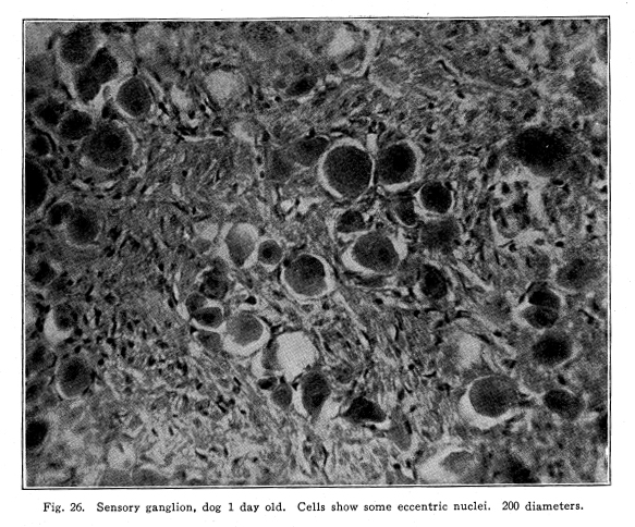
The exact relationship between the secretory influence of the cranial nerves and of the centers in the upper thoracic cord has not been demonstrated. The vaso-constrictor control of the salivary, buccal and pharyngeal glands is evidently placed in the upper thoracic cord. The physiology of the delators, and the secretory action as independent of the vascular control, is yet to be studied. The activity of the centers controlling the circulation through the cranial structures directly is influenced by impulses from the following sources: I. Descending impulses from the vaso-motor centers in the medulla control the size of the cranial vessels. (It must not be forgotten that the indirect control of the cerebral circulation by changing the general arterial pressure is probably more efficient and active than is the direct action here described.) II. Sensory impulses from all the structures innervated from the first to the fifth thoracic segments affect the activity of these centers. In this way the bony lesion, the irritation of diseased viscera, and, to a slight extent, the irritation of the skin, may interfere with the circulation through the cranial structures. III. Descending impulses from the red nucleus, etc., bring the cerebral circulation under the control of the emotional states. Pallor and blushing, the erection of the hair in fright, illustrate this reaction. Certain facts of clinical finding seem to indicate the possibility that the phenomena of hysteria and related states may depend upon the localization of excessive vaso-contriction in certain brain areas. This seems to be due to unbalanced and excessive activity of the basal ganglia under emotional storms. Fig. 26. Sensory ganglion, dog 1 day old. Cells show some eccentric nuclei. 200 diameters. The
relationship is not demonstrated, but the possibility should be borne in
mind in treating such cases.
The centers controlling the circulation through the structures of the throat lie in the second, third, fourth and fifth thoracic segments. They may intrude upon the first and sixth thoracic segments. The axons of the lateral horn cells concerned in the action of these centers leave the cord chiefly by way of the anterior roots and enter the thoracic cord. The individual fibers follow the cord until they reach the middle or superior cervical ganglion. Here they form synapses with the cells of these ganglia. The gray fibers, axons of the sympathetic cells, pass with the cranial nerves to the area of their distribution. For the most part they are associated with the sensory nerves; rarely they follow the purely motor branches. The blood vessels of the visceral muscles, the glands, the skin, and the skeletal muscles of the neck and throat are thus innervated. The centers just mentioned are influenced as follows: I. Descending impulses from the vaso-motor centers in the medulla exert the most pronounced influence upon them. II. Sensory impulses from the somatic and the visceral structures innervated from the same segments affect their action to a certain extent. This relation is especially noticeable in connection with the pharyngeal and laryngeal tissues, as they are affected by abnormal muscular contractions of the upper thoracic posterior spinal muscles, and by slight malpositions of the upper thoracic vertebrae or by the first and second ribs. Hyoid malpositions are also often concerned in affecting the activity of the centers controlling the throat structures. III.
Descending impulses from the red nucleus and associated ganglia affect
the action of these centers. Thus the emotional states may be responsible
for dilatation of the vessels of the cervical tissues, especially of the
laryngeal tissues. The thyroid seems sometimes to be greatly affected
in this way.
The
control of the secretions of the thyroid, parathyroids, and other glands
of the cervical region is not exactly known. The few facts of clinical
and experimental evidence seem to indicate that secretory nerves follow
the paths and have the central connections of the vaso-motors.
The centers for the control of the circulation through the upper limbs and the shoulder girdle are found in the gray matter of the second, third, fourth, fifth and sixth thoracic segments. The cells may extend into the first and the seventh thoracic segments. Rarely there is found evidence of cells capable of affecting the circulation through the arms as low as the eighth thoracic or as high as the seventh cervical segments. The fibers concerned in this function leave the spinal cord by way of the anterior roots of the third, fourth and fifth segments, chiefly, and enter the sympathetic cord. (Fig. 42.) They pass without relay to the ganglion stellatum, rarely to the middle cervical ganglion. They terminate by entering into the formation of the pericellular baskets of the sympathetic. The gray fibers, the axons of these sympathetic cells, pass as gray rami communicantes to the nerves of the brachial plexus. They are distributed with these nerves to the muscles, bone and bone marrow, articular surfaces, and skin of the shoulder girdle, arm, wrist, hand and fingers. No vaso-motors are found in the palmar surfaces of the tips of the fingers, and they are scantily supplied to the palms. The action of these centers is affected by impulses from the following sources: I. Descending impulses from the vaso-motor centers in the medulla. II. Sensory impulses from the structures innervated from the same spinal segments. Thus visceral irritations may somewhat affect the circulation through the hands, especially, and malpositions of the upper and midthoracic vertebrae may cause phenomena similar to the symptoms of Raynauds disease, if not identical with them. III. Descending impulses from the red nucleus and other basal ganglia affect these centers. Thus the circulation through the upper limbs may be affected by emotional states, and the hairs of the arms, especially, may be erected through the action of the pilo-motors. IV.
These centers are affected through association cells in such a manner that
during the active contraction of the muscles the circulation through them
is greatly increased. Whether this reaction is due to dilators or
the inhibition of constrictors, or whether it is caused through segmental
reflexes or through the activity of the general vaso-motor centers, is
yet doubtful.
The centers concerned in increasing the rate and force of the hearts beat are placed in the lateral horns of the gray matter of the second, third, fourth and fifth thoracic spinal segments. Perhaps the cells of the first and the fifth may be concerned in the same function to a certain extent. The fibers from these cells leave the cord chiefly by the anterior roots and pass as white rami communicantes into the sympathetic chain. The fibers pass without relay to the cervical region, where they terminate by forming synapses with the cells in the superior, or the middle, or the inferior cervical ganglion. From these ganglia the gray fibers, axons of the sympathetic cells, pass to the trunk of the vagus nerve, and with that nerve they reach the cardiac plexus. It is not probable that the fibers from the sympathetic form synapses with intrinsic cells of the cardiac ganglia, though this has not been certainly determined. The blood vessels of the heart probably receive vaso-motor fibers by the same pathway. The existence of vaso-motor fibers to the cardiac vessels has not been experimentally demonstrated, but certain clinic phenomena seem to indicate that they are present, and that they are derived from the spinal segments and carried by the path already described. The cardio-spinal centers are influenced by impulses derived from the following sources: I. Descending impulses from the chief cardiac center in the medulla. The action of the accelerators and the inhibitor of the vagus is thus coordinated. II. Descending impulses from the vaso-motor center in the medulla. In this way the arterial pressure is maintained at an equable height during health, and the tendency is to bring it to the normal height in disease. III. Descending impulses from the red nucleus and related ganglia. By this means the emotional reactions exert a very profound influence upon the hearts action. IV.
Sensory impulses from the heart itself and other structures innervated
from the corresponding spinal segments. By this means the sensory
impulses from disordered viscera innervated from the same or adjacent segments,
or from joint surfaces abnormally placed, or from abnormally contracted
muscles, may initiate abnormal functions of the cardiac muscle, and probably
at times abnormal dilatation or constriction of the cardiac vessels.
The centers governing the blood vessels of the pleura and the lungs lie in the lateral gray matter of the first, second, third, fourth, fifth, sixth, and perhaps the seventh thoracic segments. The relationships experimentally determined indicate a tendency to a segmental innervation of these blood vessels, and this segmentation is more noticeable in the pleura than in the lung substance. The axons of the cells of these centers leave the cord chiefly by the anterior roots, proceed as white rami into the sympathetic cord, and terminate within the ganglion stellatum or the lower or the middle cervical ganglion by entering into the formation of the pericellular baskets of the sympathetic. The axons of the cells thus surrounded pass in part by way of the vagus and in part by way of the aortic plexus to the blood vessels of the pleura and the lungs. The centers are under the influence of nerve impulses from the following sources: I. Descending impulses from the vaso-motor center in the medulla. II. Descending impulses from the respiratory center in the medulla. III. Descending impulses from the red nucleus and the other basal ganglia. IV. Sensory impulses from the lungs and pleura, and from the other visceral and the somatic structures innervated from the same spinal segments. In this way the bony lesions are often concerned in the initial disturbance of the circulation, and the infection of the injured area becomes probable. It is not to be forgotten that the control of the pulmonary circulation indirectly through changes in the tension of the systemic arterioles is probably more efficient than the action of the direct vaso-motor control here described. |