
The simplest method is the use of a protective shield
to encircle the tube, permitting the exit of the rays only through a small
aperture that may be regulated as required.
In addition the use of leadfoil or sheet lead to
cover all parts which are to be protected from the ray is customary.
The Fluoroscope. In order to see the findings
of the X-ray a screen is employed containing barium platinum cyanide, a
substance which shines or fluoresces when exposed to the X-ray. Interposing
an object between this screen and the X-ray tube produces a shadow on the
screen commensurate with the amount of the ray which has been prevented
from reaching the screen. We have, therefore, a shadow picture showing
the relative density of the object traversed by the X-ray.
The Skiagraph. The rays act upon the bromide
of silver gelatin coating on photographic plates in the same manner as
ordinary light. When a plate takes the relative position of the fluoroscopic
screen the resultant picture is called a radiograph, or skiagraph, and
affords a permanent record of the condition shown.
The X-ray plates have a heavier coating than ordinary
photographic plates and are enclosed in two envelopes so that they may
be handled in daylight. The flaps on the envelopes are on the nonsensitive
side of the plate. The plate is placed on the table, the part to be skiagraphed
resting on the plate and the tube some distance above, the target being
directly over the center or the part radiographed.
As the rays diverge from the point on the target
where they are produced, the image on the plate is always enlarged. The
nearer the part is to the tube the greater the magnification.
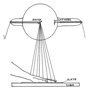
In order to obtain a picture without distortion of
the image the following rule must be kept in mind: An imaginary line
from the point on the target where the ray is generated to the center of
the plate, must be perpendicular to the plate and pass through the center
of the part skiagraphed.
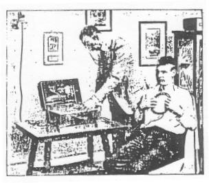
Fig. 63 show the proper relationship of the tube,
plate and hand for a skiagraph of the latter.
The length of exposure is from a few seconds to
several minutes, according to the apparatus employed and the density of
the parts. A few experimental pictures will enable the physician to determine
the approximate time for his individual outfit.
The method for developing is the same as for ordinary
photographic plates, but takes much longer, averaging about ten to twenty
minutes.
Dental Films. For skiagraphs of teeth, a
film is used. Two small films wrapped in two opaque paper coverings is
the way they are supplied to the doctor. The film is held inside the mouth,
back of he teeth, the smooth side of the paper toward the tube. Head is
adjusted so that the line from target to film is in accordance with the
rule given above. With small machines it will be necessary to experiment
to get the time of exposure. It will average 30 to 60 seconds. The films
are developed, washed and dried; the one retained by the radiographer,
the other by the patient.
Diseases Grouped According to Technique.
In treating with the X-ray the average number of treatments is three per
week. The length of exposure during the first two weeks should not be over
five minutes each time to guard against possible idiosyncrasy to the ray.
After two weeks the treatment may be lengthened to
seven, or, in some cases, ten minutes, and continued until improvement
takes place or the characteristic reaction appears.
In the former instance, the frequency of the treatment
is gradually decreased; in the latter it is suspended entirely for a few
treatments until the signs of dermatitis have subsided, when it is resumed
as before, providing the evidences of disease have not disappeared with
the reaction.
With a low tube the tube-wall is from five to eight
inches from the surface treated; medium tube eight to twelve inches; high
tube twelve to twenty inches.
A number of diseases suitable for X-ray treatment
are given herewith, grouped according to the vacuum of tube best suited
to their treatment. The lower the tube the quicker the reaction produced.
Some diseases are included under two headings., where it is a matter of
choice, either method yielding results.
X-ray Burns. An X-ray burn or dermatitis is
the result of an overdose of the ray. The earlier symptoms are itching,
redness and pigmentation. By keeping these in mind it will be possible
to avoid severe burns.
Mild burns should be let alone and they will subside
of their own accord. In severe forms the condition is an X-ray gangrene
or necrosis and calls for surgical measures.